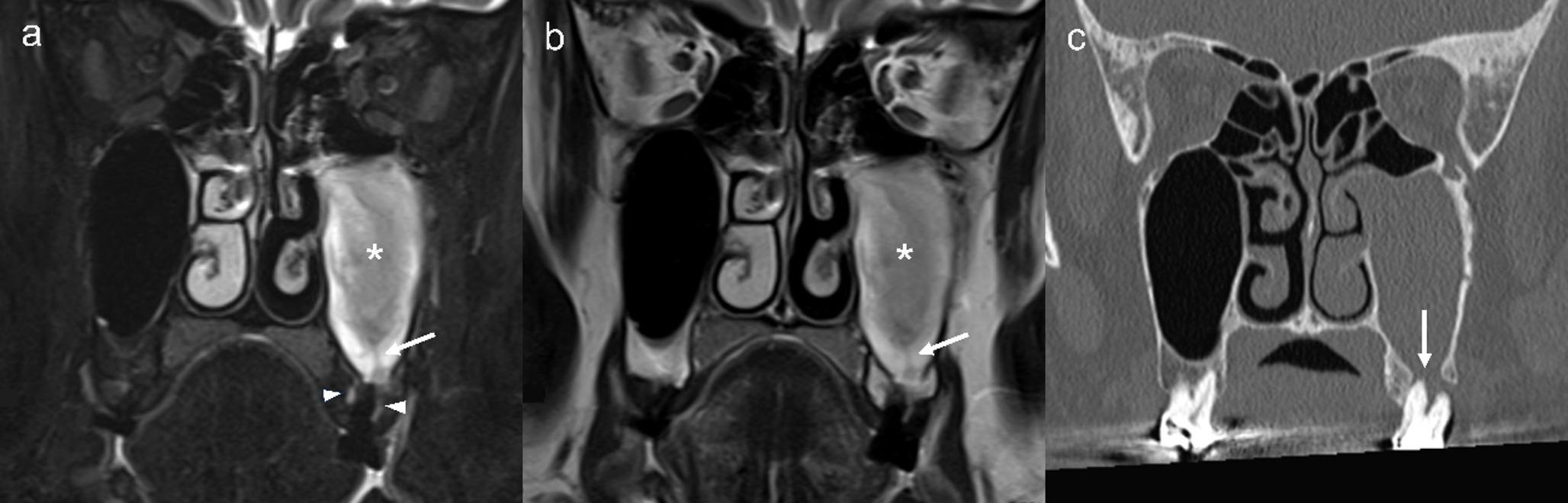
Imaging-based anatomical study of torus mandibularis: morphological features identified by computed tomography and their correlation with panoramic radiographic appearances
Objective
The anatomical characteristics of torus mandibularis (TM), particularly its vertical dimensions, remain incompletely understood. This study was performed to clarify the detailed anatomical features of TM using computed tomography (CT), including cone-beam CT (CBCT) for dental use, in relation to tooth and mandibular positioning. Additionally, it sought to clarify the radiographic appearance of TM on panoramic images.
Methods
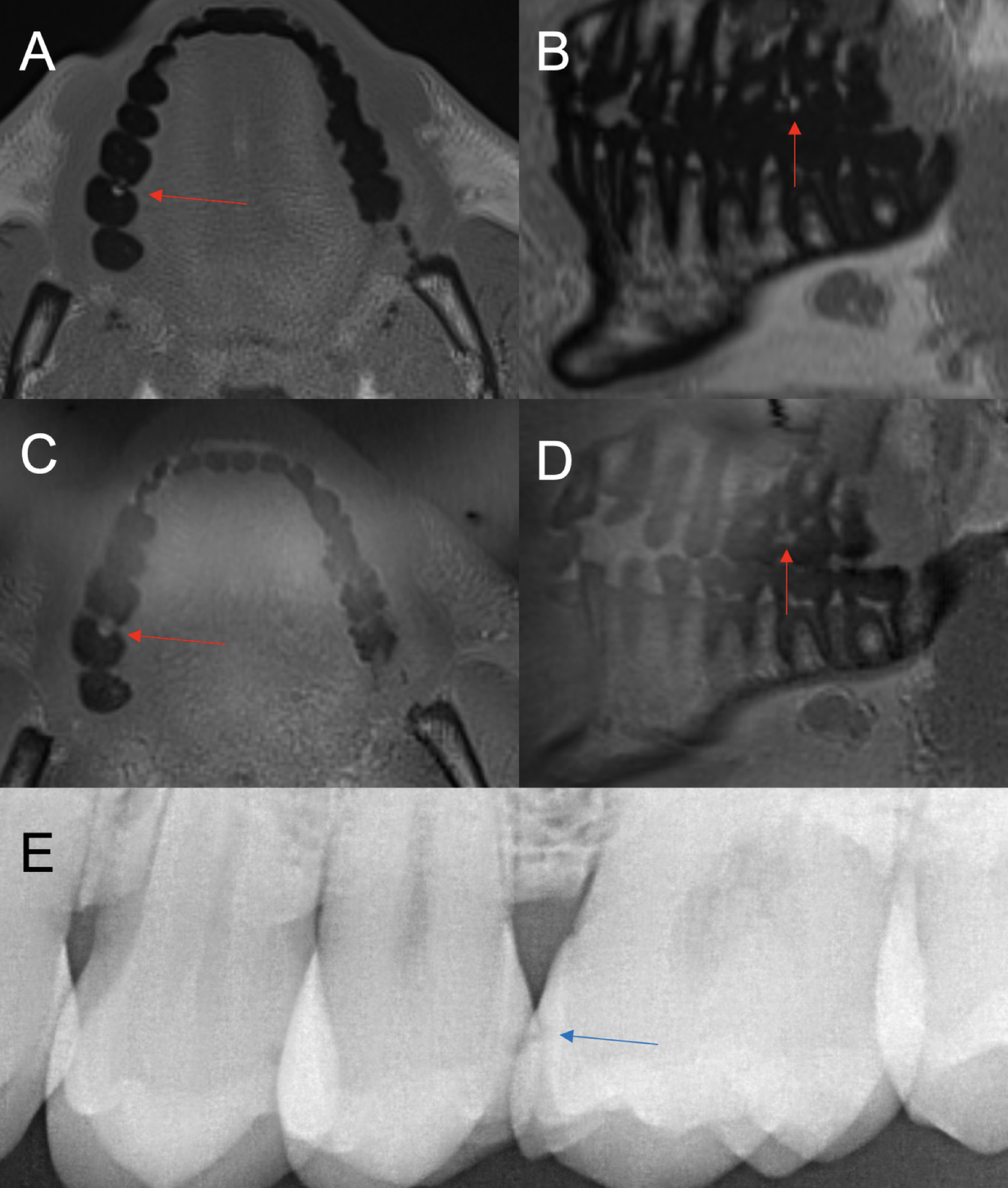
Fifty patients with TM (10 per age group: 20s, 30s, 40s, 50s, and 60s or older; 5 men and 5 women in each group) who had undergone both CT and panoramic radiography were included. Cross-sectional CT images were analyzed by dividing the mandibular area into 25 horizontal locations and 6 vertical levels (totaling 150 regions). Cortical thickness was measured using CT value profiles. On panoramic radiographs, the detectability of TM was assessed in the corresponding 150 regions. Measurement reliability was also evaluated.
Results
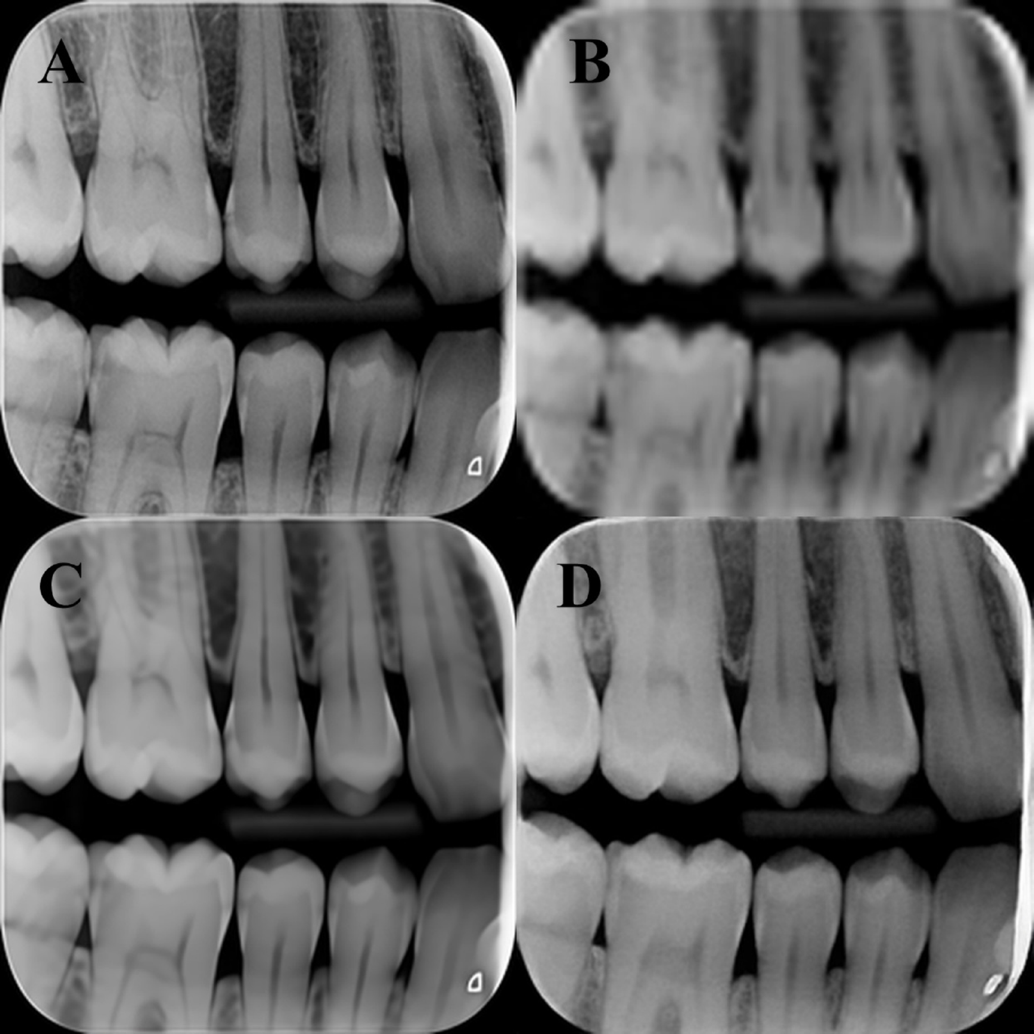
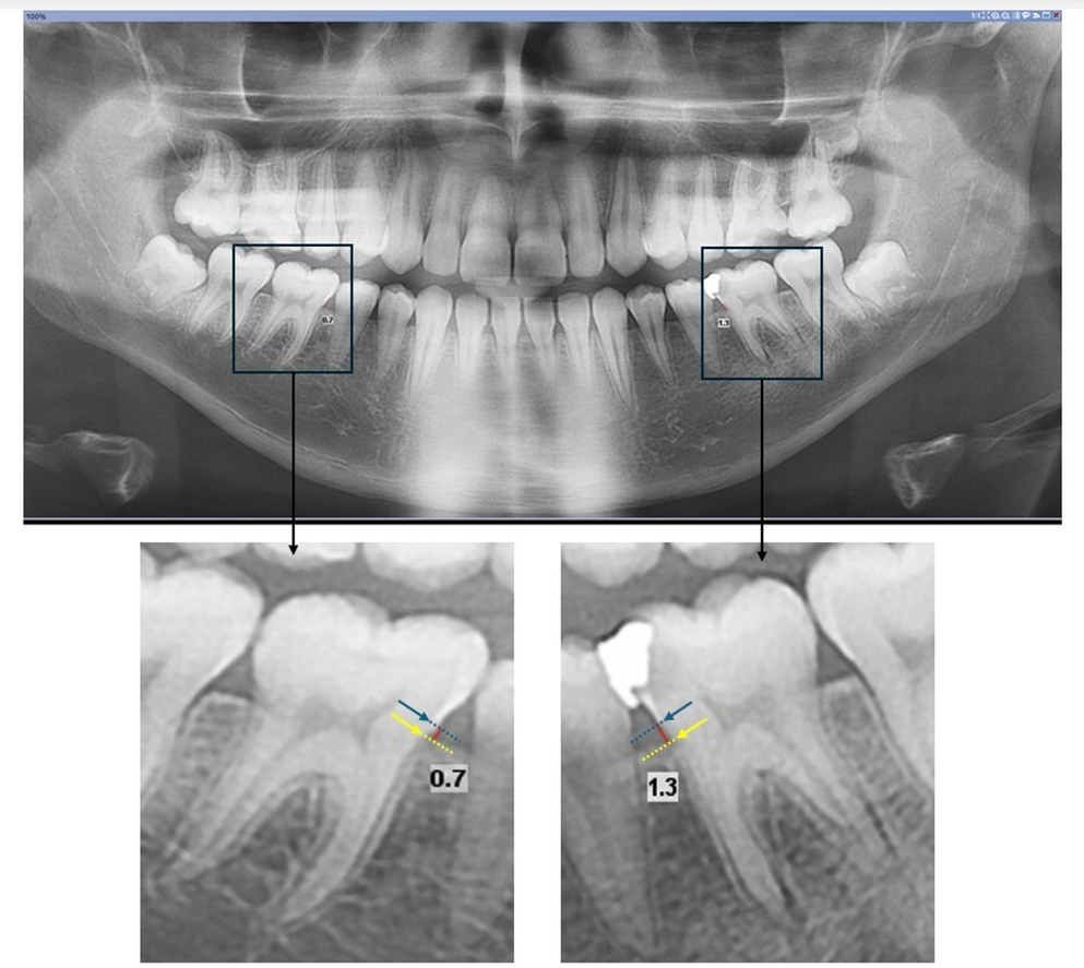
Measurement reproducibility and agreement between CT and CBCT were confirmed to be sufficient. At the 1/8 vertical level—just below the alveolar ridge—21 of 50 horizontal locations (42%) showed a mean cortical thickness of ≥ 4 mm, whereas only 6 locations (12%) at the 2/8 level met this threshold. Radiopaque findings on panoramic images were consistently located above the 3/8 level.
Conclusion
Cortical thickness exhibited symmetrical distribution, with the greatest values observed just below the alveolar ridge, particularly between the canine and second premolar. The detectability of TM on panoramic radiographs closely corresponded with its anatomical features as visualized on CT images.

Comments (0)