Establishment of the skeletal muscle dysfunction COPD models
In this study, 5-month-old male Sprague Dawley rats (SD rats, purchased from Hunan Xu Zhi yuan He Biotechnology Co., Ltd.) were used to establish chronic obstructive pulmonary disease (COPD) models of skeletal muscle dysfunction. All rats were kept in a specific pathogen-free facility where standard food and water are freely available. Before the experiment performance, the rats were maintained in a standard breeding environment (23 ± 2 ◦C, a 12:12 h light: dark cycle) for one week of acclimatization. All the research protocols of our study were approved by the Institutional Animal Care and Use Committee of the Second Xiangya Hospital, Central South University, based on NIH Guide for the Care and Use of Laboratory Animals (National Institutes of Health Publications No. 80 − 23, revised 1978), (approval No. 2021226).
SD rats were randomly divided into a negative control group (NC, n = 8) and a COPD model group (COPD, n = 24). The NC rats were exposed to fresh air, while the COPD rats were subjected to cigarette smoking exposure in an 8050 inhalation exposure system (6–8% of smog concentration, Tianjin, China). The smoke generated by commercially filtered cigarettes (trade name: Baisha, containing 0.8 mg of nicotine, 11 mg of tar, and 11 mg of carbon monoxide per cigarette) was directed into the system to achieve the condition of passive exposure. The COPD rats were daily subjected to two 1-h periods of CSE, twice per day at an interval of 6–8 h, 6 days per week for 24 weeks (Qin et al. 2022).
Lung function evaluation
The small animal lung function testing system (PFT-M, Beijing Twintech Intelligent Technology Co., Ltd., China) was used to analyze the lung function of the rats in different groups. In short, after the last CSE treatment had finished for 24 h, rats were fasted for 12 h and then anesthetized with 10% chloral hydrate. When the righting reflex and pain stimulus response disappeared, the rat trachea was cut open and connected to PFT for lung function testing after preparation and fixation. Measure forced vital capacity (FVC), forced expiratory volume at 200ms (FEV200), and maximum mid-expiratory flow rate (MMEF), and use them to evaluate lung function in the studied rats (Zhang et al. 2019; Su et al. 2020).
Implementation of HIIT and MICT training
After 5 days of treadmill adaptation training, the maximum exercise capacity test was conducted: the rats were introduced into the treadmill (tilted at a 15 ° angle and running at a speed of 15 m/min) and accelerated at a rate of 2.0 m/min every 2 min until they could not stand on the shock net even after receiving a 2-second electric shock. The training duration and maximum running speed (Vmax) were recorded. Under the same conditions (15 ° angle and running at a speed of 15 m/min), and then maintained it until the rat cannot stand on the shock net even after receiving a 2-second electric shock. Recorded the training duration. e highly selective BRD4 inhibitor JQ1 in vivo experiments which was given intraperitoneally at 30 mg/kg twice daily for 14 consecutive days, concurrently with a two-week HIIT training.
Both HIIT and MICT training were conducted 8 times within 2 weeks (Motiani et al. 2017). The HIIT training: Rats underwent 8 training sessions within 2 weeks, each consisting of 4 segments of 4-minute high-intensity training and 5 segments of 3-minute moderate-intensity training. The high-intensity training ran at 90% of maximum speed, while the moderate intensity training ran at 60% of maximum speed. Before and after the training, there was a 10-minute low-intensity training session at 40% of maximum speed as a warm-up. The HIIT single exercise volume was approximately 4 × 4 min × 90% × VO2max + 5 × 3 min × 60% × VO2max = 23.4VO2max. The MICT training consisted of 30 min of high-intensity running at 80% maximum speed. Before and after the training, there was a 10-minute low-intensity training with running at 40% maximum speed as a warm-up or relaxation. The MICT exercise volume per session was approximately 30 min × 80% × VO.max = 24.0 VO.max.
To explore if BRD4 is one of the molecular targets of HIIT in alleviating COPD-induced gastrocnemius muscle dysfunction, the highly selective BRD4 inhibitor JQ1 was given to the COPD rats intraperitoneally at 30 mg/kg twice daily for 14 consecutive days, concurrently with or without a two-week HIIT training.
Measurement of relative muscle weight and Hind limb muscle strength
After anesthesia, the rats were fixed on an anatomical plate with their abdomen facing upwards and their hind legs naturally extended. The skin and subcutaneous tissue were separated to expose the gastrocnemius muscle, which was carefully separated along the muscle fibers direction using forceps. Cut off the separated gastrocnemius muscle from the attachment point and soak it in physiological saline to keep the muscle tissue moist. After absorbing the moisture on the surface of the gastrocnemius muscle, its weight was accurately measured and recorded in a table. The relative weight of the gastrocnemius muscle (gastrocnemius muscle weight/body weight x 100%) was calculated.
After anesthetizing the rats, we fixed them on the experimental table with their abdomen facing upwards and their hind legs naturally extended. Placed the grip strength meter horizontally, with its grid facing towards the hind limbs of the rat. Gently supported the hind limbs of the rat, allowing its hind paws to naturally grasp the mesh of the grip strength meter, and ensured that the rat’s hind paws firmly grasped the grip strength meter. Grasped the end of the rat’s tail and gently pulled it back horizontally, maintaining even and increasing tension until the rat’s hind paw releasesed the grip strength gauge. Recorded the maximum force value of the rat’s hind paw grip, i.e., peak force. Performed 5 measurements on each rat and took the average as the muscle strength value of the rat’s gastrocnemius muscle.
Histological staining and immunohistochemistry
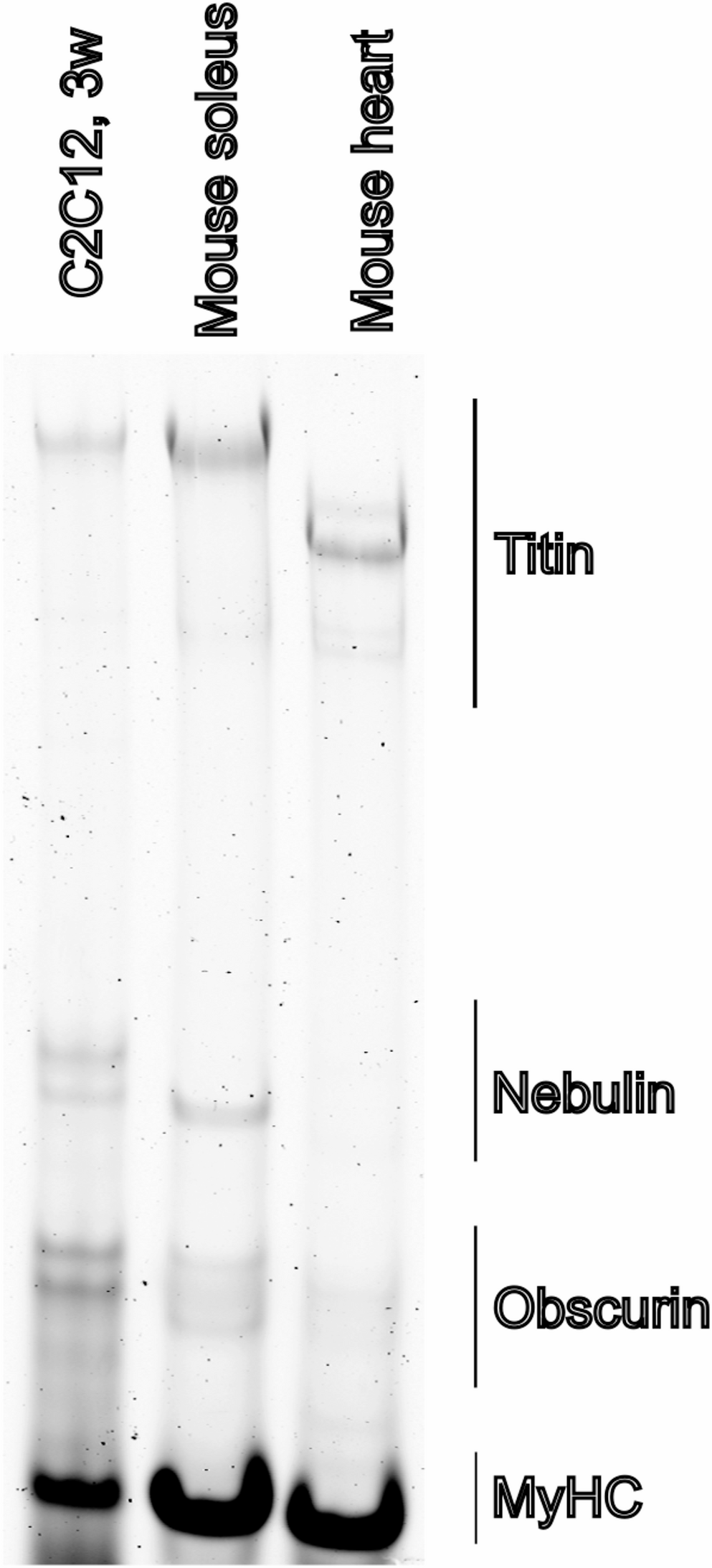
To assess pathological analysis, hematoxylin and eosin (H&E) staining was performed in the gastrocnemius tissues. The experimental steps were mainly based on the description of the previous reference literature (Su et al. 2020). Their morphologies were observed and photographed under panoramic scanning system (Panoramic MIDI, Hungary).
To investigate the changes in myofiber phenotypes in gastrocnemius muscle tissues to some extent, the immunohistochemistry (IHC) staining experiment was carried out as previously described (Ai et al. 2020). The MyHCI antibody and MyHCIIb antibody were added dropwise, and at the end of the experiment, all slides were observed and photographed under panoramic scanning system (Panoramic MIDI, Hungary).
Construction of cell models
The L6 myoblasts (CL-0136, Procell, authenticated by STR profiling) were cultured in DMEM (High glucose, PM150210, Procell), supplemented with 10% Penicillin-Streptomycin solution (PB180120, Procell), and 0.25 µg/mL of amphotericin B (no. 15240, Gibco) and 10% fetal bovine serum (FBS; no. 16420; Procell) until the cells reached about 90% confluence at 37℃(Abdelmoez et al. 2020; Gao et al. 2025).
L6 myoblasts were inoculated into a 6-well plate (2 mL/well) with a density of 2.0 × 10⁵ cells/mL and incubated overnight in a 5% CO2 cell incubator at 37 ℃. When the cells reached approximately 70–80% confluence, a gain-of-function cell model of BRD4 was achieved by transfecting 4 µg of a BRD4 overexpression synthetic plasmid (generalbiol, China) into the cells using 5 µL of Lipotectamine-3000 (L3000008, invitrogen). After 48 h, the successful overexpression of BRD4 in L6 cells was confirmed by QPCR and western blot.
To prepare CSE solution for this study, an unfiltered cigarette containing 0.8 mg of nicotine and 11 milligrams of tar was lit, and the smoke was continuously drawn into PBS solution (10 ml) using a negative pressure suction device. After purification through a 0.22 μm filter, the solution was characterized as a 100% CSE solution. In order to further construct the COPD cell models, the L6 cells (NC-Ov, BRD4-Ov) were treated with above CSE solution at a final concentration of 10% for a predetermined duration (Li et al. 2023; Liu et al. 2022b).
Bioinformatics analysis
For the sake of explore the differentially expressed genes in the gastrocnemius muscle tissues with chronic obstructive pulmonary disease (COPD) preliminarily, we initially resorted to public datasets and achieve our goals through performing an integrated bioinformatics analysis of the data from the datasets GSE197463 (RNA-Seq transcriptomic data of gastrocnemius muscle tissues from wild type and COPD C57BL/6 mice. Con: n = 5, COPD: n = 5) and GSE18033 (Expression profiling by array data of gastrocnemius muscle tissues from wild type and COPD Female C57BL/6 mice, Con: n = 6, COPD: n = 6), resulting in a combined sample size of Con: n = 11 and COPD: n = 11. The bioinformatics analysis was standardized with COPD vs. Con as the comparison. ComBat was employed to adjust for batch effects across different platforms, encompassing probe-level microarray data and FPKM-converted RNA-seq expression values. Genes that meet the criteria of a P-value < 0.05 and |LogFC| > 1.0 were considered to have significant differences between the groups.
Real-time quantitative PCR
Total RNA was extracted from rat gastrocnemius muscle tissues and L6 cells using the RNAsimple Total RNA Kit (Cat. no. PD419, Tiangen, China), and then reverse-transcribed using the RevertAid First Strand cDNA Synthesis Kit (Cat. no. K1622, ThermoFisher, USA). The QPCR was performed in the Roche Light Cycler 480 system using SYBR Green Supermix (Cat.no.FP205, Tiangen, China). 3 independent experiments were carried out, and the relative quantification comparative Ct method was used to quantify the relative mRNA levels. The primer sequences were listed in Table 1. The QPCR measurement data are presented as the means ± SD (n = 3).
Table 1 The specific primer sequencesWestern blot assay
Gastrocnemius muscle tissues and cells in each group were lysed. BCA Protein Assay Kit (Thermo Fisher Scientific, MA, USA, Cat# 23227) was used to measure the protein concentrations. Equal amounts of protein were separated with SDS-PAGE and then blotted onto PVDF membranes (Millipore, MA, USA, Cat# ISEQ00010). Next, the PVDF membranes were blocked in TBS containing 5% bovine serum albumin (BSA, Sigma-Aldrich, MO, USA, Cat# A6003) and 0.05% Tween 20 for 1.5 h, then incubated at 4℃ overnight with the relevant primary antibodies. The primary antibodies used were listed in Table 2. The amount of the protein of interest was normalized to the densitometric units of GAPDH. After washing, the PVDF blots were incubated with HRP-conjugated secondary antibodies for 1 h. Protein expression was detected with ECL (Millipore, WBULS0500) by digital imaging systems. Finally, ImageJ software was used to analyze the band density and the statistical methods employed Student’s t-test. The WB densitometry data of protein bands are shown in Fig S1.
Table 2 The information of used antibodiesMitochondrial membrane potential (MMP)
The change in mitochondrial membrane potential (ΔΨm), which is an important parameter reflecting mitochondrial activity, can be detected by the lipophilic fluorescent dye JC-1. In this study, Changes in MMP were determined using a JC-1 Mitochondrial Membrane Potential Assay Kit (HY-K0601, MCE, China) according to the manufacturer’s instructions. For cells, after JC-1 staining, cells with decreased mitochondrial activity showed green fluorescence, while normal cells showed red fluorescence. Fluorescence and red fluorescence can be detected in the FITC and PE channels, respectively. All samples were quantified for fluorescence intensity using FACSCalibur (BD Biosciences). The loss of MMP was indicated by the ratio of aggregates (red fluorescence) / monomer (green fluorescence). While for tissue samples, mitochondria were isolated from tissues and diluted to 0.1 mL (total protein 20–30 µg). JC-1 staining working solution (400-fold dilution) was prepared, and 0.9 mL was added to the mitochondrial samples. After mixing, 100 µL was added to a 96-well plate. Fluorescence was measured at Ex = 490 nm (green) and Ex = 530 nm (red) using a microplate reader. Mitochondrial membrane potential changes were analyzed using the red/green fluorescence ratio.
Statistical analysis
All experimental results listed in this research are expressed as the mean ± SD. GraphPad Prism 9.0 software (GraphPad Software, San Diego, CA, USA) was used to perform for the statistical analysis. A t-test was carried out for the comparison of two conditions. ANOVA with a Bonferroni post-test was used for multiple comparisons. Statistical significance was assigned to P values below 0.05.

Comments (0)