Preparation of probiotics
The Probiotics Library was supplied by the School of Medicine at Soonchunhyang University, along with the Human Microbiome Medical Research Center (HMMRC) and Probiotics Microbiome Commercialization Center (PMC) at the Korean Gut Microbiome Bank (KGMB), Korea. All probiotic strains were retrieved from their stock vials and stored at -80 °C. Probiotics were inoculated at 0.1% into de Man Rogosa Sharpe (MRS) broth (BD Difco) and incubated at 37 °C overnight in a shaking incubator (BioFree) or an Anaerobic microaerophilic chamber (Concept 400 M, Baker Ruskinn, UK). Post incubation, bacterial growth was confirmed by measuring the optical density at 600 nm using a portable spectrophotometer (DR 1900, Hatch). The cultures were then centrifuged at 1000 × g for 10 min (Combi R515, Hanil Scientific, Inc. Korea), the supernatants were removed, and the pellet was washed three times with a 0.85% NaCl solution, resulting in a concentrated solution of approximately 1 × 10⁹ CFU/ml. Finally, the samples were transferred to a lysing matrix B tube (MP Biomedicals, USA) containing 0.1 mm zirconia/silica beads for bacterial cell lysis through bead-beating (FastPrep-24 5G, MP Biomedicals, USA). A sterile syringe filter with a pore size of 0.22 μM (Corning, Germany) collected cell extracts devoid of visible particles.
M. tuberculosis strains and cell lines
M. tuberculosis H37Ra (ATCC 25177) and M. tuberculosis H37Rv (ATCC 27294) were procured from the American Type Culture Collection (ATCC, USA). H37Ra-GFP was developed using the prescribed methods [17]. Extensively drug-resistant (XDR) strain (KMRC 00203–00197) of M. tuberculosis was acquired from the Korean Mycobacterium Resource Center (KMRC) (Chungbuk, Korea). Experiments involving M. tuberculosis were conducted at the Soonchunhyang University Animal Biosafety Level 3 Laboratory (ABSL-3, KDCA-20–3-04). M. tuberculosis was cultivated in a shaking incubator at 37 ℃ in Middlebrook 7H9 broth (BD Difco, USA) enriched with 10% OADC (oleic acid-albumin-dextrose-catalase) (BD Difco, USA) and 0.5% Tween 80 (Sigma-Aldrich, USA). Raw 264.7 cells (KCLB 40071) were sourced from the Korean Cell Line Bank (KCLB, Seoul, Korea) and maintained in Dulbecco’s Modified Eagle Medium (DMEM, Gibco, USA) supplemented with 10% fetal bovine serum (FBS) (Gibco, USA) and 1% antibiotics (100 U/ml of penicillin and 100 µg/ml of streptomycin) (HyClone, USA) in a CO₂ incubator at 37 ℃. The anti-tuberculosis screening medium was devoid of antibiotics.
16S rRNA gene sequencing-based identification
Bacterial growth was monitored using a portable VIS spectrophotometer at an optical density (OD) of 1.0 at 600 nm. Total DNA was extracted from each sample using the QIAamp DNA Mini Kit (Qiagen, Germany) according to the manufacturer’s instructions. The extracted DNA was prepared and sent to Biofact (Korea) for bacterial species identification through 16S rRNA sequencing. The 16S rRNA gene was sequenced following standard laboratory protocols at Biofact (Korea). Genomic DNA was isolated using a protocol involving repeated heating and ice cooling. Polymerase chain reaction (PCR) was carried out using a Hushrun PCR cycler (Biofact, Korea) with primers 27F (5'-AGA GTT TGA TCC TGG CTC AG-3') and 1492R (5'-GGT TAC CTT GTT ACG ACT T-3'). The PCR products were purified and sequenced using an ABI PRISM 3730XL DNA analyzer (Applied Biosystems, USA) with the BigDye Terminator v3.1 Cycle Sequencing Kit (Thermo Fisher Scientific, USA). Sequence data were analyzed and matched to sequences in the National Center for Biotechnology Information (NCBI) GenBank database using the BLAST (Basic Local Alignment Search Tool) algorithm.
Phenotypic characterization based on metabolic profiling
The ability of the isolates to produce acid from various carbon sources was evaluated using the API 50 CHB test (BioMérieux SA, Lyon, Marcy l’Étoile, France), conducted per the manufacturer’s directions. Bacterial suspensions were prepared and inoculated into API 50 CHB strips housing 50 different carbohydrates. The strips were incubated at 37 °C for 48 h, and outcomes were recorded based on color changes that indicated acid production from each carbon source. This assessment allowed the phenotypic characterization of the isolates based on their metabolic profiles.
Whole-genome sequencing
To identify new drug candidate PMC204 at the strain level, whole-genome sequencing (WGS) analysis was performed to identify, compare, and classify microorganisms. Before proceeding with WGS sequencing, PMC204 probiotics were cultured in MRS broth at 37 °C for 24 h, centrifuged to obtain a bacterial pellet, and sent to a commercial DNA sequencing service (ChunLab) for WGS. PacBio sequencing data were assembled using PacBio SMRT Analysis 2.3.0 with the HGAP2 protocol (Pacific Biosciences). The resulting contigs derived from PacBio sequencing data were circularized using Circlator version 1.4.0 (Sanger Institute). Sequence reads and assemblies have been stored in the NCBI database under Bio-Project ID: PRJNA735892. Predicted coding sequences (CDSs) were categorized according to the Clusters of Orthologous Groups (COG) functional classification system [18].
Confocal-based screening for anti-tuberculosis efficacy
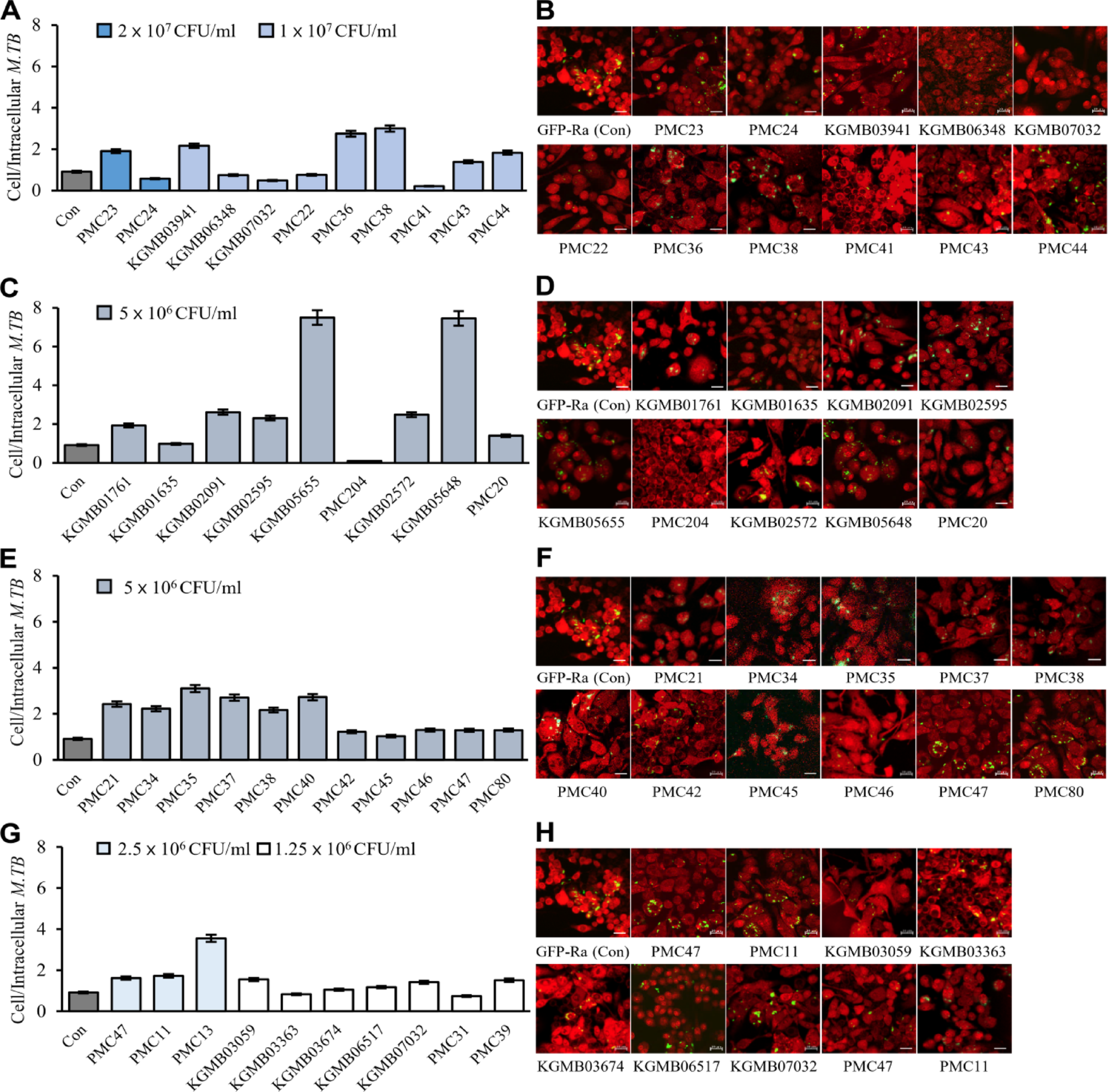
Imaging was conducted using an LSM800 confocal microscope (43X objective; Carl Zeiss, Germany). Raw 264.7 cells were cultured on a glass-bottom 96-well plate (Glass bottom black plate, Cellvis, USA) at a cell density of 5 × 10⁵ cells/ml and were infected with Green fluorescent protein (GFP)-H37Ra (MOI 1:3). Following a 2-h infection, the cells were washed thrice and treated with various concentrations of probiotic extract samples, then incubated for 72 h. Post-incubation, the cells were washed three times with 0.85% NaCl solution. SYTO59 red-fluorescent nucleic acid staining solution (5 µM/ml; SYTO59; Thermo Fisher Scientific, USA) was then added, the sample incubated for 30 min while protected from light, and subsequently, the staining solutions were removed, and the cells were rewashed thrice with 0.85% NaCl solution. SYTO59 red-fluorescent nucleic acid stain (5 µM/ml; Thermo Fisher Scientific, USA) was added, and cells were incubated for 30 min in the dark. The staining solution was removed, and the cells were rewashed three times before imaging. Two separate confocal imaging experiments were performed. The preliminary screening was designed to visually assess the probiotic candidates’ relative intracellular anti-tuberculosis efficacy. Standard antibiotic controls were not included in these experiments, as the primary goal was to compare probiotic-treated groups prior to dose–response validation. Comparative imaging and the use of antibiotics.
A parallel confocal imaging experiment was conducted using the same infection and staining protocol to provide a visual comparison with standard anti-tuberculosis drugs. Isoniazid (INH), rifampicin (RIF), and pyrazinamide (PZA), purchased from Sigma-Aldrich (St. Louis, MO, USA), were used to treat infected macrophages. Finally, the glass-bottom 96-well plate was mounted on a motorized stage.
Intracellular anti-tuberculosis activity and MIC₉₀ assay
Raw 264.7 cells were cultured overnight in a 96-well plate (Glass bottom black plate, Cellvis, USA) at a cell density of 5 × 10⁵ cells/ml and were infected with M. tuberculosis XDR (MOI 1:5) at 37 °C under 5% CO2 for 2 h. Following infection, cells were washed three times with phosphate buffer saline (PBS) to remove residual extracellular M. tuberculosis. Infected cells were then treated with various concentrations of probiotic extract samples and drugs and incubated for 72 h. Post-treatment, cells were washed three times with PBS and lysed using sterile distilled water. The number of viable bacilli was determined by plating serially diluted bacilli lysates on 7H10 agar plates. The minimum inhibitory concentration (MIC₉₀) was the lowest concentration that resulted in a ≥ 1.0 log₁₀ CFU/mL reduction compared to the untreated control. All experiments were performed in triplicate.
Isolation of membrane vesicles derived from probiotic strain PMC204
We adhered to previously established methodologies and references to isolate membrane vesicles (MVs) derived from bacteria and to study the anti-tuberculosis effects of MVs from the probiotic strain PMC204 [19, 20]. The PMC204 strain was cultured in a food-grade medium (FGM) composed of edible ingredients deemed safe for human and animal consumption. This FGM medium, modeled after standard MRS medium, included components such as soy peptone, yeast peptone, glucose, Tween 80, magnesium sulfate, and pH-adjusted conditions. The initial experiments began with a 30 mL culture, which was scaled up using a fermenter system (FMT-ST-S07, Fermentec, Korea). The cultures were incubated in a 7L fermentation system (Fermentec, Korea) at 37 °C for 24 h, connected to a gas cylinder containing 5% H₂, 5% CO₂, and 90% N₂. When the optical density at 600 nm (OD₆₀₀) reached 0.8–0.9 or entered the stationary phase, cells (≥ 7.6 × 10⁸ CFU/mL of PMC204) were harvested by pelleting via centrifugation at 3515 × g for 30 min at 4 °C, repeated twice using a general-purpose centrifuge. The culture supernatant of PMC204 (≤ 1L) was filtered through a 0.22-μm membrane filter (Bottle Top Filter, Millipore, USA). The concentrated conditioned medium was centrifuged using an ultracentrifuge (Optima MAX-XP, Beckman Coulter, Fullerton, USA) with an MLA-55 rotor at 150,000 × g for 2 h at 4 °C. The pelleted MVs were resuspended in PBS, and protein content was quantified using the Micro BCA Protein Assay Kit (Thermo Scientific, USA).
Characterization of PMC204-derived MVs
The purified MVs of B. sonorensis PMC204 were analyzed using linear gradient denaturing polyacrylamide gel electrophoresis on a 4 to 12% Bis–Tris gel (Invitrogen, USA), with all samples prepared at the same protein concentration. Before gel loading, Bacteria Whole Cell lysate (WCL) samples were denatured by boiling in a reducing antioxidant agent (Nupage). The gel was subsequently stained with Coomassie Blue Stain Reagent (SumplyBlue SafeStain, Invitrogen, USA). MVs were examined using Transmission Electron Microscopy (TEM) after staining with 2% uranyl acetate for negative staining. Uranyl acetate was applied to the grid, and the excess solution was removed with filter paper. The samples were carbon-coated on copper and plasma-cleaned for 30 s before being dried and viewed under a Transmission Electron Microscope (TEM) operated at 120kV (Bio-TEM, Tecnai G2 Spiri TWIN, FEI). Particle counts of MVs were confirmed using a Nano Sight NS300 system (Malvern Instruments, Malvern, UK), which measured the size and concentration of the particles. Samples were prepared in 1 × PBS buffer with measurements conducted at 25 °C across three 60-s sessions for each sample, using camera settings of level 16 and a threshold parameter of 5.
Proteomic analysis of MVs derived from B. sonorensis PMC 204
All analyses were repeated in three biological replicates. A total of 200 µg of MVs underwent methanol/chloroform precipitation, as described in previous research [21]. As indicated in the prior study, these precipitated proteins underwent in-gel digestion with trypsin [22]. The tryptic peptides were dissolved in 20 μL of buffer A (0.1%formic acid in water) and 5 μL was injected onto a trap column, Acclaim PepMap C18 nano Viper 100 (75 μm × 2 cm, 3 μm) (Thermo Fisher Scientific) at a flow rate of 5 μL/min with 95% buffer A for 4 min, then analyzed in LC–MS/MS with an Orbitrap EclipseTM TribridTM mass spectrometer (Thermo Fisher Scientific, San Jose, USA) coupled with an UltimateTM 3000 UHPLC system (Thermo Fisher Scientific, USA). Loaded peptides were separated on an analytical column, PepMap RSLC C18 ES803A (75 μm × 50 cm, 2 μm) (Thermo Fisher Scientific), using a 180-min gradient from 5 to 90% of solvent B (0.1% formic acid in acetonitrile) at a flow rate of 300 nL/min. The mass spectrometer was operated in a data-dependent Top 20 scans mode, alternating between MS and MS2. Analysis parameters included 10 ppm mass accuracy, 1850 V ion spray voltage, 275 ºC capillary temperature, m/z 375–1575 full scan resolution, and 120,000 higher-energy collisional dissociation activation scans with 35% normalized collision energy. The quadrupole isolation window was set at 1.4 Da. MS/MS spectra were detected on Orbitrap with a resolution of 30,000. The MS raw files were searched using the MaxQuant program (version 2.2.0.0) (Max Planck Institute, Germany) against the B. sonorensis strain’s protein database (release 2023_04) from UniProt. Tolerance settings were 20 ppm for both precursor and fragment ions. The trypsin digestion allowed for two potential missed cleavages. The employed modifications included a fixed carbamidomethylation of cysteine (57 Da) and variable modifications for deamidation of asparagine and glutamine (1 Da), oxidation of methionine (16 Da), and acetylation of the N-terminus (42 Da). All identified proteins were statistically analyzed using the Scaffold program (version 5.3.0), selecting only proteins with a Protein threshold ≥ 0.99 and a minimum of one peptide for further analysis. Normalized weighted spectra in the Scaffold program facilitated protein quantitation. Protein sequences were analyzed for interactions using String (v12.0) and Cytoscape (version 3.8.2), then mapped to the Gene Ontology (AmiGO 2) and KEGG database to predict biological processes and pathways. The results indicated significant enrichment of protein–protein interactions (PPI) (p < 1.0ⅹ10–16) and a false discovery rate (FDR) of ≤ 0.05. Protein–protein interaction networks and protein sequences were analyzed using STRING (v12.0) and Cytoscape (v3.8.2).
Detection and analysis of autophagy
Acidic vacuoles were detected using Acridine Orange (AO), a pH-sensitive dye that accumulates in acidic compartments such as lysosomes and emits red fluorescence in these environments to investigate autophagy. Confocal imaging of AO-stained cells was performed with an LSM800 microscope (43X objective; Carl Zeiss, Germany), and fluorescence was quantified using Image J software. The excitation lasers were set at 502 nm for green fluorescence and 606 nm for red fluorescence, with emission filters at 525 nm and 659 nm, respectively. Additionally, autophagy gene expression was analyzed in PMC204-treated RAW 264.7 cells. Total RNA was extracted using an RNA protection bacteria reagent kit (Qiagen, Germany), following the manufacturer’s protocol, and quantified with a Qubit Fluorometer and Qubit RNA Assay Kit (Invitrogen, USA). RNA was reverse-transcribed into cDNA using a cDNA synthesis kit (Bio-Rad, USA). Per the manufacturer's instructions, real-time PCR was conducted using the SYBR Green Supermix Kit (Bio-Rad, USA) on a CFX96 Real-Time PCR detection system (Applied Biosystems, USA). RAW 264.7 cells were treated with M. tuberculosis or PMC204 cell extract at two different concentrations, and mRNA levels of autophagy-related genes (ATG5, ATG7, ATG12, and ATG16) were measured. The comparative Ct method normalized expression levels to the endogenous control gene glyceraldehyde 3-phosphate dehydrogenase (GAPDH).
Assessment of cytotoxicity and acute toxicity of probiotic extracts in cell and animal models
Cytotoxicity and acute toxicity tests were conducted to evaluate probiotics’ safety. Cytotoxicity was assessed using the EZ-Cytox reagent (Dogen, Korea) through a Water-Soluble Tetrazolium Salt (WST) assay. Macrophages (1.5 × 10⁵ cells/ml) were seeded in a 96-well plate and incubated at 37 °C with 5% CO₂ overnight. Various probiotic extracts were added to each well and washed with 1 × PBS. EZ-Cytox reagent (20 μl) was added, incubated for 30 min, shaken for 1 min, and the absorbance was measured at 450 nm using a microplate reader. Toxicity in Animal Models was assessed by administering probiotics orally to animals once a day, 5 days a week, for 2 weeks. Animals were housed under controlled conditions (20–25 °C, 30–70% relative humidity, 12-h light–dark cycle). Two groups were compared: one treated with probiotics cultivated in MRS broth and the other with sterile saline as a control. Animals were monitored for clinical signs, mortality, and changes in body weight throughout the treatment period.
Measurement of nitrite and intracellular reactive oxygen species levels
Nitric oxide (NO) secretion was quantified by measuring nitrite (NO₂⁻) levels, indicative of NO synthesis, using the Griess reagent (Promega, USA). RAW 264.7 cells were cultured overnight at a density of 5 × 10⁵ cells/mL on a 96-well plate. The cells were exposed to M. tuberculosis H37Rv at 37 °C and 5% CO₂ for 2 h, followed by three washes with 1 × PBS and subsequent treatment with probiotic extract for 72 h. L-NG-mono-methylarginine (L-NMMA) was used as a nitric oxide synthase inhibitor. After incubation, 50 μL of the medium supernatant was transferred to new plates, mixed with 50 μL of Griess reagent solution, and the nitrite concentration was measured using a microplate reader after further incubation. Reactive oxygen species (ROS) levels were determined in RAW 264.7 cells using the ROS H₂O₂ assay kit (Promega, USA). Cells were seeded in 80 μL monolayers on a 96-well plate (SPL Life Sciences, Korea) and incubated overnight at 37 °C in a humidified 5% CO₂ atmosphere. Upon reaching 75 – 85% confluency, the cells were washed with warmed phosphate-buffered saline (1 × PBS) and exposed to M. tuberculosis H37Rv for 2 h. Menadione (Sigma Aldrich, USA), used as a positive control, was applied to the cells. The infected cells were then washed, treated with fresh medium containing probiotic extract, and incubated for 18 h. Subsequently, 20 μL of H₂O₂ substrate solution was added and incubated for 2 h. Finally, 100 μL of ROS-Glo detection solution was added to each well and incubated for 20 min at room temperature, and luminescence was measured using a Victor Nivo Multiplate reader (PerkinElmer, USA).
Statistical analysis
All experiments were performed in triplicate (n = 3), and data are presented as mean ± standard deviation (SD). Statistical significance was evaluated using one-way analysis of variance (ANOVA) followed by Tukey’s post hoc test for multiple comparisons or Student’s t-test for pairwise comparisons, as appropriate. A p-value less than 0.05 was considered statistically significant.

Comments (0)