Search results
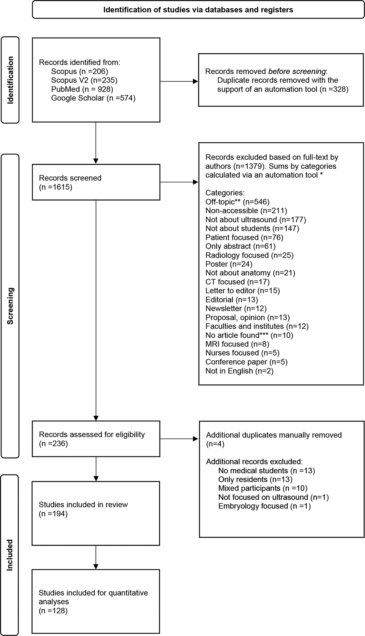
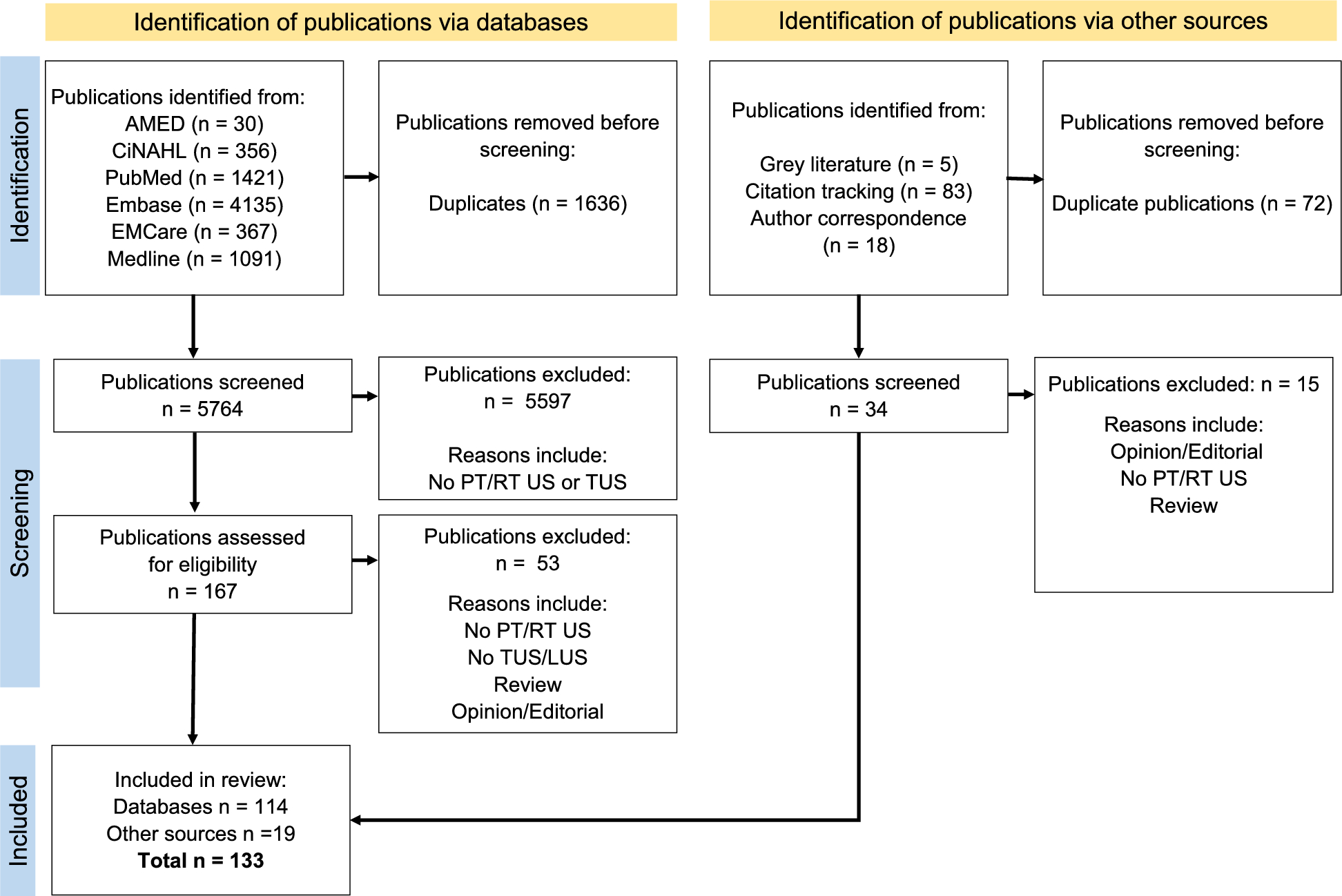
The literature search resulted in a total number of 1,943 records, of which 328 were removed after identification of duplicates (Fig. 1). From 1615 potential full-text-records screened, 1379 were categorized based on specific exclusion criteria (Fig. 1) and excluded. Four additional duplicates were detected and removed after further screening. A group of 38 records were excluded for not complying with all inclusion criteria. A total of 194 studies were considered relevant to the topic and were included in the literature review (Fig. 1).
Study characteristics
Among the 194 included studies, more than half were original articles as shown in Table 2 Different types of reports (technical, medical, educational and brief) were published on the subject, indicating a variety of methods to evaluate and record teaching outcomes.
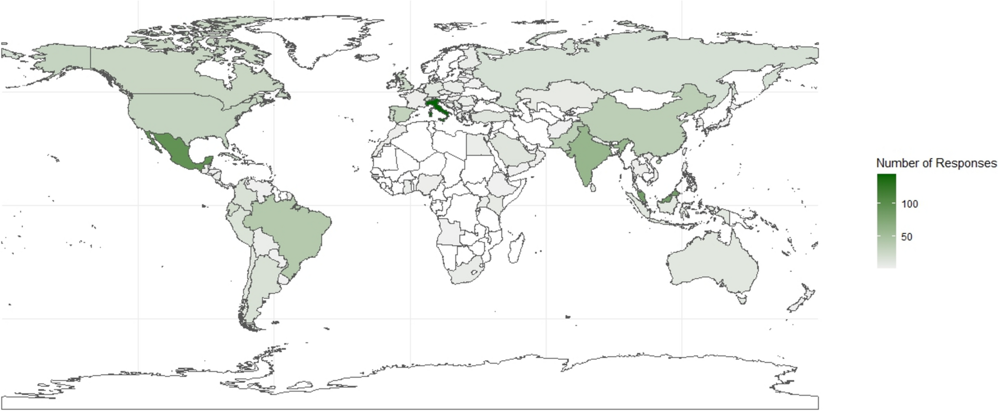
Half of the studies included in the review were conducted in the USA (50.5%), and to a lower degree in Germany (7.2%), UK (5.2%) and Canada (4.1%). Based on the affiliation information, the majority of countries across the world published their results as an individual university/institute/center, and few countries conducted collaborative work (6.2%).
Table 2 Characteristics of all studies selected in the systematic literature review (N = 194)Research study designs
The classification of original studies based on criteria from MERSQI showed that medical students were in majority enrolled from a single institution at 90.6% (item 2, Table 3). One reason could be the need to apply for ethical approval at the Institutional Review Board for each institution as seen in [19] and [20]. The recruitment of medical students from different programs has shown benefits with the possibility to compare an innovative curriculum from one program with a traditional curriculum from the other program [21]. The selected original studies were mainly conducted as a single group of students evaluated with post-tests (35.9%) or as a single group of students assessed with pre- and post-tests (28.9%) (item 1, Table 3), indicating a lack of a separate control group. The motivation of students after agreeing to participate was high with 64.8% response rate equal to or above 75% (item 3, Table 3).
The validity of evaluation instruments´ score was very low for all items (items 5, 6 and 7, Table 3), indicating low report of internal consistency between the assessment and the construct intended to evaluate, low report of content validity to assess the construct and little comparison between variables or a single variable to explain learning outcomes of medical students.
The statistical analysis of data (items 8 and 9, Table 3) was predominantly appropriate for the study design or the type of data and beyond descriptive analysis as many studies compared the student performance, skills and knowledge in identifying anatomical regions with ultrasound before and after teaching. Descriptive statistical analyses were often presented as percentages from surveys and questionnaires to illustrate satisfaction, perception and confidence levels of medical students.
Close to a third of the original articles measured satisfaction, perceptions, attitudes of medical students (item 10, Table 3) while behavior was the least studied (6.25%). In addition to the self-assessment of students, many of the publications evaluated the knowledge and skills assessments of medical students by teachers and trainers (43.8%).
Table 3 Study designs of original articles according to criteria from the MERSQI score [13]Participants and training characteristics
The population of medical students presented in the original articles was mainly first- and second-year students (Table 4). The percentage of students evaluated in anatomy skills with ultrasound decreased with increasing education (Table 4). These results should be considered as trends since 14.1% of the studies have not clearly specified the level of education of medical students. Nevertheless, these results support the idea that most anatomical courses are basic requirements and planned at the beginning of the medical curriculum. The duration of anatomy training with clinical ultrasound is mainly in hours with almost half of the studies including a study time between 60 min and 23 h while close to a third of the studies have not specified (Table 4).
Table 4 Learning parameters of the original articles selected (n = 128)The average group size of students during an ultrasound training was 139 individuals (Table 5), with a minimum group of 6 students [22] and a maximum sample size of 1260 learners in a study conducted for 6 years [23]. Ultrasound sessions in small groups of students (4 to 8 students) were in general preferred by the teachers and the students as they allow guidance in the proper probe placement, imaging acquisition and accurate identification [24].
Table 5 Descriptive data of medical student groups involved in anatomy learning sessions with ultrasoundFollowing the description of publication characteristics and student demographics, the type of anatomical regions taught with ultrasound were identified (Table 6). Ultrasound training on veins, the carotid artery, and major vessels were detected in the original studies and added to the vascular category. Ultrasound-guided injections were classified by authors as pertaining to both vascular and musculoskeletal (MSK) regions. The head and neck category involved carotid artery assessments and thyroid screenings. The renal region encompassed kidneys and bladder. The aorta category covered studies referencing the term “aorta” broadly, often implicating the abdominal aorta.
The obstetrics and gynecology category comprised studies focused on female pelvic organs and breast biopsy, while male reproductive anatomy was represented under a separate prostate and testicular category. The lung category incorporated training on pulmonary anatomy, pleural ultrasound, and pneumothorax identification. Cardiac-focused areas encompassed ultrasound of the parasternal long axis, inferior vena cava, and apical four-chamber view. The upper abdomen category included stomach, liver, gallbladder, and pancreas. Studies covering tendons, joints, ligaments, and muscles were grouped under MSK.
Vascular and cardiac anatomy were the most frequently targeted regions for ultrasound instructions(Table 6). The upper abdomen was addressed in nearly 40% of the studies, while MSK, head/neck, renal, and aorta regions were considered in approximately 30% of the studies. The least represented regions were the lungs, ocular, and prostate/testicular anatomy (Table 6).
Table 6 Specific area of anatomy instructed during the ultrasound sessionsStudy and learning outcomes
Learning outcomes in the original studies were identified based on the interpretations of results and conclusions [25]. A positive learning outcome was defined in our study as a significant improvement in skills and knowledge related to ultrasound use, high student satisfaction and confidence to perform ultrasound examinations and learn anatomy, increased motivation to use ultrasound for future practice and potential long-term retention. Mixed outcomes represented progress in the learning of students for some points and an essential need to improve other points with further studies. Neutral outcomes were characterized as no shown effect or difference between groups and/or no main conclusion based on the learning outcomes of medical students. A negative outcome was defined as a decrease in skills and knowledge, increased confusion or a lack of interest in ultrasound to learn anatomy. Close to 80% of the studies reported a positive outcome when teaching anatomy with clinical ultrasound to medical students (Table 7). A negative outcome was described in two studies [26, 27], in which the ultrasound scan was the least preferred method to learn anatomy compared to lectures, textbooks, dissections, lab videos, 3-dimentional radiology [26] or other radiological imaging techniques [27].
Table 7 Study outcomes in anatomy learning with ultrasound (original studies, n = 128)According to level 1 of the Kirkpatrick´s model, the reaction to the training experience was globally positive with increased confidence and comfort levels in identifying anatomical landmarks with ultrasound [28,29,30]. Interpretations on the satisfaction, perception and confidence of students using ultrasound during their medical education have been broadly studied, and additional information can be found in other systematic reviews [10, 12, 25].
The reaction to the learning experience or learning outcome as level 2 of the Kirkpatrick´s model in the original studies showed mixed results. Knowledge and skills of students were generally evaluated within the same group of students at post-test or in the interval between pre- and post-test as seen in the results of this review and as explained in other systematic reviews [12, 18, 25]. Within the selected original publications in this review with a second group or a control group of students, one study reported a non-significant difference in written anatomy final examination and anatomy practical scores between the experimental group of first- and second-year medical students participating in a practical ultrasound workshop on neurologic disorders and the control group not participating in the workshop [31]. Another study with first-year medical students compared the degree of deviance in palpation of shoulder anatomical areas between a student group receiving ultrasonography instructions, materials, equipment, anatomical atlas and a control student group given the anatomical atlas only [32]. There were no significant differences in the degree of deviance between the student groups for three out of four anatomical landmarks to identify [32]. However, one study showed an improvement in the identification of cardiovascular structures for first-year medical students with ultrasound images between pre- and post-training and between the first-year medical student group with ultrasound training (intervention) and second-year medical student group without training as control [33]. In the same study, the ability to recognize anatomical structures was greater in cadaveric images compared to ultrasound images. The skills acquired by the students during the study decreased after 6 months, indicating impaired long-term retention of skills and a need for regular training [33].
The behavior defined by the authors of this review as the transfer of skills and knowledge as well as long-term retention (level 3) was demonstrated in a few original publications. Students from one study [34] agreed to the proposition that the workshop on sonographic anatomy and guided-injection with ultrasound will change how they handle the pain management of patients in their future medical practice [34]. In a similar way, another study indicated that students significantly agreed to the statement: “ultrasound will play a significant role in their future medical practice after participating in this workshop” on neurological disorders [35]. Long-term retention was rarely evaluated in the selected studies (10/128), and the results were mixed. Knowledge retention in knee anatomy with ultrasound was partially lost 9 weeks after the training in first-year medical students as students could not identify and differentiate knee pathologies in ultrasound images after 9 weeks as well as they did between pre- and post-tests [36]. The studies assessing skills or hands-on retention in identifying anatomy landmarks with ultrasound demonstrated greater long-term retention. The ability to identify fetal heart, head, placental location and vertical pocket of amniotic fluid was improved with students who participated in an obstetric workshop and were tested in their skills after 3 months compared to students who did not participate in the workshop, indicating a good retention of obstetrics hands-on ultrasound skills [37]. An improved skills retention was observed in a study conducting a flipped-classroom with a workshop for POCUS learning in cardiac anatomy [38]. Three weeks after the workshop, 26 out of 32 students could generate images of all 4 cardiac views (100% score) and label structural areas in an appropriate way during an OSCE follow-up [38]. A different study evaluated the student skills acquisition after multiple dyspnea teaching sessions and compared the student skills retention with the same instructors after 8 months [39]. There was no significant difference in the skills of students between the end of the teaching session and the retention test 8 months later, indicating good retention of skills [39]. Altogether, these results could indicate that practical knowledge of anatomy with ultrasound is better retained than theoretical knowledge. The repetition of training sessions is necessary to retain skills and knowledge [40].
Among the original studies selected for the literature review, some have implemented a standardization of educational anatomy teaching with ultrasound within an institution after years of experience [23, 41]. In both studies, courses on human anatomy with clinical ultrasound were organized for first-year medical students with anatomy blocks including practical ultrasound sessions. There is no standard method to teach anatomy with ultrasound or implement anatomy ultrasound in the medical curriculum at a national level [42]. Organizations such as the Society of Ultrasound in Medical Education and the EFSUMB exist to monitor and update POCUS knowledge as well as provide recommendations on ultrasound training in medical education [8, 9, 23].
The costs (level 4) related to the application of ultrasound in anatomy courses for medical curriculum vary to a large extent depending on the objective and design of the course. Ultrasound machines, high technology portable ultrasound devices connected to tablets and annual subscription cloud storage were estimated to several thousands of dollars [43]. Costs could be reduced with specific activities such as the small-scale production of gelatin models. For example, gelatin models can be developed to simulate breast biopsy with ultrasound and the current published recipes cost less than 5 dollars (USD) per model [44]. A study calculated the cost of implementing an anatomy and ultrasound surgical training program with embalmed cadavers and surgery supplies [45]. The total cost amounted to 716 dollars (USD) per student per year for the specific study [45].
Despite potentially high initial costs, the application of clinical ultrasound in anatomy teaching offers multiple opportunities such a direct application on real or standardized patients [22, 23, 30, 37, 39, 46], gamification and virtual/mixed reality options [43, 47, 48] and a diversity in teaching types such as lectures with PowerPoint, manual reading, online content, 3-dimensional models, near-peer teaching, team-based learning and flipped-classroom [38, 43, 48, 49]. With portable ultrasound devices, anatomy teaching can be applied in low-income countries, difficult to reach or rural areas and improve the detection rate of pathologies in remote places [46].

Comments (0)