El-Naggar AK, Grandis JR, Takata T, Slootweg PJ, editors. WHO classification of head and neck tumours. 4th ed. Lyon: International Agency for Research on Cancer Publications; 2017.
Google Scholar
Chrcanovic BR, Gomez RS. Odontogenic myxoma: an updated analysis of 1,692 cases reported in the literature. Oral Dis. 2019;25:676–83. https://doi.org/10.1111/odi.12875.
Article
PubMed
Google Scholar
Saalim M, Sansare K, Karjodkar FR, Farman AG, Goyal SN, Sharma SR. Recurrence rate of odontogenic myxoma after different treatments: a systematic review. Br J Oral Maxillofac Surg. 2019;57:985–91. https://doi.org/10.1016/j.bjoms.2019.09.005.
Article
CAS
PubMed
Google Scholar
Takahashi Y, Tanaka K, Hirai H, Marukawa E, Izumo T, Harada H. Appropriate surgical margin for odontogenic myxoma: a review of 12 cases. Oral Surg Oral Med Oral Pathol Oral Radiol. 2018;126:404–8. https://doi.org/10.1016/j.oooo.2018.06.002.
Article
PubMed
Google Scholar
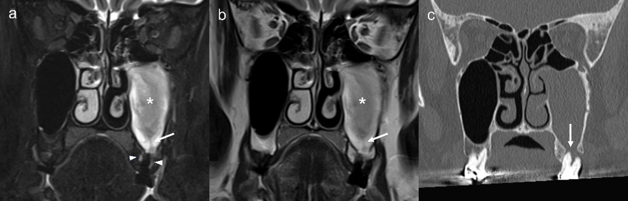
Sumi Y, Miyaishi O, Ito K, Ueda M. Magnetic resonance imaging of myxoma in the mandible: a case report. Oral Surg Oral Med Oral Pathol Oral Radiol Endod. 2000;90:671–6. https://doi.org/10.1067/moe.2000.108917.
Article
CAS
PubMed
Google Scholar
Kheir E, Stephen L, Nortje C, van Rensburg LJ, Titinchi F. The imaging characteristics of odontogenic myxoma and a comparison of three different imaging modalities. Oral Surg Oral Med Oral Pathol Oral Radiol. 2013;116:492–502. https://doi.org/10.1016/j.oooo.2013.05.018.
Article
PubMed
Google Scholar
Kawai T, Murakami S, Nishiyama H, Kishino M, Sakuda M, Fuchihata H. Diagnostic imaging for a case of maxillary myxoma with a review of the magnetic resonance images of myxoid lesions. Oral Surg Oral Med Oral Pathol Oral Radiol Endod. 1997;84:449–54. https://doi.org/10.1016/s1079-2104(97)90047-0.
Article
CAS
PubMed
Google Scholar
Hisatomi M, Asaumi J, Konouchi H, Yanagi Y, Matsuzaki H, Kishi K. Comparison of radiographic and MRI features of a root-diverging odontogenic myxoma, with discussion of the differential diagnosis of lesions likely to move roots. Oral Dis. 2003;9:152–7. https://doi.org/10.1034/j.1601-0825.2003.01802.x.
Article
CAS
PubMed
Google Scholar
Giallonardo P, Cutilli T, Masciocci C, Appia F, Corbacelli A. [Role of NMR in staging and treatment of extensive myxoma of the upper jaw] (article in Italian). Stomatol Mediterr. 1990;10:263–8.
CAS
PubMed
Google Scholar
Asaumi J, Matsuzaki H, Hisatomi M, Konouchi H, Shigehara H, Kishi K. Application of dynamic MRI to differentiating odontogenic myxomas from ameloblastomas. Eur J Radiol. 2002;43:37–41. https://doi.org/10.1016/s0720-048x(01)00453-3.
Article
PubMed
Google Scholar
Asaumi J, Konouchi H, Hisatomi M, Kishi K. Odontogenic myxoma of maxillary sinus: CT and MR-pathologic correlation. Eur J Radiol. 2001;37:1–4. https://doi.org/10.1016/s0720-048x(00)00229-1.
Article
CAS
PubMed
Google Scholar
Yabuuchi H, Matsuo Y, Kamitani T, Setoguchi T, Okafuji T, Soeda H, Sakai S, Hatakenaka M, Nakashima T, Oda Y, Honda H. Parotid gland tumors: can addition of diffusion-weighted MR imaging to dynamic contrast-enhanced MR imaging improve diagnostic accuracy in characterization? Radiology. 2008;249:909–16. https://doi.org/10.1148/radiol.2493072045.
Article
PubMed
Google Scholar
Yabuuchi H, Fukuya T, Tajima T, Hachitanda Y, Tomita K, Koga M. Salivary gland tumors: diagnostic value of gadolinium-enhanced dynamic MR imaging with histopathologic correlation. Radiology. 2003;226:345–54. https://doi.org/10.1148/radiol.2262011486.
Article
PubMed
Google Scholar
Lam PD, Kuribayashi A, Imaizumi A, Sakamoto J, Sumi Y, Yoshino N, Kurabayashi T. Differentiating benign and malignant salivary gland tumours: diagnostic criteria and the accuracy of dynamic contrast-enhanced MRI with high temporal resolution. Br J Radiol. 2015;88:20140685. https://doi.org/10.1259/bjr.20140685.
Article
CAS
PubMed
PubMed Central
Google Scholar
Thoma KH, Goldman HM. Central myxoma of the jaw. Oral Surg Oral Med Oral Pathol. 1947;33:B532–40. https://doi.org/10.1016/0096-6347(47)90315-3.
Article
CAS
PubMed
Google Scholar
Martínez-Mata G, Mosqueda-Taylor A, Carlos-Bregni R, de Almeida OP, Contreras-Vidaurre E, Vargas PA, Cano-Valdéz AM, Domínguez-Malagón H. Odontogenic myxoma: clinico-pathological, immunohistochemical and ultrastructural findings of a multicentric series. Oral Oncol. 2008;44:601–7. https://doi.org/10.1016/j.oraloncology.2007.08.009.
Article
CAS
PubMed
Google Scholar
Zhang J, Wang H, He X, Niu Y, Li X. Radiographic examination of 41 cases of odontogenic myxomas on the basis of conventional radiographs. Dentomaxillofac Radiol. 2007;36:160–7. https://doi.org/10.1259/dmfr/38484807.
Article
CAS
PubMed
Google Scholar
Kaffe I, Naor H, Buchner A. Clinical and radiological features of odontogenic myxoma of the jaws. Dentomaxillofac Radiol. 1997;26:299–303. https://doi.org/10.1038/sj.dmfr.4600261.
Article
CAS
PubMed
Google Scholar
Peltola J, Magnusson B, Happonen RP, Borrman H. Odontogenic myxoma–a radiographic study of 21 tumours. Br J Oral Maxillofac Surg. 1994;32:298–302. https://doi.org/10.1016/0266-4356(94)90050-7.
Article
CAS
PubMed
Google Scholar
MacDonald-Jankowski DS, Yeung R, Lee KM, Li TK. Odontogenic myxomas in the Hong Kong Chinese: clinico-radiological presentation and systematic review. Dentomaxillofac Radiol. 2002;31:71–83. https://doi.org/10.1038/sj.dmfr.4600678.
Article
CAS
PubMed
Google Scholar
Koseki T, Kobayashi K, Hashimoto K, Ariji Y, Tsuchimochi M, Toyama M, Araki M, Igarashi C, Koseki Y, Ariji E. Computed tomography of odontogenic myxoma. Dentomaxillofac Radiol. 2003;32:160–5. https://doi.org/10.1259/dmfr/16752462.
Article
CAS
PubMed
Google Scholar
Iwasaki T, Harazono Y, Fukawa Y, Kaida A, Kayamori K, Sasaki Y, Harada H, Yoda T. Retrospective analysis of odontogenic myxoma and odontogenic myxofibroma in the oral and maxillofacial region: a fibrous tissue-related differentiation. Br J Oral Maxillofac Surg. 2024;62:464–70. https://doi.org/10.1016/j.bjoms.2024.02.003.
Article
PubMed
Google Scholar
Sumi M, Ichikawa Y, Katayama I, Tashiro S, Nakamura T. Diffusion-weighted MR imaging of ameloblastomas and keratocystic odontogenic tumors: differentiation by apparent diffusion coefficients of cystic lesions. AJNR Am J Neuroradiol. 2008;29:1897–901. https://doi.org/10.3174/ajnr.A1266.
Article
CAS
PubMed
PubMed Central
Google Scholar
Sakamoto J, Kuribayashi A, Kotaki S, Fujikura M, Nakamura S, Kurabayashi T. Application of diffusion kurtosis imaging to odontogenic lesions: analysis of the cystic component. J Magn Reson Imaging. 2016;44:1565–71. https://doi.org/10.1002/jmri.25307.
Article
PubMed
Google Scholar

Comments (0)