
Remember me
Complications following thoracic surgery can be broadly classified based on their timing: early complications, which manifest within the first 30 days postoperatively (e.g., persistent air leak, atelectasis, and pneumonia); late complications, which develop beyond this period (e.g., bronchial anastomotic stricture, lung herniation); and complications that may arise at any stage, such as bronchopleural fistula and empyema [14] (Table 2).
Table 2 Postoperative complications after lung cancer surgeryPersistent air leakPersistent air leak occurs when air continues to escape from the lung parenchyma into the thoracic cavity beyond the expected timeframe for resolution. While some degree of air leakage is common following lobectomy, it typically resolves within 24–48 h as the remaining lung expands to occupy the pleural cavity. Air leaks persisting beyond 5–7 days are classified as persistent and may be associated with increased morbidity and prolonged hospitalization [8, 14].
CXRs and CT scans often reveal residual pneumothorax or subcutaneous emphysema [14, 15] (Fig. 3).
Fig. 3
Persistent air leak following left lower lobectomy. CXR taken 1 week postoperatively appears unremarkable (a). However, a pneumothorax in the left lower lung field is evident 2 weeks after surgery (b)
AtelectasisAtelectasis is the most frequently encountered complication after thoracic surgery, typically arising during the early postoperative period [9]. It is most often caused by airway obstruction from retained secretions, leading to collapse of segments of the remaining lung. In some cases, superimposed infection may develop [9, 16]. Radiographic features vary depending on the location and extent of the lung involvement [8] (Fig. 4). On CT, atelectasis appears as increased parenchymal opacity with displacement of adjacent fissures and the hemidiaphragm [16].
Fig. 4
Atelectasis following right upper lobectomy. CXR reveals atelectasis of the right middle lobe, manifesting as opacification along the mediastinal aspect of the right upper lung field
PneumoniaPostoperative pneumonia is frequently associated with aspiration, mechanical ventilation, and inadequate pain control, all of which contribute to impaired pulmonary clearance and infection risk. On CXRs, pneumonia typically presents as patchy areas of consolidation, while CT provides better characterization, particularly in cases of aspiration pneumonia [9] (Fig. 5).
Fig. 5
Pneumonia following left lower lobe wedge resection. The patient developed oxygen desaturation on postoperative day 1. Initial CXR was suggestive of pneumonia, and same-day CT imaging confirmed the diagnosis of aspiration pneumonia. CT revealed bilateral consolidation and ground-glass opacities predominantly in the lower lobes, consistent with aspiration pneumonia
HemothoraxPostoperative hemothorax arises from residual bleeding within the thoracic cavity, often due to persistent leakage from systemic thoracic vessels (e.g., bronchial and intercostal arteries), suture dehiscence of a pulmonary artery, or venous injuries. Hemorrhage from systemic arterial sources is the most frequent cause.
Radiographic findings on CXR include rapid opacification of the postoperative cavity. On non-contrast CT, hemothorax typically appears as a high-attenuation pleural effusion (~ 50 HU), often heterogeneous or with a fluid–fluid level [6] (Fig. 6). High attenuation within the effusion is strongly suggestive of a hematoma. However, findings may vary based on the timing of the bleed.
Fig. 6
Hemothorax after right lower lobectomy. The patient developed hemorrhagic shock following the removal of the drainage tube. CXR after tube removal demonstrated a sudden decrease in tight-sided translucency (a, b). Urgent non-contrast CT revealed a pleural effusion containing high-attenuation areas, indicative of hemorrhagic fluid (c)
In cases where active bleeding is suspected, contrast-enhanced CT angiography can identify the source, and transcatheter embolization may be considered a therapeutic option.
Acute respiratory distress syndrome (ARDS)Postoperative ARDS is most commonly observed following pneumonectomy and has historically been associated with poor prognosis [8]. However, recent studies indicate a reduced mortality rate of 40% to 45% [14].
Imaging findings include rapidly progressive bilateral opacities on CXR, while CT imaging reveals dorsal-predominant consolidation and ground-glass opacities (GGOs). These imaging features correspond to diffuse alveolar damage (DAD), the histopathologic hallmark of ARDS. During the organizing phases, traction bronchiectasis may also be present [8, 17, 18]. Unlike other causes of ARDS, postoperative ARDS may demonstrate asymmetric distribution, with relative sparing of the resected lung and predominant involvement of the contralateral lung [18] (Fig. 7). Radiologists play a critical role in identifying these characteristic imaging features, facilitating early detection of DAD and its distinction from other postoperative complications, such as infection or cardiogenic edema. Precise interpretation is vital for prompt management and improved clinical outcomes [8, 17].
Fig. 7
Acute respiratory distress syndrome (ARDS). Twenty-two days following right middle and lower lobe resection, follow-up CXR reveals diffuse ground-glass opacities in the left lung (a). CT imaging demonstrates extensive ground-glass opacities predominantly in the contralateral left lung rather than the postoperative right lung (b, c). Two days earlier, new opacities were noted in the left lung on CXR and CT (images not shown), prompting the initiation of antibiotic therapy. Despite oxygen administration at 8 L/min via face mask, the patient’s hypoxemia persisted, and the left lung opacities progressed. Given the clinical and imaging findings, a diagnosis of ARDS was established
Postoperative acute exacerbation (AE) of interstitial lung disease (ILD)ILD, particularly idiopathic pulmonary fibrosis (IPF), is frequently associated with lung cancer. In patients with pre-existing ILD, surgical resection is often selected with curative intent and to reduce complication risk despite available alternatives such as chemotherapy and radiotherapy. However, postoperative acute exacerbation (AE) of ILD is a well-documented complication.
With an incidence of 9.3% and a mortality rate of 43.9%, AE of ILD represents the leading cause of lung cancer surgery-related mortality [19]. The risk of AE increases with the extent of lung resection (bi-lobectomy/pneumonectomy > segmentectomy/lobectomy > wedge resection) [19, 20].
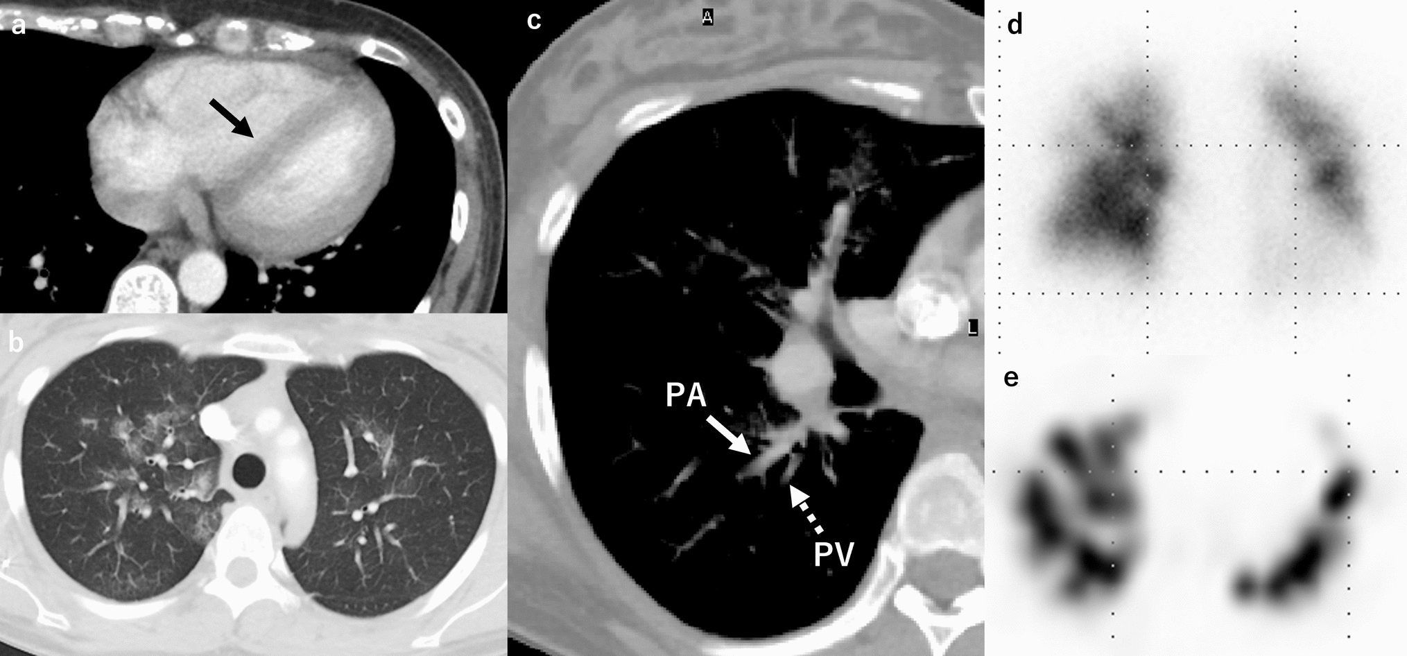
Imaging predictors for AE, such as preoperative CT findings of GGOs, consolidation, and pulmonary trunk enlargement, have been associated with an increased risk of AE [20]. The role of honeycombing on CT in predicting AE remains controversial [19,20,21]. Postoperatively, AE manifests as new GGOs and reticular opacities superimposed on pre-existing fibrosis (Fig. 8), resembling AE of ILD findings. Traction bronchiectasis and other contraction-related changes may also be present [22].
Fig. 8
Postoperative acute exacerbation of idiopathic pulmonary fibrosis. Preoperative CT showed interstitial lung disease with the usual interstitial pneumonia pattern, including honeycombing (a). Four days after right upper lobectomy, the patient developed significant hypoxemia, prompting CT imaging. High-resolution CT reveals newly developed diffuse ground-glass and reticular opacities (b)
Late postoperative complications (> 30 days post-surgery)Bronchial anastomotic strictureBronchial anastomotic stricture is a late complication and the most frequently encountered complication following sleeve lobectomy, with an incidence of 18% [23]. The degree of stenosis and the presence of airway secretions determine whether distal lung collapse occurs.
Thin-section CT and multi-planar reconstructions are the most effective imaging modalities for evaluating airway narrowing and luminal obstruction [8] (Fig. 9).
Fig. 9
Bronchial anastomotic stricture. Four months after a right upper sleeve lobectomy for lung cancer, CT in the lung window demonstrates marked narrowing at the bronchial anastomotic site
Lung herniationPostoperative pulmonary hernia is a rare late complication in which the lung protrudes through a weakened region of the chest wall. While often asymptomatic, some patients present with a palpable or visible chest wall mass, which may become more prominent during coughing [24]. CT demonstrates lung parenchyma extending beyond the thoracic cavity, confirming the presence of the herniation (Fig. 10).
Fig. 10
Postoperative lung hernia. Two years after left upper partial lobectomy, CT demonstrates lung parenchyma protruding through a chest wall defect at the left lung apex
Chronic expanding hematomaChronic expanding hematomas are uncommon but clinically significant late postoperative complications. They may present as slowly enlarging, space-occupying lesions that persist over months to years and can mimic malignancy on imaging. These lesions are frequently associated with a history of thoracic surgery for conditions such as tuberculosis, pneumothorax, trauma, or tuberculous pleurisy [25].
CT typically reveals a well-defined, encapsulated mass with peripheral calcification. Contrast-enhanced CT may demonstrate scattered small nodular enhancement at the lesion margins. On T2-weighted MRI, the “mosaic sign” has been reported as a characteristic feature, reflecting heterogeneous signal intensities due to the presence of both acute and chronic hemorrhagic components [26] (Fig. 11).
Fig. 11
Chronic expanding hematoma. Five years following left lower lobectomy, non-contrast CT (a) reveals a mass with a focal calcified component (arrowhead) within the capsule. Contrast-enhanced CT (b) shows a small nodular enhancement at the lesion margin (arrow). T2-weighted MRI (c) demonstrates a mosaic pattern with areas of high and low signal intensity, characteristic of the “mosaic sign”
Local tumor recurrencePostoperative local recurrence may involve bronchial stump recurrence, staple line recurrence within the lung parenchyma, mediastinal lymph node metastasis, pleural dissemination, and seeding along the surgical tract. These recurrences are most frequently observed within 2 years postoperatively [6]. Limited resections, such as segmentectomy or wedge resection, are associated with a higher recurrence rate than lobectomy.
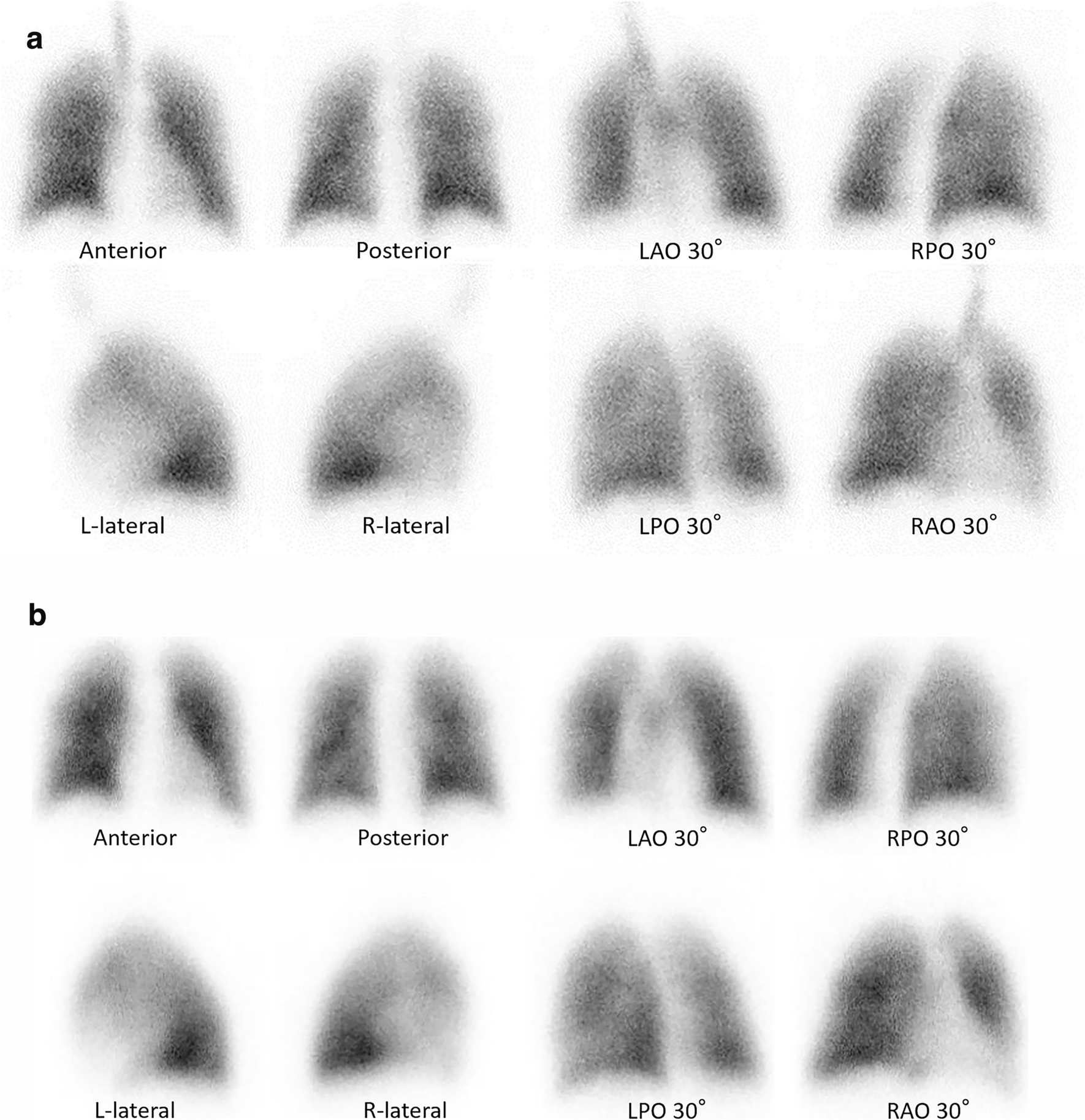
On CT, parenchymal recurrence near the resection margin is more frequently encountered after limited resection, such as segmentectomy or wedge resection, compared to lobectomy. Soft tissue growth on follow-up CT suggests recurrence at the bronchial stump, while staple line recurrence appears as focal soft-tissue enlargement along the resection margin. FDG-PET remains a valuable tool for detecting recurrent disease, with a reported accuracy of 94% (Fig. 12) [27].
Fig. 12
Local tumor recurrence. Bronchial stump recurrence after right upper and middle lobectomy (a–d). FDG-PET/CT performed 7 months postoperatively demonstrates increased FDG uptake in a small nodular lesion at the bronchial stump (a), which shows further progression at 13 months (b). CT reveals a soft-tissue mass protruding into the right bronchial lumen (c), and bronchoscopy confirms local recurrence (d). Staple line recurrence after partial resection of the right upper lobe lung cancer (e–g). Serial CT scans at 3 years (e) and 4 years (f) postoperatively demonstrate progressive soft-tissue enlargement along the staple line. FDG-PET/CT at 4 years shows abnormal FDG uptake consistent with tumor recurrence (g)
However, postoperative inflammatory changes may also exhibit FDG uptake, complicating differentiation from malignancy. While inflammatory uptake typically subsides within 3 months and is rarely seen beyond 6 months, prolonged or worsening FDG accumulation has been documented, making definitive diagnosis challenging [28,29,30] (Fig. 13).
Fig. 13
Staple line granuloma. FDG-PET/CT performed 3 years postoperatively shows increased FDG uptake at the bronchial stump (a). Bronchoscopy confirms no evidence of tumor recurrence. At the 5-year follow-up (b), FDG uptake persists but is slightly reduced, consistent with granulation tissue
Unilateral pleuroparenchymal fibroelastosis (PPFE)Unilateral PPFE is an infrequent but emerging and clinically relevant late complication following lung cancer surgery [31, 32], characterized by progressive fibrosis predominantly affecting the operated upper lung lobe. The incidence is approximately 4.3%, with a 10-year cumulative incidence of 5.3% [33]. Several risk factors have been identified, including male sex, lobar resection, the presence of a preoperative pulmonary apical cap, and reduced vital capacity (VC < 80%) [33].
Postoperative pleural effusion at 6 months has been recognized as a key early finding, often preceding the development of fibrotic changes [33]. As PPFE progresses, patients may experience worsening respiratory function and become more susceptible to chronic infections, such as pulmonary aspergillosis and nontuberculous mycobacterial infection. In some cases, this complication can contribute to significant morbidity and mortality, making early recognition crucial for distinguishing it from disease recurrence or other postoperative complications [33].
On CT, PPFE manifests as pleural thickening with adjacent sub-pleural fibrosis, predominantly affecting the operated upper lung field. As the disease advances, progressive volume loss, cystic changes, and thoracic deformity may develop, further compromising lung function (Fig. 14). The presence of a preoperative pulmonary apical cap and a persistent postoperative pleural effusion has been proposed as a potential predictor of this condition [33].
Fig. 14
Unilateral pleuroparenchymal fibroelastosis. Seven years after right upper lobectomy, coronal CT images at postoperative year 1 (a) and year 7 (b) show progressive thickening of the apical cap, cystic changes in the right upper and middle lung fields, volume loss, and thoracic cage contraction. Axial CT at year 7 (c) reveals a nodule suspected to be a fungal ball within a sub-pleural cyst, leading to a diagnosis of aspergillosis
Complications occurring at any stageBronchopleural fistula (BPF)A BPF represents an abnormal communication between the bronchial tree and pleural space, occurring in early and late postoperative periods. BPFs are more frequently encountered following right pneumonectomy than left pneumonectomy, likely due to anatomical features of the right main bronchus—specifically, its shorter length, larger diameter, and reduced mediastinal coverage [6, 14]. These anatomic factors may also explain the increased use of bronchial stump reinforcement with fat tissue on the right side as a preventive measure against BPF formation.
CXR findings on imaging include decreased pleural fluid, persistent pneumothorax, or subcutaneous emphysema. CT is particularly valuable in identifying air-fluid levels within the pleural cavity, and in approximately 50% of cases, direct visualization of communication between the bronchus and pleural space can be identified [6] (Fig. 15a).
Fig. 15
Bronchopleural fistula (BPF). Following right lower lobectomy (a, b), BPF with aspiration pneumonia and empyema are observed. CT (a) demonstrates a bronchial stump fistula (arrow) with adjacent air and fluid accumulation within the pleural cavity. The lung parenchyma shows ground-glass opacities (circle), consistent with aspiration pneumonia. An open-window thoracostomy was performed to manage the empyema (b)
When empyema develops secondary to BPF, prompt management is critical. Initial treatment typically involves chest tube drainage and antimicrobial therapy to control infection and prevent contralateral contamination. In persistent or complex cases, advanced interventions such as open-window thoracostomy (Fig. 15b) or closure of the residual space with a muscle flap may be required [34]. These procedures aim to eradicate infection and eliminate the pleural dead space to prevent recurrence.
EmpyemaPostpneumonectomy empyema is a severe complication and is associated with high mortality, with BPF as its primary etiology [15]. Early postoperative empyema is often due to residual pleural infection or intraoperative contamination, while late cases may develop months after surgery [9, 14].
Radi
Comments (0)