This retrospective study was approved by our institutional review board; prior informed consent was waived.
Subjects
Between July and December 2023, we initially enrolled 66 patients who underwent U-HRCT for lung cancer or suspected lung cancer. We then excluded three patients who underwent neoadjuvant chemotherapy due to morphological nodal changes. Consequently, the study population consisted of 63 patients (35 males, 28 females; median age 74 years, range 41–88 years); 58 patients were pathologically diagnosed with lung cancer (n = 52), metastatic tumor (n = 3), mycobacterial infection (n = 1), granulomatosis with polyangiitis (n = 1), and organizing pneumonia (n = 1) by surgery. One patient was diagnosed with bronchoscopy and underwent chemoradiation therapy. One patient underwent radiation therapy without pathological examination. Although one patient was diagnosed with lung cancer, he was followed up due to severe liver dysfunction. Two other patients were followed up without treatment because inflammation was suspected. There were 12 patients with emphysema and 3 patients with interstitial pneumonia.
U-HRCT scanning
All subjects were scanned with a 160-detector U-HRCT scanner (Aquilion Precision, Canon Medical Systems) using super-high-resolution mode without contrast material. The parameters for helical scans were tube voltage 120 kV, tube current automatic exposure control, preset noise 25 Hounsfield units (HU), rotation time 0.5 s, beam collimation 0.25 mm × 160 rows, focus size 0.6 × 0.6 mm, pitch factor 0.806, matrix size 512 × 512, field of view (FOV) 500 × 500 mm. The images were reconstructed at a slice thickness/interval of 5/5-, 1/1-, and 0.25/0.25-mm. Hybrid iterative reconstruction (AIDR 3D Standard FC52) was applied. The reconstructed FOV depended on the patient’s body size. For 63 patients, the mean was 365 × 365 mm (SD: 30). The mean computed tomography dose index and the dose length product on the 63 U-HRCT scans were 17.8 mGy (SD: 6.9) and 945.5 mGy・cm (240.9), respectively.
Reference standards
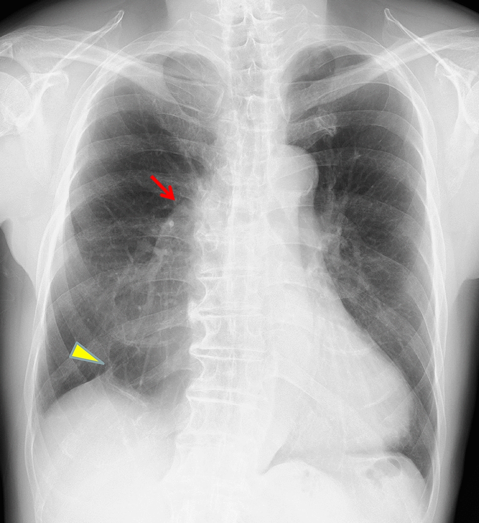
Two board-certified radiologists identified nodules more than 4 mm in diameter on 1-mm HRCT slices independently and set the reference standard consensually. The 4-mm cutoff size was set based on Lung-RADS and earlier studies [1, 13]. The nodule diameter and subtype were also recorded. For the measurement of nodules less than 10 mm, the average of long-axis and short-axis diameters was adapted, and for those greater than 10 mm, the maximum diameter was adapted based on the recommendations for measuring pulmonary nodule at CT from the Fleischner Society [14]. The nodules were classified as solid-, part-solid-, and ground-glass nodules (GGNs).
The board-certified radiologists reviewed all nodules detected by the DL-LND system and unidentified nodules whose diameter exceeded 4 mm were recorded; nodules that were missed by the radiologists and detected by the DL-LND system were also included in the reference standard.
Deep-learning-based lung nodule detection
The DL-LND system attached to the SYNAPSE SAI viewer V2.4 (FUJIFILM Medical Co., Ltd) was employed to inspect 5-, 1-, and 0.25-mm-thick slice images. The vendor recommends non-contrast chest CT images of adults (matrix size 512 × 512, slice thickness and interval 5 mm, lung kernel). The DL-LND instrument was programmed to identify solid nodules larger than 3 mm and sub-solid nodules (part-solid nodules and GGNs) larger than 5 mm using a green square.
Evaluation of the DL-LND system
The total and mean number of lesions detected by the DL-LND system per CT scan on 5-, 1-, and 0.25-mm slices were recorded. Differences in the number of lung nodules detected by the DL-LND system according to the presence or absence of underlying emphysema and interstitial pneumonia were investigated between the same slice thickness. The system’s sensitivity and the PPV, the total- and the mean number of FP nodules per CT scan were evaluated at the three slice thicknesses. Its sensitivity for nodules and the PPV were determined based on their percentage in the reference standards. To examine the utility of thinner slice thickness images, the sensitivity for small nodules (4–10 mm) and sub-solid nodules were also investigated. The processing time of the DL-LND system to detect lung nodules on 5-mm, 1-mm, and 0.25-mm slices was manually measured.
Statistical analysis
Statistical differences between the number of nodules and FP nodules and the image noise on 5-, 1-, and 0.25-mm slices were determined with the paired t test. Student's t test was used to evaluate the differences in the number of lung nodules detected by the DL-LND system according to the presence or absence of underlying emphysema and interstitial pneumonia. The chi-squared test was applied to assess statistical differences in the sensitivity and PPV of the three slice thicknesses. All analyses were with a statistical software package (JMP Pro 18.1.0); p values less than 0.05 were considered statistically significant.

Comments (0)