
Remember me

All methods and procedures were approved by the Institutional Animal Care and Use Committee at the University of California at Irvine in accordance with the NIH Animal Welfare Guidelines and are reported here following the ARRIVE (Animal Research: Reporting of the In Vivo Experiments) guidelines. Domestic shorthaired cats (Felis catus) were obtained from a breeding colony at the University of California at Davis. The animals were group-housed and given free access to dry food and water in the housing area.
Data are presented from ten cats (two male, eight female) that were chronically implanted with an animal version of a clinical CI electrode array (described in the following section). Three of the cats received only the CI array. An additional seven cats were implanted with a dual-electrode device that had the same CI array, supplemented by a single-shank electrode that was positioned in the auditory nerve as part of a separate ongoing study. The present study reports only data for stimulation by the CI arrays; there was no indication that the presence of the unstimulated single-shank devices influenced the responses to the CI arrays. On the day of implantation, the cats ranged in age from 10.2 to 79.1 months (median = 23.2 months), including six cats < 24 and four cats ≳36 months, and weighed from 2.8 to 4.7 kg (median = 3.1 kg).
Scalp recordings were obtained regularly at ~ 2–3-week intervals after implantation. Here, we focus on recordings generally taken on sessions nearest to 10 weeks after implantation, which was regarded as a benchmark for stability of chronic implantation. Exceptions to that included two CI-only cats, NU and MO. Those cats were implanted at an earlier stage of the study and were tested with a more limited eFFR stimulus set before we changed to the present protocol at their post-implantation weeks 18 and 24, respectively; their data from those weeks are presented here as we observed no functional changes in neural responses over those longer post-implantation durations (addressed in the “Results” section). Additionally, data for cat CL are from week 6 as that cat exhibited hardware failure of the implant device at week 8 but showed robust scalp-recorded responses prior to the failure. Any data reported other than at the ~ 10-week session are noted in the “Results” section.
Deafening and Chronic ImplantationAll cats were deafened bilaterally ~ 2 weeks prior to implantation according to a protocol that has been previously described [13, 26]. Briefly, each cat was sedated (see details below, Scalp Recordings) and auditory brainstem responses (ABR) were recorded in response to broad-band noise bursts, ≥ 10 ms in duration, at varying sound intensities to obtain thresholds of elicited neural responses; noise bursts were presented through a horn tweeter placed 20 cm to the left of the cat’s left pinna. An ototoxin was then administered (kanamycin; 300 mg/kg dissolved in saline; sub-cutaneous) and, after a delay of ~ 30 min, the diuretic ethacrynic acid was infused intravenously (1 mg/ml dissolved in saline) in doses of 10–20 mg at 10-min intervals until the ABR threshold was elevated beyond the maximum equipment level, typically > 70 dB above the initial threshold. For ~ 1 h thereafter, ABR responses were monitored for any recovery of hearing thresholds and subsequent doses of ethacrynic acid doses were given as needed; the total ethacrynic acid amount ranged from 10.7 to 22.6 mg/kg, median 15.2. In three of the nine cases, residual hearing was detected on the day of implantation ~ 2 weeks after the original deafening, and it was necessary to repeat the deafening procedure.
Each cat was implanted chronically with a CI consisting of an eight-channel lateral-wall research electrode array manufactured by Advanced Bionics, LLC (Valencia, CA). The electrodes were platinum half cylinders, each with a geometric surface area of 1.1 × 105 µm2 and separated center to center by 0.75 mm. The electrical return electrode was a platinum cylinder, 1.5 mm in diameter and 1.6 mm long. The CI array and return electrode were each attached to flexible coiled cables that led to a multi-contact percutaneous connector.
The implantation procedure was conducted in aseptic conditions while the cats were anesthetized with isoflurane gas; anesthesia and vital signs were monitored by a university veterinarian. The tympanic bulla was first exposed and then opened with a carbide burr to visualize the round window. The electrode array was inserted through the round window as far as possible in the scala tympani so that the distal, most apical, electrode was about halfway around the basal turn. That apical electrode was 8.75 mm distal to the base of the array, which typically fell at or just deep to the round window margin. The electrode array was then secured in place by filling the bulla with acrylic dental cement. The return electrode was positioned deep in the neck musculature caudal to the bulla. The coiled cables from the array and return electrodes were led under the temporal muscle to the percutaneous connector where it was placed on the dorsal midline of the skull. The connector was set inside a protective stainless-steel cylinder, and both were fixed in place with skull screws and acrylic cement. The surgical incision was closed, and the cylinder was covered with a screw-on cap that shielded the connector.
The continuity of the CI device was measured by the electrode impedance during each recording session. The impedance was computed from the voltage needed to pass a ± 5-µA current of a 6-kHz sinusoidal probe, which had a period (166.7 µs), similar to that of the electrical stimulation biphasic pulses (164 µs). Impedances of the most apical CI electrodes of each cat ranged from 6 to 14 kOhm (median = 6.4 kOhm) for the recording sessions reported here.
Electrical Stimulation and Scalp RecordingsStimulus generation and data acquisition used System III hardware from Tucker-Davis Technologies (TDT; Alachua, FL) controlled by custom MATLAB software (The MathWorks, Natick, MA) on a Windows-based desktop computer. The electrical stimuli were generated by an optically isolated current-controlled IZ2H stimulator from TDT. The output sample rate was 24,414 samples/s. Scalp recordings were made with a TDT Medusa4Z amplifier also having a sample rate of 24,414 samples/s; the amplifier had an onboard analog filter with a bandpass of 0.3 to 10,986 Hz.
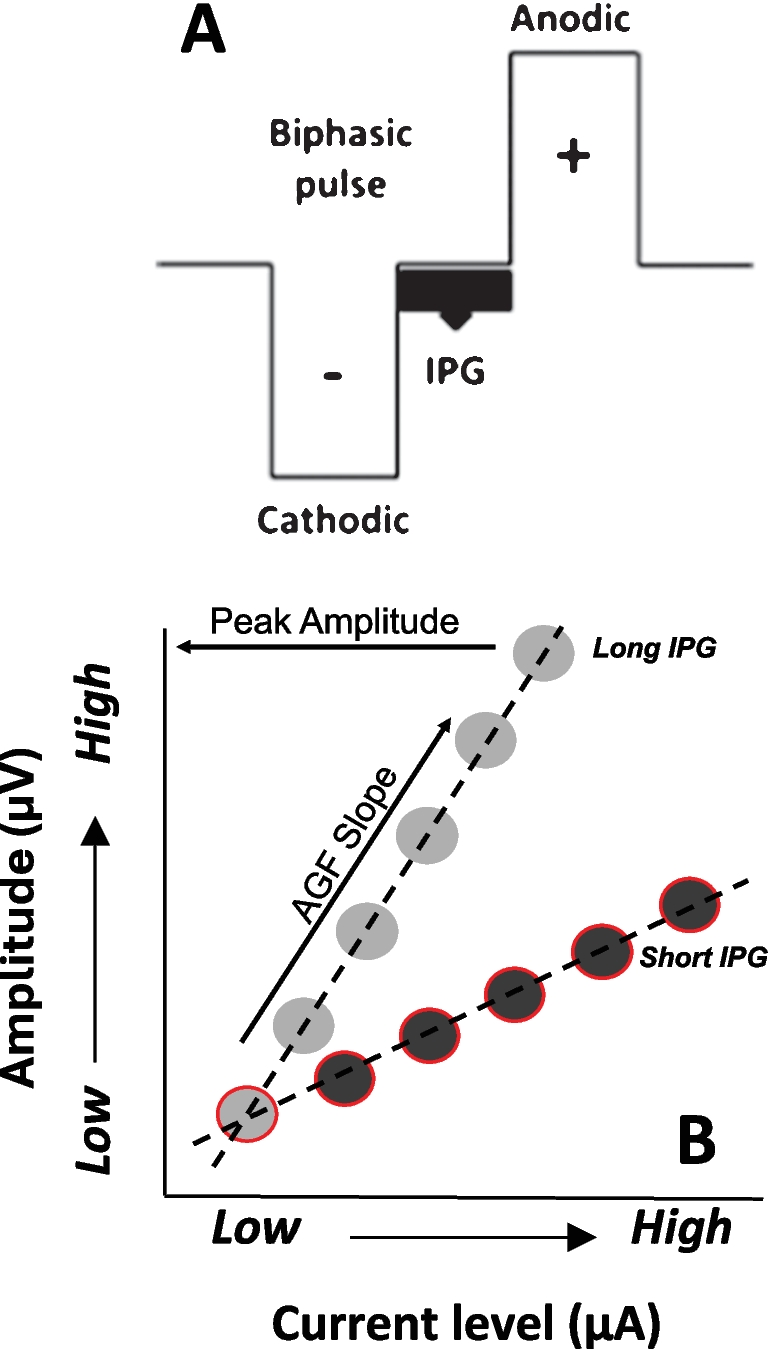
Electrical stimuli consisted of symmetrical biphasic pulses, 82-µs per phase in duration with no inter-phase gap (164 µs total duration). Pulses with cathodic- or anodic-leading polarity were alternated across trials for the purpose of reducing electric artifact after trial averaging. Stimulation was always delivered to the most apical CI electrode in the results reported here. That electrode had the tightest fit in the scala tympani so that it generally produced the lowest neural activation thresholds and it stimulated auditory-nerve fibers with the lowest characteristic frequencies accessible to the CI array, which potentially provided the greatest temporal acuity [16].
Stimuli for the eFFR recordings were 500-ms trains of electric pulses, fixed in current level, that varied across trials in the rate of pulses per second (pps). Trains were repeated at 1200-ms intervals, onset-to-onset, with the inter-train interval jittered by 5% intended to mitigate entrainment with intrinsic oscillatory activity. The pulse rates ranged from 43 to 642 pps and were restricted to integer divisors of the output sample rate. All cats were tested with a sample of 31 pulse rates with increments ranging from 5 to 32 pps. Pulse trains with fixed polarity of pulses were presented so that each rate was presented once in a random order, then again in a new order with the opposite polarity, and so on, until 40 repetitions were presented for each rate, including 20 trials each of the anodic- and cathodic-leading polarity. The recorded epoch for each trial included 100 ms before to 650 ms after the pulse-train onset.
Current levels for the eFFR stimuli were determined by measuring the growth of electrically evoked ABR (eABR) responses to single pulses at 1-dB increments of current. At higher current levels, an electromyographic (EMG) response often was elicited, sometimes accompanied by visible movement of the pinna, and was attributed to activation of the facial nerve [27]. The EMG was identified by marked changes in waveform morphology and a steep growth of amplitude, i.e., 10 to 100 times increases in magnitude over a 2-dB increase in current level. The eFFR stimulus level for each cat was set at a high point of the eABR growth function that was at least 2 dB below the lowest level at which the EMG was noted. Those eFFR current levels ranged from + 5.9 to + 10.2 dB (median = 8 dB, interquartile range = 2.3 dB) relative to each cat’s eABR threshold level at which a neural response could be detected compared to a no-stimulus condition.
Electrophysiological recordings began approximately 2 weeks after implantation (i.e., ~ 4 weeks after deafening). In each session, cats were sedated using a light level of anesthesia induced with an intramuscular injection of ketamine (25 mg/kg) and acepromazine (1 mg/kg). At those doses, the cats were typically areflexic with no spontaneous movement, although eye-blink or limb-withdrawal reflexes sometimes could be elicited at later times in the recording session. Supplemental doses of ketamine alone were given when necessary to maintain an immobile state.
Scalp potentials were obtained with subdermal electrodes consisting of hypodermic needles. The electrodes were rubbed with an abrasive cloth to remove any silicone coating, re-sterilized, and inserted through the skin. The reference electrode was placed on the midline of the head, rostral to the percutaneous connector and approximately 1–2 cm above the supraorbital margin of the eyes. One active electrode was placed over the left mastoid, ipsilateral to the CI, and a second active electrode was placed over the right mastoid, contralateral to the CI. The symmetric configuration of the two active-electrode channels on the head permitted a general characterization of responses at the two hemispheres, each having varying proximity to neural generators of the auditory pathway. A ground electrode was placed on the cat’s back, caudal to the scapulae. The recorded trial data were high-pass filtered at 3 Hz to eliminate DC voltage and then stored to the computer. Trial-averaged and filtered data were displayed for online monitoring. Note, because the recordings were referenced with respect to the midline scalp electrode, we reversed the polarity of all waveform data in the following eFFR analyses, which conforms with the more conventional practice of using the mastoid(s) as the reference site in electrophysiological recordings. That is, midline-positive waveform deflections are drawn as upward going and deflections from predominantly unilateral sources (e.g., the cochlear nucleus) appear opposite in sign when recorded on ipsi- versus contralateral channels.
eFFR Data AnalysisAll signal processing and statistical analyses were performed in MATLAB (version 2023b). The eFFR was analyzed in terms of the frequency-domain amplitude and phase values of synchronized responses at each pulse rate. Electric pulse artifacts pose a particular challenge to eFFR analysis because the periodic artifact has similar spectral characteristics to that of the FFR, and can superimpose on the ongoing neural responses, making it difficult to isolate from the waveform. To address these challenges, we adapted strategies previously used in human eFFR studies in which the trial waveforms were first submitted to a multi-step artifact reduction procedure [28,29,30,31,32]. That procedure is described and evaluated in the following “eFFR Artifact Reduction” section.
After artifact reductions, the trial waveforms were sorted with respect to pulse rate and stimulus polarity. Segments from 0 to ~ 50 ms re pulse-train onset were excluded from the analysis to omit any onset-specific responses, yielding an ~ 450-ms waveform duration. Trial waveforms for each pulse rate were screened for excessive noise assessed by the variance computed over time. Outlier trials were excluded if the variance exceeded the 75th percentile of variances across trials by more than 1.5 times the interquartile range, which on average amounted to rejection of 4.2 ± 2% (± standard deviation) of trials. The remaining trial waveforms were reconstituted by pairwise averaging of trials with anodic- and cathodic-leading stimulus polarity; that procedure provided the cancellation of any residual opposite-phase artifact to facilitate trial-level statistical analysis. Averages were then formed from all trial waveforms of each pulse rate, referred to here as the full-length waveforms. Additionally, the waveforms were segmented into ~ 23-ms intervals as adjusted to the nearest integer multiple of each pulse-rate period. Averages formed from those segments are referred to as the folded waveforms. The folding interval was based on the 23.3-ms period of the lowest pulse rate, 43 pps. For example, the waveform for 152.6 pps was folded in time over intervals of 26.2 ms (i.e., its period of 6.55 ms × 4), which yielded 17 waveform segments across the 450-ms waveform duration. Across all pulse rates, folded waveforms were formed from 12 to 20 segments (median = 19). All illustrated waveforms in the results were bandpass filtered from 40 to 1500 Hz; the filter order was set to 1, which was doubled by the bandpass design and doubled again by use of the MATLAB zero-phase filtfilt function, resulting in a fourth-order filter.
The fast Fourier transform (FFT) was computed for the full-length and folded waveforms, and eFFR transfer functions were obtained by extracting the spectral amplitude and phase at each pulse-rate frequency (F0). Prior to the FFT, the waveforms were tapered by Hann-windowed onset/offset ramps having durations of 100 ms for the full-length waveforms and 10 ms for the folded waveforms. All waveforms then were zero-padded so that the FFT output had 0.5-Hz frequency bins. The analysis of eFFR amplitudes utilized the full-length waveform spectra because of the greater inherent frequency resolution as compared to the shorter folded waveforms. For evaluating the artifact reduction procedures, additional amplitude values were taken at 2 to 4 multiples of the F0; those higher harmonics generally showed greater sensitivity (i.e., change in magnitude) than the F0 to the presence of the highly non-sinusoidal artifact. The noise floor level was estimated for each amplitude value by averaging 12 adjacent spectral bins (6 on each side) with 2-Hz spacing between bins to match the original frequency resolution of the ~ 500-ms pulse trains. The analysis of eFFR phase utilized the folded waveform spectra because the additional averaging constituting those waveforms was found to provide smoother progressions of phase lag as function of the pulse rate.
A Delay-and-Add Model of Amplitude Transfer FunctionThe amplitude transfer functions typically contained local minima and maxima (“dips” and “peaks”) across the tested pulse rates. Those spectral amplitude features presumably reflected the frequency-dependent destructive and constructive sums of multiple neural generators recorded simultaneously at the scalp [24, 25, 33]. To test whether that sum-of-generators hypothesis could account for the present amplitude features by electric stimulation, we adapted a delay-and-add modeling approach that was developed initially for FFR in normal-hearing cats [24] and expanded upon for normal-hearing humans [33].
The periodic responses of scalp-recorded neural generators were simulated by sets of four sinusoids, 500-ms in duration, all equal in frequency to the F0 of each stimulus. For each F0, the four sinusoids were generated having relative delays corresponding to biologically plausible latencies of particular generators: cochlear nucleus (fixed at 1.2 ms), other early brainstem generators including superior olivary complex (2.0 to 4 ms), inferior colliculus (4 to 8 ms), and thalamic/cortical generators (8 to 24 ms); an initial version of the model used separate thalamocortical and cortical generators, but that yielded only qualitatively, not statistically improved results. The fixed cochlear-nucleus latency was based on the first response after pulse onset in the eFFR waveforms, which was prominent and consistent across cats. The ranges of latency for the three subsequent generators were based on published measures of electrically evoked first-spike latencies and group delays of spiking activity of the respective auditory nuclei [9, 10, 14, 16, 34,35,36,37]. Values of latency within the three ranges were tested exhaustively by summing the sinusoids with particular sets of latencies and computing the resulting amplitude transfer functions across the range of stimulus rates; to reduce the search space, candidate latencies were restricted within each range by increments of ~ 0.5, ~ 0.5, and ~ 1.25 ms, respectively, for the brainstem, inferior-colliculus, and thalamic/cortical generators. In addition, following Tichko and Skoe [33], we scaled the relative magnitudes of the sinusoids to account for differences in proximity of generators to the recording electrodes (e.g., larger cortical than brainstem responses) as well as anatomical or physiological variations among cats. Exploratory testing revealed generally good model fits to empirical amplitudes with the following scalars: cochlear nucleus (fixed at 1), brainstem (fixed at 1), inferior colliculus (varied from 2 to5), and thalamic/cortical (varied from 5 to 10). Also, to account for declining phase locking at higher levels of the auditory neuroaxis, the inferior colliculus and thalamic/cortical generators were linearly tapered from full to zero amplitude, starting from the respective cut-off frequencies of 250 and 90 Hz and extending an octave above those cut-offs. The various model transfer functions were compared with empirical transfer functions by finding the sets of latencies and generator scalars that minimized the frequency differences of peaks and dips and overall amplitude differences between the modeled and empirical transfer functions.
Group Delays Computed from Phase Transfer FunctionsThe phase lags of the empirical transfer functions were unwrapped by accumulating phases across increasing pulse rates, adding 2π radians wherever there was a difference greater than π between phases of adjacent rates (unwrap function in MATLAB). The eFFR response latencies were estimated by the group delays, which are given by the slopes of portions of phase transfer functions that are linearly related to pulse rate [38]. We generally observed systematic transitions from steeper (e.g., longer-latency) to shallower (e.g., shorter-latency) phase slopes with increasing pulse rates (cf. [25, 39]). We employed two methods to evaluate slopes of transfer functions across limited ranges of pulse rate: piecewise linear regression and 3-point running group delay. The piecewise regression method partitioned the pulse rates into 5 linearly fitted segments that taken together minimized the sum of squared errors among all possible combinations of 5 ranges of pulse rates. Here, the 5 segments were fit to all statistically significant cumulative phase values (a maximum of 31), with each segment consisting of a minimum of three or more pulse rates. Only 4 segments were used in some cases that had 20 or fewer significant pulse rates. Note, the number of segments was based on fitting considerations and assumed neither the number of generators contributing to the scalp-recorded responses nor the independence of generators in the group-delay latency estimates. After fitting the 5 (or 4) segments, adjacent segments that did not differ significantly in slope (p > 0.05, Analysis of Covariance) were fused into a single segment. The result of each piecewise regression analysis was a set of 2 to 5 line segments (median = 5, 25th–75th percentile = 4–5), each segment restricted to a range of pulse rates and yielding a group delay. The 3-point running group delay method provided a more continuous estimate of latency as a function of pulse rate. Here, group delays were computed for successive sets of three consecutive pulse rates, so that linearly fitting started with the lowest three rates ranking 1–3, followed by ranks 2–4, then 3–5, and so on until the highest three rates. Given up to 31 pulse rates, that analysis could yield a sequence of as many as 29 group delays. Those group delays then were linearly interpolated to provide 1-pps resolution.
Both methods for analysis of phase slopes had two additional constraints. First, to maintain relatively localized estimates of latency, fitting did not occur over gaps in pulse rate wider than 60 pps (80 pps in cases of < 20 significant pulse rates). Second, fitting was applied only to cumulative phases that increased monotonically, while allowing only for phase reductions ≥ -π/20 between adjacent pulse rates. That tolerance accounted for possible noise in the phase measurement and was based on the 95th percentile of residual errors taken from fitted segments restricted to purely monotonic cumulative phases, aggregated across all cats and recording sessions.
StatisticsThe statistical significance of the eFFR for individual cats at each pulse rate was obtained by the Rayleigh test for non-uniformity of phase values across trials, assessed at the level of p < 0.01 [40]. Group-level analyses were based on a repeated measures experimental design with each animal as the experimental unit. Repeated measures analysis of variance (RMANOVA) tested for main effects and interactions for the factors of pulse rate, recording channel (ipsilateral vs. contralateral), and phase-lag analysis method (i.e., piecewise linear regression vs. 3-point running group delay). Significance was assessed at the level of p < 0.05 with the Greenhouse–Geisser (G–G) correction applied for non-sphericity, although the original degrees of freedom are reported. Post hoc pairwise comparisons used the non-parametric two-tailed Wilcoxon signed-rank test. Whenever appropriate, a 5% false discovery rate (FDR) correction was applied to account for multiple comparisons [41].
eFFR Artifact ReductionThe eFFR trial waveforms for each recording channel were submitted to an artifact reduction procedure that consisted of the following steps: (1) template subtraction, (2) blanking and linear interpolation, and (3) averaging across trials that alternated in stimulus polarity. Figure 1a shows that the electric pulse artifacts had maximum amplitudes from 0 to ~ 600 µs after pulse onset, followed by amplifier-induced ringing with diminishing amplitudes up to ~ 1400 µs; see × 100 higher gain in the Fig. 1a inset. Blue and red lines indicate anodic- and cathodic-leading pulse polarities, respectively. Figure 1b shows examples of the artifact for segments of eFFR pulse trains and Fig. 1c–f shows the artifact reduction steps.
Fig. 1
eFFR Artifact reduction procedures. a An example of artifact by a single biphasic pulse, from 0 to 2 ms re pulse onset, for both the anodic- (blue) and cathodic-leading (red) pulses, 82 μs/phase. The inset shows a higher-gain view (×100) of the artifact from 0.8 to 2 ms. b Examples of artifact recorded for eFFR pulse trains for three experimental pulse rates. The dashed box indicates the time window of 0 to 1.4 ms used to construct the artifact template. c Examples of the artifact templates for anodic- and cathodic-leading pulses. d The recorded eFFR waveform for three stimulus cycles of a single pulse rate (290 pps) after the template was subtracted from each pulse artifact. Note the ×100 higher gain compared to panels a-–c. The CN label indicates the earliest neural response peak identified, attributed to the cochlear nucleus. e The same eFFR waveform as panel d, after blanking and interpolating over three possible durations relative to pulse onset. The asterisk after 800 μs indicates the blanking length that was selected for routine artifact reduction. f The same eFFR waveforms as panel e, after averaging across the two stimulus polarities
In the template method, a model of the artifact was constructed and then the model was subtracted from the recorded waveform. As a first step, the recorded waveforms were up sampled by 10 times the original sample rate. That served to reduce any aliasing effects and to homogenize the shape of the artifact, thereby providing both a more accurate template model and greater precision in aligning the templates to the recorded waveforms prior to subtraction. Separate templates of single electric pulses were formed for each of the anodic- and cathodic-leading stimulus polarities. As depicted by the dashed rectangle in Fig. 1b, the templates were derived from the first pulse of each of the pulse trains for a given polarity, regardless of pulse rate, and then averaged together. The template interval started at 0 µs and ended at 1400 µs re the pulse-train onset. Importantly, the template interval for the first pulse in each train was absent of any ongoing neural responses to preceding pulses because of the 700-ms no-stimulus interval between subsequent trials. Nevertheless, the later portion of the template interval could contain a short-latency neural response to the first pulse, occurring as early as 1 ms after the pulse onset. To isolate that neural response from the templates, we exploited the opposite phase polarities of the anodic and cathodic templates, which could be averaged together to yield primarily the equal-phase neural activity. That average waveform trace was then subtracted from the separate anodic and cathodic templates. That yielded the single-pulse templates for the two polarities (see Fig. 1c). Those were concatenated into a sequence of templates that were matched in time to recorded pulse artifacts for each pulse rate. Time alignments were achieved by cross correlating the single-pulse template and FFR waveforms and computing the peak lag indices; that procedure accounted for occasional stimulating or recording asynchrony, which was never more than ± 41 µs (i.e., ± 1 sampling point). Last, to account for any amplitude fluctuations over the recording duration, each single-pulse template in the sequence was scaled to have the same peak-to-peak amplitude as its assigned pulse artifact in the recorded waveform.
The artifact template sequences were subtracted from the respective trial waveforms for each pulse rate and polarity. Figure 1d depicts a waveform segment after template subtraction and demonstrates that pulse artifacts could be effectively reduced, often to magnitudes near or below the neural response amplitude; note the × 100 difference in amplitude scales between Fig. 1a–c and d. We found that the first neural response following the largest portion of pulse artifact could be attributed to the cochlear nucleus (CN), which occurred at ~ 1.2 ms after pulse onset. For our purposes, the time points before the CN response were considered irrelevant; that included the far-field compound action potential from the auditory nerve, which in cat has electrically evoked latencies of ~ 500 µs and could be difficult to isolate from artifact [27, 42]. In Fig. 1d, the positive-going CN response is distinguished by having similar phase characteristics regardless of stimulus polarities; as described in the results, that response also shows longer latencies and reduced amplitudes with increasing stimulus rate, which is expected for a neural response but not an electric artifact. Nevertheless, remaining artifact effects could often be detected at time points before the CN response. For that reason, a segment of the template-subtracted intervals was blanked, and the data were linearly interpolated across the blanked samples. Figure 1e shows the blanking and interpolation procedure for 3 blanking lengths (dashed lines), which always started at 50 µs after the pulse onset. We evaluated a range of blanking lengths from 200 to 1600 µs to find the minimal duration necessary that showed evidence of reducing the influence by residual artifact, that yielded amplitude and phase responses characteristic of neural activity, and that was stable among adjacent blanking lengths, indicating low volatility to small manipulations. Figure 1f shows the averaging across stimulus polarity for the 3 example blanking lengths, which almost entirely eliminated residual effects of the opposite-phase artifacts. Asterisks indicate the 800-µs blanking length chosen for the main analysis as described in the following section.
Figure 2 shows in one example cat a representative pattern of amplitude and phase responses that resulted from various conditions of artifact reduction. Compared to no artifact reduction (black squares, “Artifact”), Fig. 2a shows that the initial step of template subtraction (black circles, “T.S. Only”) markedly reduced both the response (solid lines) and noise (dotted lines) amplitudes. Those amplitude reductions were largest at pulse rates above ~ 350 pps, presumably due to the increased number of the artifacts being subtracted per unit of time. By examining the highest pulse rate, 642 pps, one can see that applying interpolation over short blanking lengths (400–600 µs) reduced the amplitude only slightly compared to template subtraction alone. There was another intermediate amplitude reduction for lengths of 800 to 1000 µs, but at yet longer lengths the responses were highly diminished or became non-significant (open symbols), likely because of excessive blanking that could extend over 90% of the stimulus period at the highest pulse rates.
Fig. 2
Evaluation of the artifact reduction procedures in one representative cat. Colors indicate the separate artifact removal conditions, including no artifact reduction (black squares, “No Reduction”), template subtraction alone (black circles, “T.S. Only”), and various blanking lengths applied after template subtraction. a Solid lines with symbols depict the spectral amplitudes summed for each pulse-rate F0 and its harmonics 2-–4 for the artifact reduction conditions. Dashed lines are the noise levels estimated at each pulse rate from the spectral bins adjacent to the F0 or harmonics frequencies and summed. b Cumulative phase values for pulse rates greater than 350 pps for each artifact reduction condition. The vertical positions of the various curves are arbitrary. The dashed-line box indicates the range of pulse rates (>500 pps) over which the slopes of the phase progressions yielded group delays that were used to evaluate the effects of artifact reduction in each condition
Figure 2b shows the cumulative phase lags over a range of the higher pulses rates, > 350 pps; the vertical positions of curves are arbitrary. We computed group-delay latencies from the slopes of cumulative phase only for the highest of those rates (> 500 pps; see dashed-line box), which were expected to show the strongest influence from the artifact. Indeed, the group delay computed prior to artifact reductions was 0.4 ms, corresponding with the latency of maximum amplitudes of the artifact (see Fig. 2a). By reducing the influence of the artifact, however, template subtraction and interpolation over increasing blanking lengths tended to produce longer-latency group delays, here up to 2.5 ms for a blanking length of 800 µs, consistent with response latencies of the auditory brainstem. At longer blanking lengths, the group delays began to decrease again, or phase progressions became unstable by including non-significant responses; note the aberrant phase values at the highest pulse rate for blanking lengths > 1000 µs, indicative of excessive blanking. Based on those amplitude and phase results, which were comparable across cats and recording channels, a blanking length of 800 µs, extending from 50 to 850 µs after pulse onset, was applied to all eFFR waveforms. That blanking length was ~ 3 to ~ 51% of the inter-pulse interval for the lowest to highest tested pulse rate, 43 to 642 pps, respectively.
Comments (0)