Medical literature describing the association between atopic dermatitis and neuropathy is relatively sparse. While not often observed, particularly in Western cultures, atopic myelitis is a well-documented condition linking atopy and neuropathy. Given that neurologic symptoms such as pruritus and dysesthesia are commonly encountered in atopic conditions, their evaluation remains relevant to dermatologic practice. DP is an approved treatment option for controlling inflammatory disorders such as atopic dermatitis and prurigo nodularis. This subjective study has indicated amelioration of patient-reported peripheral neuropathy symptoms. Herein, among all associated symptoms of peripheral neuropathy, tingling, pins and needles sensation, and hypothesia to prick showed the most significant improvement, whereas itching and painful cold showed the least improvement.
In contrast, itching—commonly associated with peripheral neuropathy and atopic dermatitis—showed the least improvement. However, this outcome may be influenced by the lower baseline severity of itching prior to treatment, though some degree of symptom relief was still observed following DP administration. Notably, no symptom exhibited a worsening effect, further supporting the potential therapeutic benefit of DP in this context. Interestingly, several patients were unable to walk prior to therapy and were able to report that, with neuropathic symptom relief, daily ambulation was achieved with the DP regimen.
Our research suggests that Th2 modulation by DP may be attributable to its therapeutic benefit. However, the precise mechanisms underlying these effects remain incompletely understood, preventing a definitive explanation of the mode of action. Nonetheless, this study represents an initial step toward understanding the relationship between immune responses, biological mechanisms of action, and peripheral neuropathies.
DP is an IL-4R alpha inhibitor that targets the Th2 pathway implicated in highly pruritic inflammatory diseases such as atopic dermatitis, prurigo nodularis, and urticaria [7]. The primary cytokines involved in this pathway include IL-4, interleukin-5 (IL-5), IL-13, and IL-31 [8]. Upregulation of these cytokines contributes to the various symptoms observed in these conditions. Specifically, IL-5 is associated with edema and blister formation, whereas upregulation of IL-4 and IL-13 impairs the barrier function of the epidermis, thereby leading to xerosis. Moreover, these cytokines inhibit cutaneous innate immunity and facilitate skin colonization by common microorganisms such as Staphylococcus aureus [9].
Pruritus in the Th2 pathway is associated predominantly with the upregulation of IL-31. IL-31 mediates pruritus by activating the IL-31 receptor and oncostatin M receptor (OSMR) receptors in keratinocytes and the dorsal root ganglia of cutaneous sensory nerves [10]. Activation of IL-31R receptors on the dorsal root ganglia leads to the sensation of itch [11], by triggering transient receptor potential cation channels (transient receptor potential cation channel subfamily V member 1 (TRPV1)/transient receptor potential cation channel subfamily A member 1 (TRPA1)) [11]. An in vivo study on mice has demonstrated significantly diminished IL-31R-induced pruritus in TRPV1+-deficient mice, thus developing a strong correlation between IL-31 and pruritus [9]. Notably, IL-31 upregulation is not confined to the Th2 pathway but also occurs in T-helper 1 (Th1) reactions in the presence of IL-4. IL-4 induces the upregulation of IL-31, thus suggesting a direct relationship between these cytokines. Genetic studies have shown that inhibiting IL-4 decreases IL-31 levels within 48 h [12].
Our literature search did not yield specific studies on the modus operandi of DP in PN and the Th2 pathway. This is partly because peripheral neuropathy is an umbrella term encompassing multiple pathological subtypes with distinct and, in some cases, unknown pathophysiologies. Interestingly, several reports and studies have already explored the association between the development of peripheral neuropathy and Th2-associated interleukins such as IL-4, IL-5, IL-13, and IL-31.
A commonality among studies is that increased atopy and eosinophilia have been found to lead to mast cell destruction of the blood–brain barrier and the induction of spinal cord lesions [13]. Th-2 mediates this response, thus potentially explaining why DP ameliorated the neuropathies in our patients. Another prospective observational study on 13 subjects showed that using mepolizumab, an IL-5 inhibitor, significantly improved treatment-resistant peripheral neuropathy for pain and numbness [14].
However, in studies on chemotherapy-induced peripheral neuropathy, IL-13 administration has been reported to induce IL-10 production and to cause a shift from inflammatory M1 macrophages to reparative M2 macrophages in patients with neurological damage from cisplatin [15]. Similarly, studies in mice models have shown that Il-13 decreases tactile allodynia after partial sciatic nerve ligation [16]. Another notable finding is that IL-4 is the primary cytokine that stimulates immune cells, including Schwann cells, macrophages, fibroblasts, and neurons, to adopt a healing phenotype in nerve regeneration. This makes IL-4 a promising target for the treatment of peripheral nerve injuries [17]. An additional study demonstrates that IL-4 can alleviate neuropathic pain by promoting the accumulation of macrophages, which exert a therapeutic effect [18].
DP additionally acts as an indirect IL-31 modulator through the IL-4 pathway, demonstrating significant effects on inflammation. Given that IL-31 is considered the primary cytokine driving pruritic conditions, IL-31 targeted therapy might also offer substantial benefits for patients with peripheral neuropathy. However, the potential risk of neuromodulation warrants further investigation; incorporating a direct IL-31 modulator such as nemolizumab might potentially decrease these risks [19]. The results across many studies have led us to understand that further investigation of biologics as neuropathic therapies is warranted to develop new treatment protocols for patients with peripheral neuropathies or subtle signs of neuropathy.
Although our study demonstrated that DP might serve as a possible novel therapy for peripheral neuropathy, it is important to highlight the potential risks of immune modulation for neurological disorders, particularly in patients without atopic root causes. Because DP decreases Th2-mediated inflammation, it might shift toward a Th1- interferon gamma (IFN-γ) response, thus further damaging neurological pathways and potentially leading to permanent damage. Two cases have been described in which IL-13/IL-4 modulation has led to either chronic inflammatory demyelinating polyneuropathy or acute inflammatory polyneuropathy after DP treatment onset [20, 21]. After discontinuing DP, the numbness resolved in the latter case after 6 months. Furthermore, DP has also been associated with Th1/17 immune dysregulation in a patient with multiple sclerosis (MS) [22]. In contrast, reports have indicated amelioration of MS lesions in a single patient receiving DP and teriflunomide therapy [23].
Adverse reactions to DP specifically are relatively uncommon and usually mild. An adverse event of interest is the development of arthritis after DP therapy. One possible mechanism is a shift from a Th2-heavy immune response to a Th1 response, leading to inflammatory arthropathies. In some patients, underlying arthralgias masked by chronic and longstanding neuropathy may become unmasked by DP therapy.
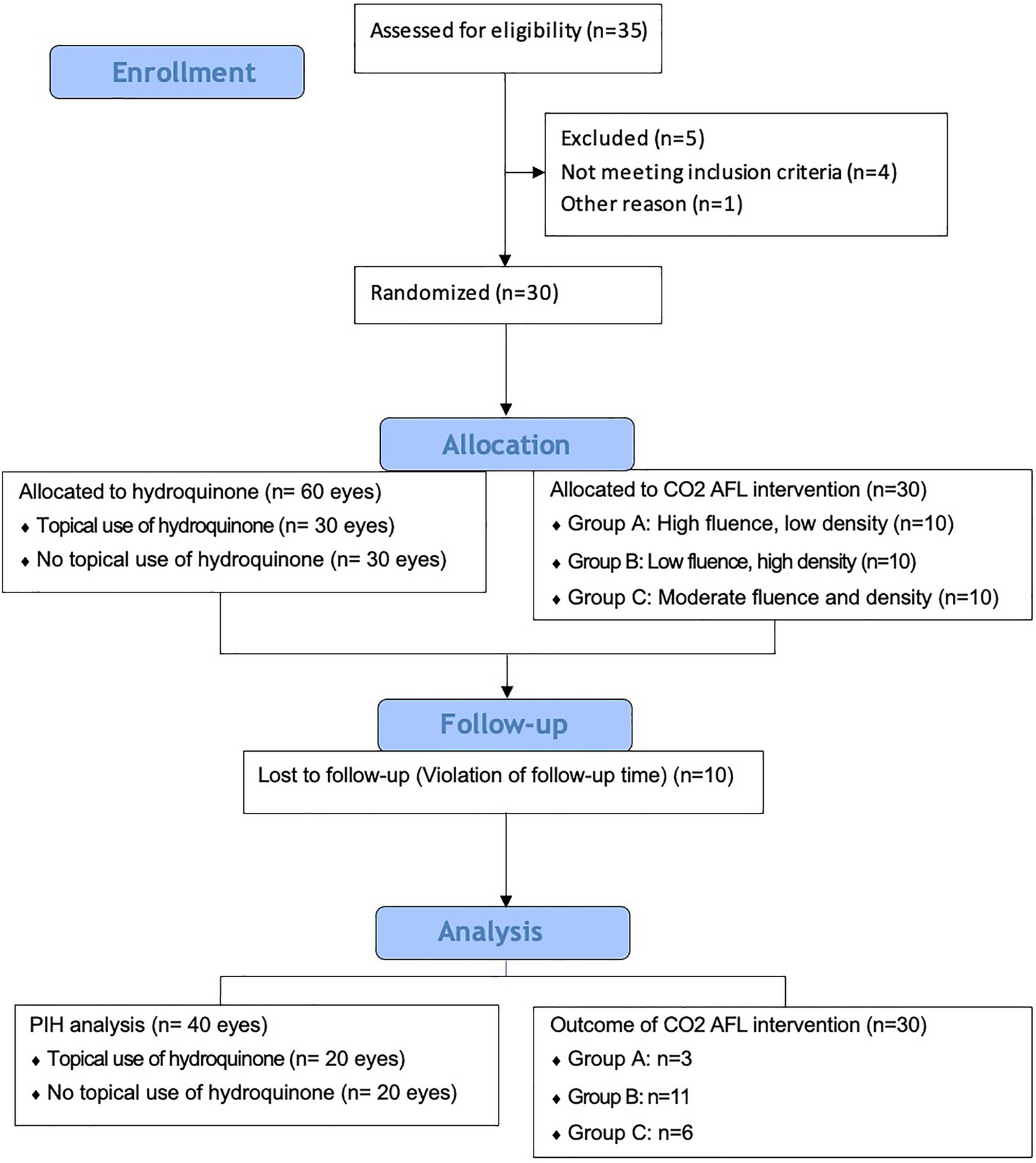
Our study provides insight into the potential off-label use of DP for patients with peripheral neuropathy. Given our smaller sample size and subjective measurements of improvement, observing the effects of DP therapy and the amelioration of peripheral neuropathy in our patients has provided the notion to gather quantitated studies as to the mechanism DP is a possible treatment to PN. We propose that further investigation of the use of biologic agents in neuropathic therapies should be encouraged further to elucidate the therapeutic potential of DP in this context. We propose a study incorporating a larger sample size and standardized EMG throughout the treatment course to monitor symptomatic changes objectively. Skin and nerve biopsies should also be performed to assess histopathological alterations in response to DP therapy. In patients with comorbid atopic disorders, further investigations—including IgE levels, cerebrospinal fluid (CSF) analysis, and spinal cord MRI—may also provide critical evidence supporting the role of biologic agents in the management of peripheral neuropathies. We encourage the discussion to develop new treatment protocols for patients with peripheral neuropathies to recognize, treat, and/or prevent secondary damage, further improving patient prognosis.


Comments (0)