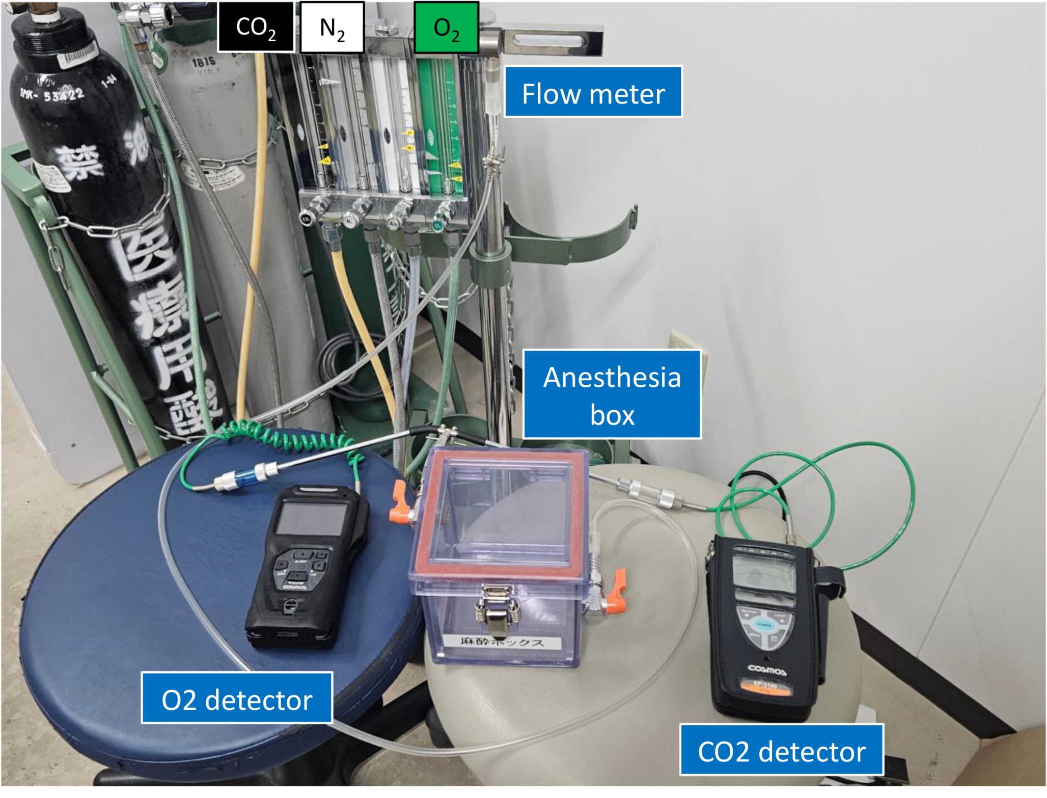
In the present study, we developed animal models to identify genetic markers for the differential diagnosis between CO2 intoxication and asphyxia due to oxygen deficiency. For Group D, the composition of the gas inhaled was 5% O2 and 95% N2. N2 is non-toxic and, like helium gas, is known to cause death by asphyxia due to oxygen deficiency when an enclosed space is filled with N2 [12]. In qRT-PCR analyses, Egr1 expression levels were significantly increased in Group B compared to Groups A and C, but there was no significant difference between Group B and Group D. Therefore, Egr1 is not a candidate for a specific diagnostic marker of CO2 intoxication. On the other hand, Nrgn expression levels were significantly increased only in Group B compared to the other three groups, suggesting that Nrgn may be a candidate diagnostic marker for CO2 intoxication. Since Ttr expression levels were significantly increased only in Group D compared to the other three groups, we consider Ttr to be a potential candidate for a specific diagnostic marker of hypoxia.
Neurogranin (Nrgn) is expressed abundantly in brain tissue and acts as “third messenger” substrate of protein kinase C (PKC)-mediated molecular cascades during synaptic development and remodeling [13]. The substrate, which is encoded by the Nrgn gene, binds calmodulin in the absence of calcium [14]. In the brain, Nrgn is expressed abundantly in the cerebral cortex, basal ganglia, hippocampus, thalamus, and hypothalamus, but is also expressed in the nuclei in the pons, such as lateral parabrachial nucleus, Kolliker-Fuse nucleus, and Solitary tract nucleus in the medulla oblongata, where the respiratory centers are located [15]. In recent years, Nrgn has been implicated in a variety of neurodegenerative diseases such as Alzheimer’s disease and Parkinson’s disease, psychiatric disorders such as schizophrenia and depression, and infectious diseases such as Creutzfeldt-Jakob disease, neuro-HIV, and neurosyphilis, and has been reported as a potential biomarker candidate for these conditions [16,17,18,19,20]. Among such studies, Hagihara et al. [18] reported that activity is increased, while working memory, prepulse inhibition, social behavior, and anxiety-like behavior are decreased in Nrgn knock-out (KO) mice compared to control mice, suggesting a schizophrenia model. They also observed increased brain lactate and lower brain pH in Nrgn KO mice. They hypothesized that the low brain pH is involved in the expression of various pre- and postsynaptic genes, including Nrgn, and causes schizophrenia-like symptoms. Therefore, we consider that the significantly increased Nrgn expression specific to the CO2 intoxication group (Group B) (Fig. 3c) is due to respiratory acidosis based on CO2 intoxication. On the other hand, there was no significant increase in Nrgn expression in Group C, despite the CO2 concentration (30%) being greater than that in the atmosphere (0.04%). As mentioned above, the induction of Nrgn expression may be caused by respiratory acidosis associated with increased CO2 concentration in the blood. In a previous study [21], there was a positive correlation between atmospheric CO2 concentration and the partial pressure of CO2 in the blood. The CO2 concentration in Group C (30% CO2) was lower than that in Group B (70% CO2), and it is thought that Nrgn expression did not increase because the concentration was not high enough to cause respiratory acidosis. Furthermore, even when the O2 concentration in the air is low, the CO2 concentration in the blood does not increase because CO2 is expelled as exhalation through breathing [22]. Therefore, we considered that there was no significant increase in the Nrgn expression in Group C.
Early growth response protein 1 (Egr1) is an immediate early gene (IEG) that regulates the transcription of various target genes and plays an important role in regulating responses to growth factors, DNA damage, and ischemia [23]. Egr1 is expressed widely in the brain, not only in the brainstem regions such as the midbrain, pons, and medulla oblongata, but also in the cerebral cortex, olfactory bulb, hippocampus, basal ganglia, thalamus, and hypothalamus [24]. In an in vitro model, Wen et al. [25] showed that Egr1 expression is increased in primary hippocampal neurons under hypoxic conditions. Egr family members, including Egr1, are upregulated in response to cellular stresses such as hypoxia and are involved in neuroprotection through the expression of target genes involved in cell growth, differentiation, and apoptosis. In our study, the significantly increased Egr1 expression in the CO2 intoxication group (Group B) compared to the control group (Group A) (Fig. 3b) may be due to respiratory center depression based on hypercarbonemia, resulting in cerebral hypoxia. In the hypoxia group (Group D), Egr1 expression levels did not increase significantly, but there was a trend toward an increase. Therefore, Egr1 cannot be a candidate as a postmortem diagnostic marker of CO2 intoxication, as it is thought to increase even in hypoxia.
Transthyretin (Ttr) is synthesized mainly by hepatocytes and epithelial cells of the choroid plexus (ChP), which are the sources of Ttr in plasma and cerebrospinal fluid, respectively [26]. Ttr is the carrier protein for thyroid hormones such as T3 and T4. Recent studies clarified that Ttr production by epithelial cells of the ChP is up-regulated to promote neuroprotection in the acute phase of ischemic stroke [27, 28]. Most studies using RT-PCR and in situ hybridization (ISH) indicate that Ttr is produced only in epithelial cells of the ChP in the brain and not in neurons and other resident brain cells. In our study, Ttr expression levels were significantly increased only in Group D, which may be a biological response to protect neurons from hypoxia. On the other hand, there was no significant increase in Ttr expression in Group C, despite the lower O2 concentration compared to the atmosphere. We assume that the hypoxia was not sufficient to cause a significant increase in Ttr expression in Group C, but was sufficient to increase Ttr expression significantly in Group D. In both humans and mice, the ChP consists of the left and right lateral ventricular ChP (LVChP), the third ventricular ChP (3rdChP), and the fourth ventricular ChP (4th ChP). Since the brainstem was used as the sample in this experiment, it is thought that Ttr production increased in epithelial cells of the 4th ChP contained in the brainstem. These results suggest that Ttr expression may be a candidate postmortem diagnostic marker for hypoxia.
Acid-sensing ion channel 4 (Asic4) is one of at least eight different Asic subunits (including Asic1a, 1b, 1b2, 2a, 2b, 3, 4, and 5) that are proton-gated voltage-independent ion channels [29]. Asics are expressed widely in the peripheral and central nervous system in both humans and mice. In the central nervous system, Asics are expressed in the pituitary gland most abundantly, but is also well expressed in brainstem regions, such as the pons and medulla oblongata, with roles in various important physiological functions and pathological states [30]. Although the role of Asic4 investigated in our study remains to be clarified, Asic1a has been found to exacerbate infarction by functioning to induce acid-induced neurotoxicity, i.e., neuronal cell death, during cerebral infarction [31]. Therefore, in the present study, the significant decrease in Asic4 expression in the experimental groups compared to controls may be a biological response to O2 deprivation in the brain. However, since Asic4 expression was decreased in all experimental groups compared with control, it is not a useful indicator for differentiating between CO2 intoxication and asphyxia due to oxygen deficiency, which was the main objective of this study.
Among other indicators we examined, Oprd1, which encodes a type of opioid receptor, and Sema3f, which encodes a receptor for the angiogenic factor Vascular endothelial growth factor, are both expressed in the central nervous system, and their expression has been reported to increase in response to hypoxia in the brain and to act in neuroprotection [32, 33]. Moreover, Tph2 encodes the rate-limiting enzyme for serotonin synthesis in the central nervous system and is expressed predominantly in serotonergic neurons in the raphé nucleus of the midbrain [34]. The elevation of CO2 concentration in the blood stimulates serotonergic nerves in the raphé nucleus, which in turn stimulates the motor nuclei of the diaphragmatic nerves in the spinal cord and promotes respiratory movement [35, 36]. However, contrary to our expectations, there was no significant variation in the expression of Oprd1, Sema3f, or Tph2 in experimental groups compared to control in this study. It is likely that the differences in O2 and CO2 concentrations and the time between inhalation of the gases and death prevented any significant variation.
Although few studies have investigated molecular diagnostic markers of CO2 intoxication, Sato et al. [37] reported that Alkylglycerone phosphate synthase (Agps) and Heat shock protein beta2 (Hspb2) expression in the frontal lobe and Pancreatic polypeptide (Ppy) and Corticotropin releasing hormone receptor 2 (Cchr2) expression in the hypothalamus were significantly increased in a rat model of CO2 intoxication and may be candidates for adjunctive diagnostic markers of CO2 intoxication. They concluded that the fluctuations in expression of those four molecules were caused by cognitive dysfunction and respiratory depression resulting from CO2 intoxication. Therefore, their research focuses on the symptoms of CO2 intoxication and targets the molecules involved. On the other hand, we focused on the pathogenesis of CO2 intoxication and used the brainstem region, where the respiratory center is located. Incidentally, neither Agps, Hspb2, Ppy, nor Crhr2 showed expression variation in their study, while, in this RNA-Seq study, we observed significant variation.
Limitations
In this study, RNA was extracted from the entire brainstem and mRNA expression was examined (Bulk analysis). Cells that showed significant expression changes in Nrgn, Egr1, and Ttr were only discussed based on previous literature and were not identified here. We intend to consider single cell analysis in future studies.
Although we identified candidate genes as potential markers for postmortem diagnosis of CO2 intoxication and hypoxia using murine models, it is necessary to conduct similar studies using human autopsy samples to determine whether they can be applied to actual forensic diagnosis.
The composition of the gases used in this study was adjusted to allow survival for 10 to 30 min after exposure to search for gene expression that varies significantly with CO2 intoxication and asphyxia due to O2 deficiency. However, in actual cases, the gene expression changes found in this study may not be observed because death occurs in a shorter period of time when exposed to extremely high concentrations of CO2 or low concentrations of O2. Future studies should examine whether the increase in gene expression observed here is also observed in models of short-term mortality from exposure to even higher concentrations of CO2 or lower concentrations of O2. In addition, the changes in gene expression over time after exposure to the gases in the present model should be examined.


Comments (0)