
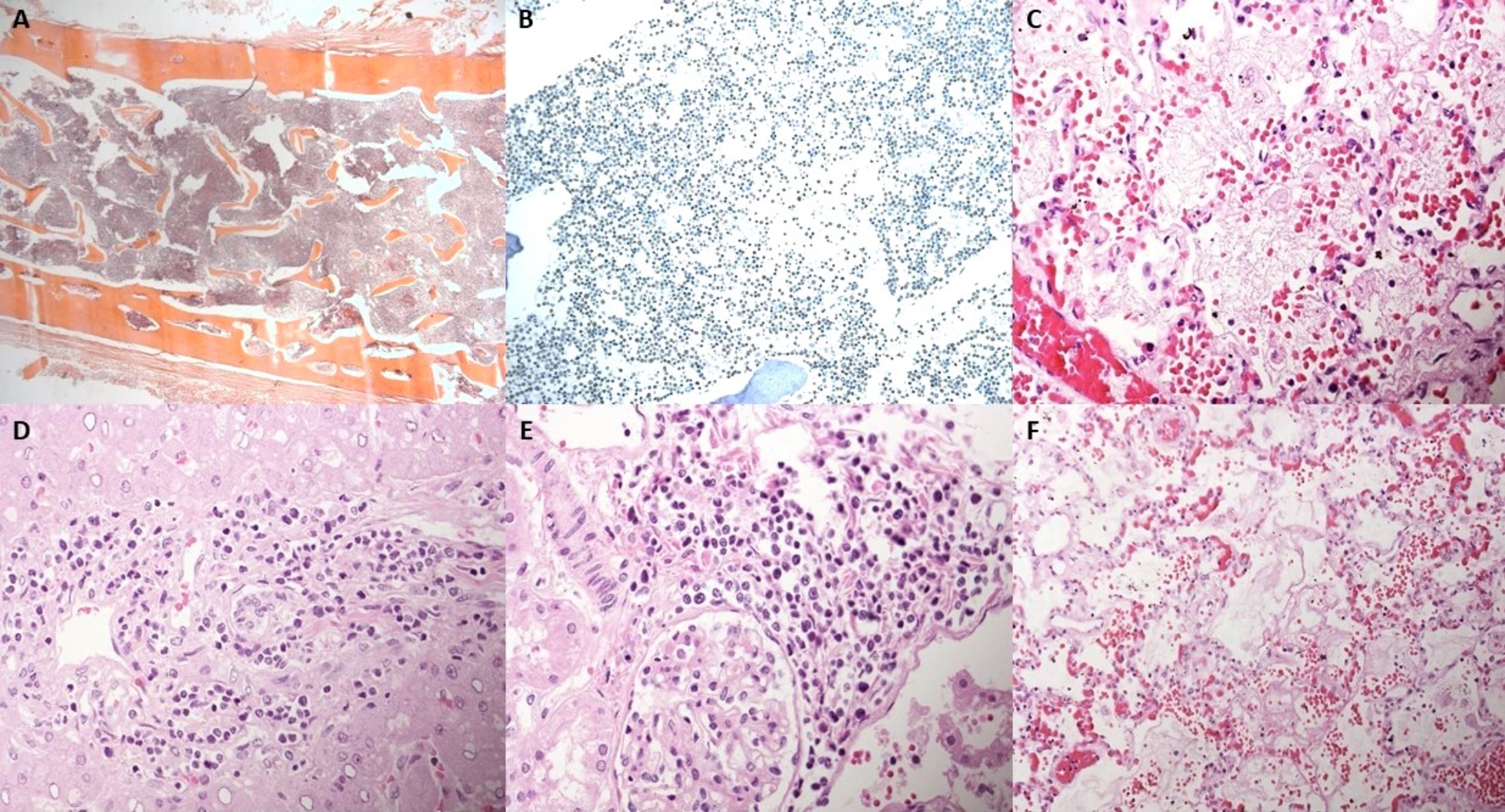
Remember me
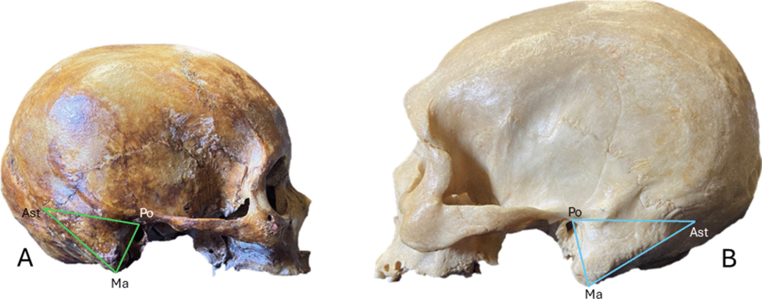

The European brown bear (Ursus arctos arctos) belongs to the extant family Ursidae and is characterized as a robust plantigrade carnivorans, with a body mass ranging from 150 to 250 kg [1, 2]. In addition to having a heavyset body profile, the dentition of the European brown bear consists of 42 teeth, including elongated tapering canines used for tearing and gripping, cuneiform incisors used for cutting, and carnassial teeth used for crushing and processing meat [2] (Fig. 3a). The forefeet (Fig. 3b) have slightly curved claws that are approximately twice as long as those on the hindfeet [3]. During attacks on humans, bears rear onto their hind legs and strike indiscriminately, with their forelimbs and teeth targeting the nearest body parts, primarily the neck, nape, and head regions [2, 4, 5]. The brown bear subsequently subdues its prey by holding it down with one forelimb while using the other forelimb to inflict injuries [5].
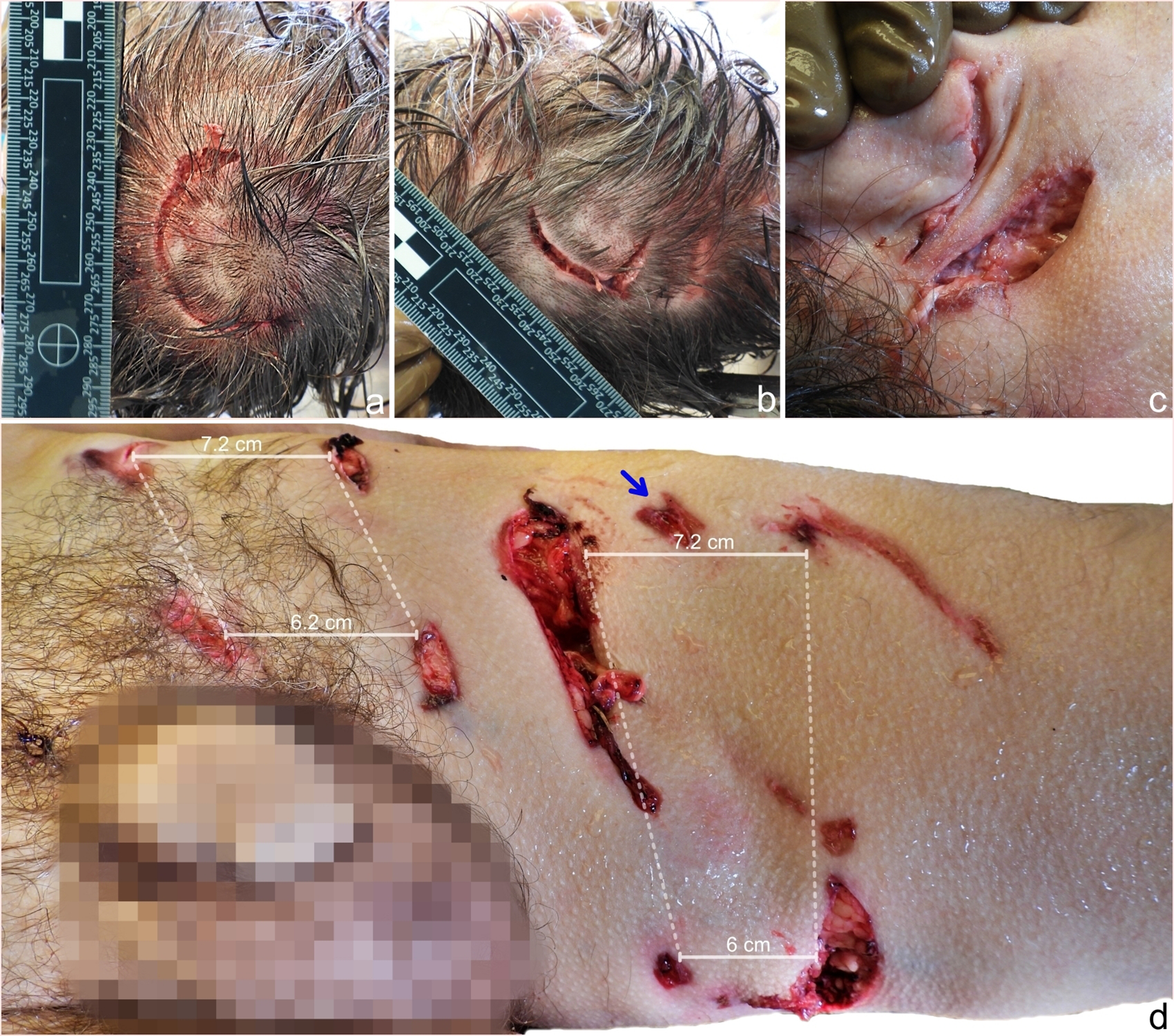
Fig. 3
a– Dentition of a brown bear, consisting of six incisors (black asterisk) between each pair of canine teeth (white asterisk). Carnassial teeth are located behind the canine teeth (white arrow) b– Forefoot of a brown bear with five nonretractile claws
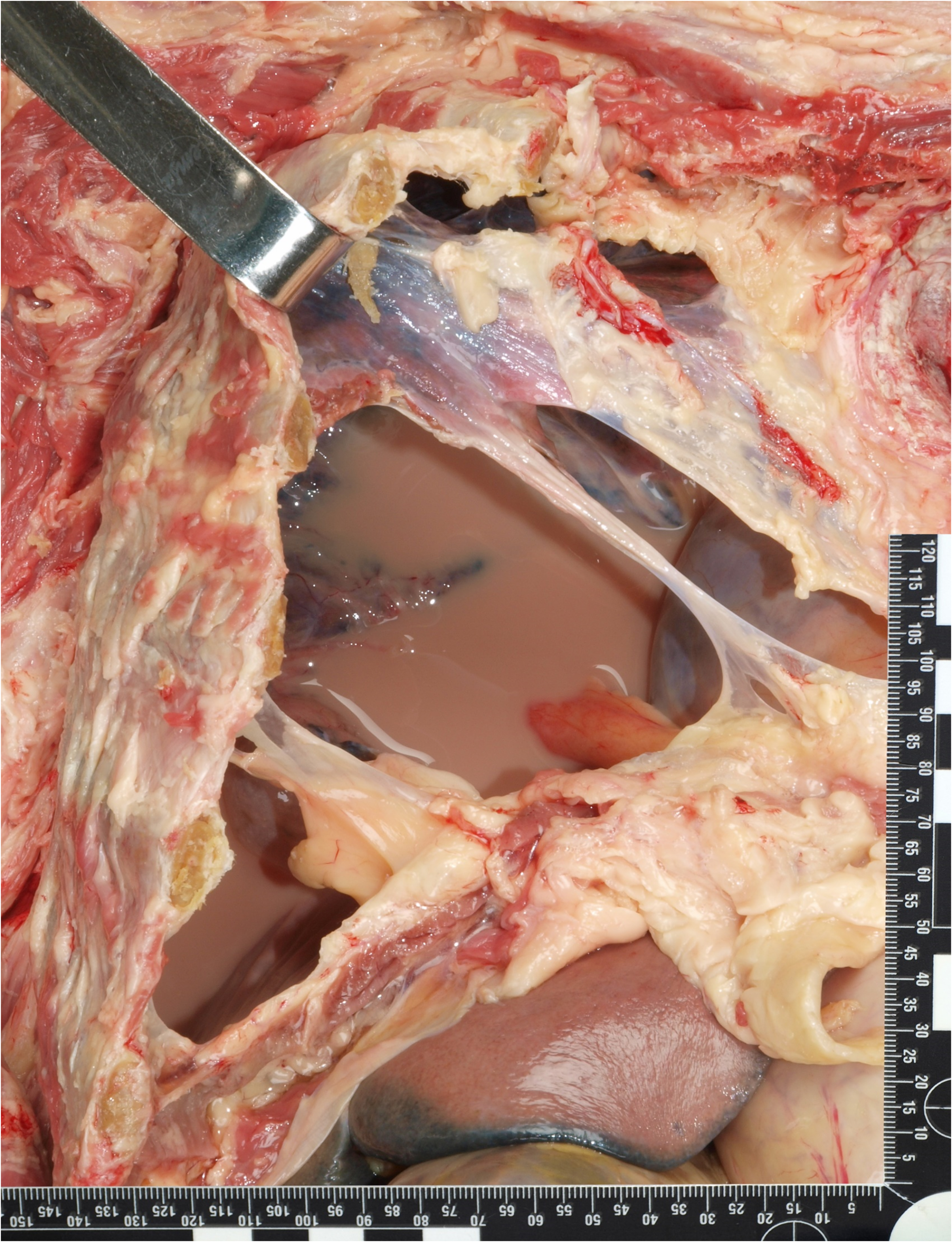
The appearance of a bite mark corresponds to the morphology of the teeth, their arrangement within the dental arch, the bite force applied and the movement of the jaws. The average length of the brown bear’s canines ranges between 3.4 and 4.3 cm and rarely exceeds 5.5 cm [6]. On the basis of the tapered ends, conical shape, and lateral placement of these canines within the jaw, the injury pattern consists of four circular puncture wounds in a rectangular arrangement, with the diameter of the punctures not exceeding 2.3 cm. The maxillary intercanine width is typically greater than the mandibular intercanine width, with the difference varying from a few millimeters to 1.5 cm [6]. The difference in the intercanine width reflected in the wounds also depends on the canine width and bite depth. The injury pattern formed by cuneiform incisors manifests as six adjacent abrasions among the canine wounds. The semi-arched arrangement of the maxillary incisors can be distinguished from the more linear arrangement of the mandibular incisors. In superficial bites, the diameter and depth of canine-inflicted wounds are reduced, and incisor marks may not be present [6]. Bite force is directly correlated with the severity of bite injuries, which primarily contribute to blunt trauma components, such as contusions and bone fractures. The average canine and molar bite forces of brown bears are 1627 N and 3175 N, respectively [7]. The force tolerance of adult facial bones (zygomatic bone and maxilla) ranges from 490 to 1800 N (50–183 kg), which are well within the bite force ranges of brown bears [8]. When ripping flesh from bones, the bear moves its jaws, resulting in torque, leading to the laceration and avulsion of superficial soft and deep tissues and altering the morphology of primary bite marks. In these two cases, the morphology of the puncture wounds on the left thigh differed due to the violent torque movement of the jaws, which led to ruptures of the underlying soft tissues, as shown in Fig. 1d. The limbs of brown bears include five nonretractile claws with lengths up to 8 cm, which are used for digging up tubers and small prey [3]. Injuries incurred by clawing and mauling range from superficial linear abrasions to avulsed lacerations with decollement [4]. Grievous injuries, such as neurocranial fractures and brain evisceration attributed to mauling, have also been described in the literature [9, 10].
Most large carnivore attacks in Europe involve brown bears [11]. The injuries caused by bear assaults predominantly occur in craniofacial regions or as defensive wounds on the upper extremities [4]. Dog attacks are associated with similar wound topographies, including soft tissue trauma; however, claw-inflicted injuries are absent [12]. The most frequent cause of death in bear attacks is exsanguination [5]. The less frequent causes include cervical spine injuries, brain evisceration and air embolism [5, 9, 10, 13]. In contrast, cervical trauma is the leading cause of death in attacks by large felids, such as lions, tigers, and leopards, due to their distinct hunting behavior [14,15,16]. The forensic relevance of deaths due to animal attacks lies in differentiating the observed injuries from homicidal and self-inflicted wounds [17]. The presented cases highlight the importance of meticulous external examination with morphometry of the injuries and site observation. Additionally, biological samples can be used to identify the animal responsible for the attack and improve investigative processes.
Comments (0)