Chronic obstructive pulmonary disease (COPD) is a relatively common progressive respiratory condition primarily linked to smoking. In 2022, approximately 2.5% of Australians were living with COPD which contributed to 4% of deaths that year [10]. Chronic pulmonary inflammation leads to excess mucus production, airway narrowing, and loss of alveolar structure. Over time, these processes result in chronic scarring and irreversible airway obstruction [11]. Hyperinflation, a feature of emphysema (a COPD subtype), occurs when elastin in airway walls is destroyed reducing recoil. This results in airway collapse during exhalation and air trapping distally. Additionally, destruction of alveolar septa leads to larger alveoli and increased lung volume. Lungs with increased residual volume have reduced inspiratory capacity, as they are already close to their maximum level. Larger lung volumes may also flatten the diaphragm, reducing its ability to in generate ventilatory forces [12]. These changes significantly impact upon quality of life causing dyspnea and productive coughs.
Endobronchial valves are relatively recent bronchoscopically-inserted devices that are designed to specifically treat hyperinflation resulting from emphysema. They function as one-way valves, blocking air from entering diseased tissue causing atelectasis of the emphysematous regions, allowing healthier lobes to expand and relieve pressure on the diaphragm, improving ventilation [13, 14]. Prior to the use of endobronchial valves surgical removal of hyperinflated and dysfunctional lobes had been shown to enhance ventilation [15, 16]. Endobronchial valves, however, improve spirometry-measured lung function without the need for surgery, increasing forced expiratory volume in the first second (FEV1) and decreasing residual volume [17].
The most common complication of endobronchial valves is pneumothorax, which occurs in approximately 20% of cases, although the range is 4.6–26%. The mechanism behind pneumothorax is believed to be increased negative pressure in the pleural space after atelectasis of contralateral lung lobes, leading to bullae disruption and pneumothorax. Other complications include infection-related issues such as pneumonia and more frequent COPD exacerbations [18, 19].
Failure or ineffectiveness of the valve, if not due to migration or incorrect placement, may result from disease progression with mucus or bacterial aggregation preventing valve closure. Valve migration occurs in approximately 1.9% of cases and may lead to foreign-body airway obstruction [19, 20].
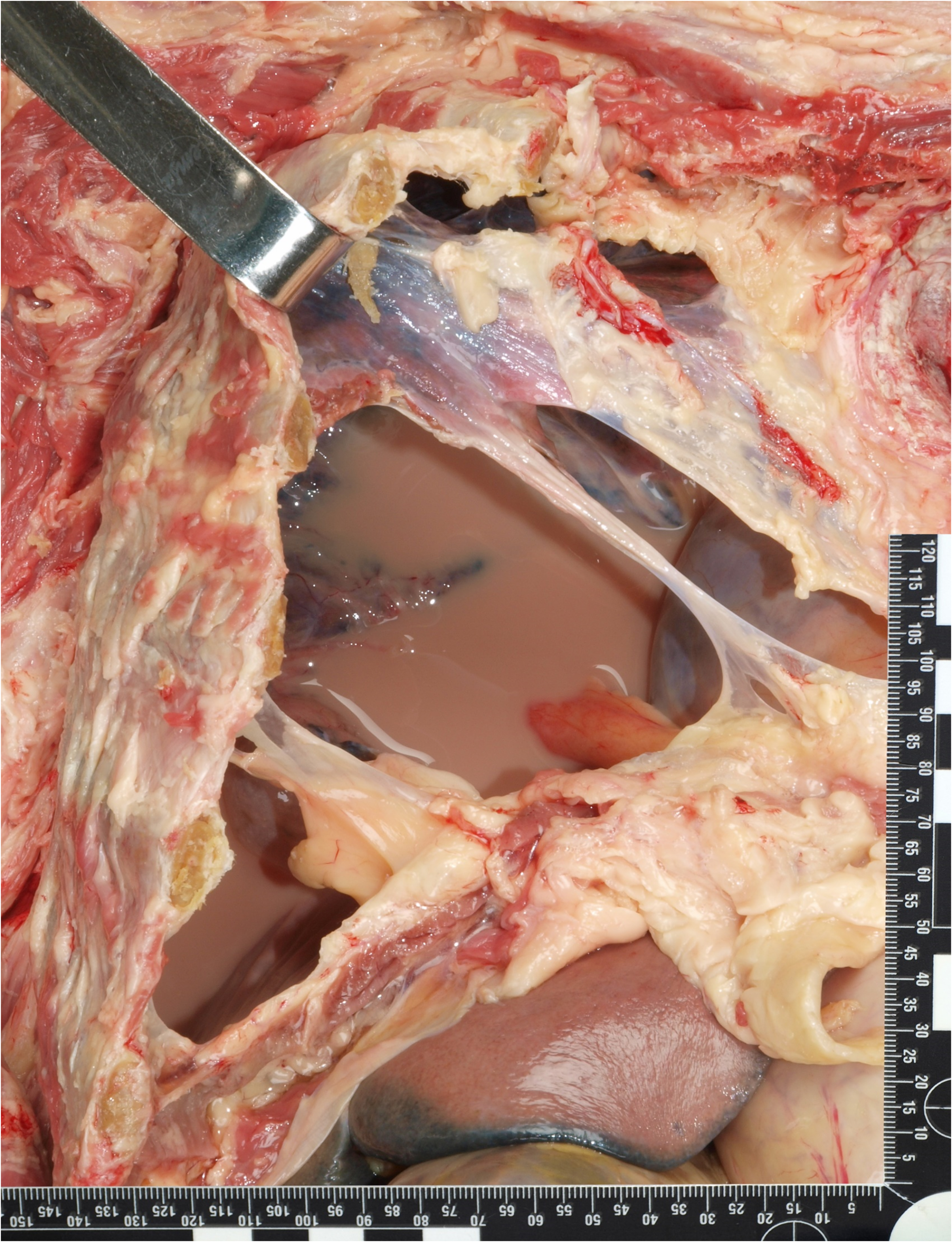
As was observed in the reported case endobronchial valves may also be associated with hemoptysis, although this is uncommon and usually not medically significant. Minor hemoptysis has been reported in 1.9–5.6% of cases and is often associated with granulation tissue formation, as in the current case [18,19,20,21]. It should be recognised therefore that more recent endobronchial treatments for emphysema may be responsible for hemoptysis in decedents with COPD rather than this finding raising the spectre of potentially more sinister inflammatory or neoplastic lesions. These should, however, still be excluded at autopsy. As there may be no history of endobronchial valve insertion provided at the time of autopsy PMCT scanning provides a very useful screening tool for such devices.

Comments (0)