
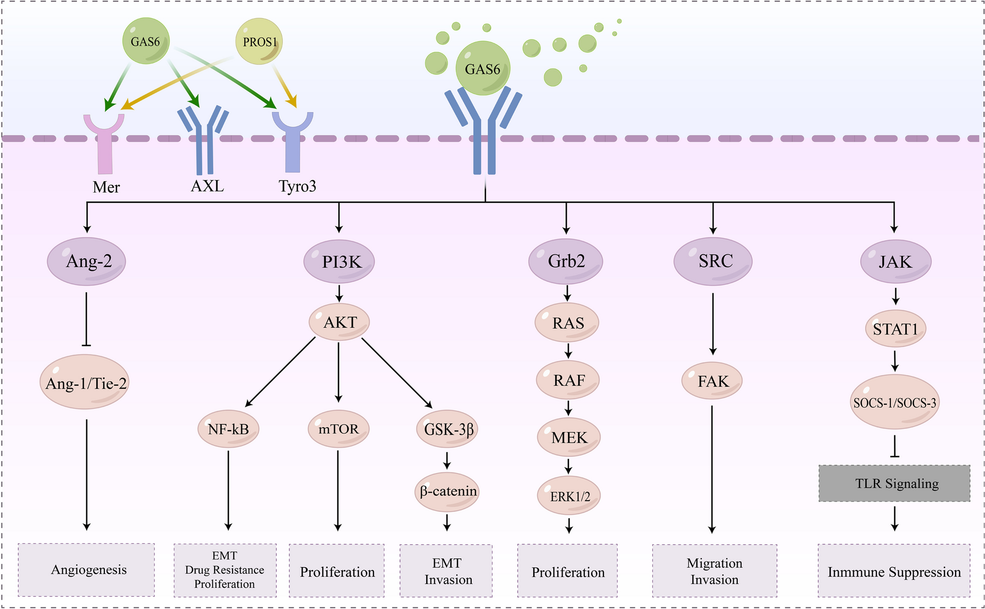
Remember me


Please refer to Additional File 1, Supplementary Methods for the materials and methods of this study. To explore the effects of DLA for PDAC CTCs, we followed the workflow illustrated in Fig. 1A. DLA with sufficient MNC collection efficacy (median: 49%; range: 8–71%; Fig. 1B-C) for this purpose was performed in 60 PDAC patients (DU-DLA-cohort) without technical issues or notable adverse effects. Although lower efficacies also produced CS-CTC positive samples, DLAs with a collection efficacy equal to or above the median resulted in a higher CS-CTC-detection frequency (11/30 vs. 21/30; Fisher’s exact test: p = 0.02, Fig. 1C). From the processed median blood volume of ~ 2.8 L (2,795mL, range: 769-5,765mL) we generated DLA products with a median volume of 45mL (range: 10-100mL). The capacity of the CS-assay required limiting the sample input to around 200*106 WBC [11], which represented approximately 5% of a DLA product. The remaining material was cryopreserved as aliquots. The calculated CS-input corresponded to a median blood volume of ~ 70mL (70.6mL, range: 20.50mL-181.03mL) (Additional File 1, Supplementary Fig. 1). Applying this process, we observed a significantly increased detection frequency of 53% (32/60) in DLA compared to 19% in matched PB samples (11/58; Fisher’s Exact Test, p < 0.001). With 1.05 (±1.51) CTC/mL the mean CS-CTC concentration was 10-times higher in DLA compared to 0.10 (±0.37) CTC/mL in PB (Fig. 1D). To estimate the overall CTC-load in DLA products, we extrapolated the detected CS-CTCs to the entire DLA product, revealing a calculated median of 71.5 CTCs (range: 0-218) per DLA product and a 60-fold (mean) increase in CTC numbers compared to PB. This enrichment suggests that PDAC-CTCs fall into the density window targeted by our DLA settings. Next, we aimed to determine the DLA-product volume, required to analyze for detecting at least one CS-CTC in every PDAC patient. Fitting our PB and DLA data to a distribution function described previously for PB [9], we ascertained that the distribution function also holds for the larger volumes of the DLA product. Furthermore, we extrapolate that analyzing a DLA product with a corresponding median blood volume of 1000mL is expected to yield 1 CTC in 96% of samples, 3 CTC in 88% of samples and 10 CTC in 67% of samples (Fig. 1E). This volume is equivalent to approximately 76% of the DLA product (34mL). The 30% discrepancy between the volumes derived from PB samples (7.5mL) and DLA samples (~ 70mL) indicates the CTC loss incurred in the DLA sample preparation process as compared to PB. However, it is important to stress that forthcoming technological advancements [10, 17], proficient in managing the designated DLA volumes, should feasibly enable processing of the required DLA-product volume in an imminent time frame.
Uncovering the genomic alterations of PDAC CTCs through DLA-enabled profilingOwing to their rarity in PB, genomic data on CS-CTCs in PDAC have remained unavailable until now. To demonstrate the feasibility of employing DLA-enriched CS-CTCs as a surrogate for molecular profiling and to validate their malignant character, we re-analyzed cryopreserved material from CS-CTC-positive DLA samples. Notably, the quantity of CS-CTCs in the frozen DLA samples was not significantly deviant from those freshly and prospectively analyzed (Additional File 1, Supplementary Fig. 2). These results affirm that processing the entire DLA product could further elevate the sensitivity of our approach, as was suggested by our extrapolation. After isolation, we conducted genomic profiling of 11 single CS-CTCs and matched tumor tissue of four patients (#2, #7, #17, #22), including one patient (#22) in whom relevant copy number alteration (CNAs) were not detected in the tumor tissue (Fig. 1F-G; Additional File 1, Supplementary Table 4). In two patients (#2 and #22), KRAS and TP53 mutations were not detected in either the CTCs harboring CNAs or the matched tumor tissue. Patient #7 exhibited consistent detection of a G12V mutation in both CTCs and tumor tissue that displayed an additional G12D mutation (Fig. 1F-G; Additional File 1, Supplementary Table 4). In patient #17, CTCs showed a G12V mutation while a G12R mutation was observed in the matched tumor tissue (Additional File 1, Supplementary Table 4). As a control, we analyzed 26 matched and non-matched white blood cells isolated from CS cartridges of thawed PDAC DLA samples and did not detect any pathogenic mutations (Additional File 1, Supplementary Table 4). The different hot-spot mutations between tumor tissue and CTCs prompted us to perform subsequent short tandem-repeat analysis (STR), which authenticated the sample identity (data not shown). Collectively, this exemplary genomic CTC-profiling not only confirmed the malignant nature of DLA-enriched CS-CTCs but also demonstrated the feasibility of performing comprehensive genomic analysis on CS-CTCs from DLA-products, providing additional information compared to PDAC tissue. Considering the recognized intra-patient molecular heterogeneity, employing their versatile analysis could offer substantial advantages over classical tissue biopsies in metastatic PDAC. This is particularly pertinent in the context of emerging molecular therapies, which will necessitate diagnostic molecular tumor profiling [2, 18, 19].
DLA enriched CS-CTCs are linked to higher tumor stage and poor survivalTo validate the clinical relevance of CS-CTCs enriched by DLA, we examined their association with clinical parameters. Given the individual variations in assay input (Additional File 1, Supplementary Fig. 1), CTC counts were normalized to the median analyzed blood-equivalent of ~ 70mL, with resulting decimal numbers rounded to the nearest integer (range: 0–40 CTCs). Although CTC count normalization was crucial for accurate concentration and survival analyses, notable differences in detection frequencies between M0 and M1 patients were evident without this adjustment. For DLA samples, detection rates were 18/41 for M0 patients and 14/19 for M1 patients (Fisher’s exact test: p = 0.0507, see Fig. 2A). Meanwhile, for PB samples, the detection rate was 4/41 for M0 patients and 7/17 for M1 patients (Fisher’s exact test: p = 0.0099). Notably, only DLA samples demonstrated a significantly higher CS-CTC concentration in M1 patients compared to M0 patients (median: 3.0 vs. 0.0, Mann-Whitney U test: p = 0.024, Fig. 2B). Subsequently, we tested the prognostic value of DLA-enriched CS-CTCs and adopted a cut-off of ≥ 1CTC per ~ 70mL, aligning with thresholds used in existing PDAC studies involving CS [7, 8]. Additionally, to determine a more robust threshold for DLA, we incorporated ≥ 3CTC/~70mL, considering a ~ 5% probability of detecting 0 CTC with an average detection frequency/threshold of 3 CTCs per sample versus a ~ 36% probability at a mean detection frequency/threshold of 1 CTC per sample, due to the Poisson distribution. Strikingly, DLA-derived CS-CTC revealed prognostic significance at both thresholds, with one CTC per sample reducing median overall survival (OS) to 8.5 months, compared to 28.6 months without CS-CTC (log-rank test: p = 0.002; Fig. 2C), and ≥ 3CTCs reducing OS to 10.2 months vs. 25.6 months below this threshold (log-rank test: p = 0.033; Fig. 2D). Upon multivariable analysis, the adverse effect of the threshold of ≥ 1CTC retained its significance, underscoring prognostic relevance. The prognostic effects of DLA CS-CTC outperformed those observed for CS-CTC in matched PB samples (Fig. 2C and D, Additional File 1, Supplementary Table 5), especially concerning sensitivity and accuracy in identifying patients at high risk for short OS (Additional File 1, Supplementary Table 8). The superior prognostic impact of DLA CS-CTCs was also evident in M0 patients and those treated with curative intent (Additional File 1, Supplementary Fig. 3, and Supplementary Tables 6–8). Notably, a relatively high OS-rate was apparent for M0 patients without DLA CS-CTC (e.g., at 20 months), while DLA CS-CTC-positive M0 patients exhibited similarly poor OS compared with CS-CTC-negative M1 patients (Fig. 2E). As anticipated, CS-CTC-positive M1-patients displayed the worst prognosis. Interestingly, an analysis for CTC-cutoff values, which considered both hazard ratios and AUC values collectively (Fig. 2F and G), confirmed our tested cutoffs as optimal. Expectedly, elevating CTC-cutoff values enhanced specificity while diminishing sensitivity. Further validation of the prognostic value of CS-CTCs in PDAC was sought through analyzing PB samples in an independent, large cohort (HE: n = 170) and in combination with the DU-PB-cohort (n = 228; Fig. 2H, Additional File 1, Supplementary Table 9), affirming the significant and independent prognostic impacts of CS-CTCs across various subgroups (Additional File 1, Supplementary Fig. 5 and Supplementary Tables 9–11), albeit at low detection rates. Intriguingly, even with the low detection frequency, the stage-dependent effects of CS-CTCs were congruent with the observations made in DLA CS-CTCs (Fig. 2E and H). Taken together, the prognostic data suggest that CS-CTCs could serve as a useful biomarker to select M0-patients for intensified multimodal treatments and to guide cancer-directed surgery in the challenging situation of oligometastatic PDAC [20]. For CTC + M1-patients, tumor resection may be contraindicated, whereas for M1-patients negative for DLA-CS-CTCs resection may be beneficial if technically feasible. Nonetheless, this premise requires further validation through prospective clinical studies.
Fig. 1
Study overview and DLA findings: A Experimental setup (created in part with BioRender.com); B MNCs per mL in peripheral blood samples (D-PB, n = 60) versus matched DLA samples (D-DLA, n = 60); C Collection efficacy achieved for the targeted MNC population (n = 60). The filled circles indicate CTC-positivity (≥ 1 CTC detected); D CTC-concentration per mL in PB samples (light grey circles) and matched DLA samples (dark grey circles); E. Extrapolation of log-logistic fit curves for peripheral blood samples (PB) and DLA samples (DLA). Stair plots show the empirical CDF and the continuous line the fit. Extrapolation indicates probability (Cumulative Distribution) for a certain analyzed sample volume to have at least 1 CTC or 3 CTCs in every sample; F Genomic analysis of individual CS-CTCs isolated from DLA products with panel sequencing of KRAS and TP53 genes (T: tumor tissue); G Plots of copy number profiles along the 22 autosomes expressed as log2 ratios. Significant copy number gains and losses are highlighted in red and blue, respectively (M: FFPE material from metastatic tissue)
Fig. 2
Association of PDAC CS-CTC with clinical parameters. A Frequency of CTC-positivity (≥ 1 CTC detected) in DLA products according to the metastatic stage; B Normalized CS-CTC-concentration in DLA samples according to the metastatic stage; C-D OS according to status of DLA CS-CTC (n = 60, CTC/~70mL) in green/red compared to PB CS-CTC (n = 58, CTC/7.5mL blood) in grey. C Cutoff for CTC positivity: ≥1 CTC; D Cutoff for CTC positivity: ≥3 CTC; E OS according to the CTC-status separated by metastatic stage in DLA products (optimal cutoffs: M0 = 3CTC/~70mL blood; M1 = 23CTC/~70mL blood, see Additional File 1, Supplementary Fig. 4); F ROC curves and corresponding AUC values for the top 10 DLA CS-CTC cutoffs normalized to ~ 70mL of blood. These cutoffs were selected based on their combined highest hazard ratio and AUC value in the DLA cohort of 60 PDAC patients (ref: reference); G Parameters for all significant (p < 0.05) DLA CS-CTCs cutoff values calculated in the cohort of 60 PDAC patients (Cutoff: CTCs/~70mL); H OS according to CS-CTC status (≥ 1CTC/7.5mL blood) in PB pooled HE/DU-PB cohorts separated by metastatic stage (M0 vs. M1)
Comments (0)