Peptide Synthesis
All peptides were synthesized using an Fmoc/tBu protection strategy on Rink amide resin (0.68 meq/g, 100–200 mesh). All Fmoc-amino acids were sourced from Chem-Impex (Wood Dale IL, USA), as follows: Ala, Cys(Trt), Asp(OtBu), Glu(OtBu), Gly, His(Trt), Leu, Lys(Boc), Lys(N3), Met, Phe, Pro, Ser(tBu), Thr(tBu), Tyr(tBu). 5-hexynoic acid was purchased from Combi-Blocks (San Diego CA, USA).
Rink amide resin was swelled for 10 min in N, N-dimethylformamide (DMF). A PS3 peptide synthesizer (Gyros Protein Technologies, Uppsala, Sweden) was used to assemble linear peptide chains by iteration of the following steps: deprotection with 20% (v/v) piperidine in DMF (2 × 5 min); DMF wash (3×); agitation for 50 min in activated amino acid solution comprising Fmoc-amino acid (3 eq.) and O-(1H-6-chlorobenzotriazole-1-yl)-1,1,3,3-tetramethyluronium hexafluorophosphate (HCTU, 3 eq.) in 7% (v/v) N, N-diisopropylethylamine (DIPEA) in DMF; DMF wash (3×). 5-hexynoic acid was manually conjugated to each shuttle peptide after the final Fmoc deprotection step, under the same conditions used for amino acid coupling. The completed peptide resins were washed with DMF (3×), methanol (3×) and diethyl ether (3×), then dried using a vacuum pump.
A cleavage solution comprising 85% (v/v) trifluoroacetic acid (TFA), 5% H2O, 5% thioanisole, 2.5% triisopropylsilane (TIPS) and 2.5% ethane-1,2-dithiol (EDT) was used to remove sidechain protecting groups and cleave the peptide from the resin. The cleavage cocktail was incubated with the resin for 2 h with agitation at ambient temperature (note that longer incubation times led to increased reduction of the azide to the primary amine). The resin was removed from the mixture by filtration, and TFA was evaporated using a nitrogen stream, followed by precipitation of the peptide with excess cold diethyl ether. The peptide was collected by centrifugation (3000×g, 4 min) and the pellet washed again by resuspension in ether followed by centrifugation. The final crude material was dissolved in 50% H2O/50% acetonitrile (ACN) and lyophilized.
Reversed-Phase High-Performance Liquid Chromatography
Crude peptides were resuspended in 95% buffer A (0.1% v/v TFA in H2O)/5% buffer B (0.1% v/v TFA in ACN), then filtered using a 0.22 μm syringe filter. Samples were separated using a Vydac C18 reversed-phase high-performance liquid chromatography (RP-HPLC) column (250 × 10 mm, 10 μm) operated on an Agilent 1260 Infinity II liquid chromatography system. The column was eluted at a flow rate of 4 mL/min using linear gradients as follows: KN3-HsTX1[R14A] (reduced), 15–30% buffer B over 15 min; Hex-MTf, and Hex-Ang2, 15–45% buffer B over 30 min. Collected fractions were analyzed by liquid chromatography-mass spectroscopy (LC-MS) for identity and purity on a Phenomenex Luna C8(2) reverse-phase column (100 Å, 3 μm, 100 × 2.0 mm) fitted on a Shimadzu 2020 LC-MS, using a flow rate of 0.2 mL/min and a gradient of 0–60% B (buffer A: 0.05% v/v TFA in H2O; buffer B: 0.05% v/v TFA in ACN) over 15 min unless otherwise stated. Fractions containing pure peptide with the correct mass were lyophilized and stored at -20 °C.
Oxidative Refolding of Peptides
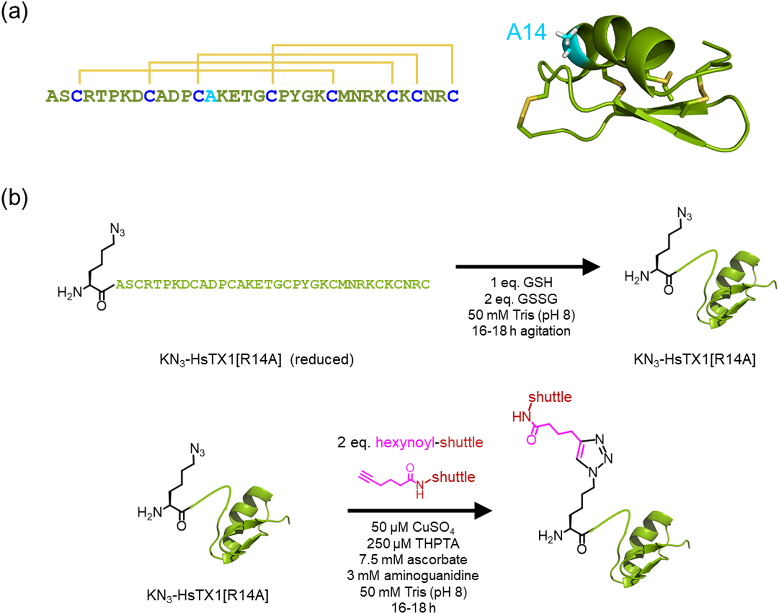
HPLC-purified KN3-HsTX1[R14A] (reduced) was dissolved (1–2 mg/mL) in 50 mM Tris pH 8 containing reduced glutathione (1 eq. relative to peptide) and oxidized glutathione (2 eq.), and air oxidized overnight with gentle agitation. The reaction mixture was purified by RP-HPLC as described above, using a linear gradient of 15–23% buffer B over 16 min at a flow rate of 4 mL/min, and fractions containing pure oxidized KN3-HsTX1[R14A] were lyophilized.
Copper-Catalyzed Azide–Alkyne Cycloaddition (CuAAC)
CuAAC was conducted under conditions adapted for bioconjugation (Hong et al. 2009). Purified oxidized KN3-HsTX1[R14A] was dissolved in 50 mM Tris (pH 8) to a final concentration of 0.1–0.3 mM, and 2 eq. of the alkyne-BBB shuttle and aminoguanidine (3 mM) was added. Tris(3-hydroxypropyltriazolylmethyl)amine (THPTA) was pre-combined with CuSO4 in a 5:1 molar ratio before addition to the reaction mixture. Finally, sodium ascorbate (5–7.5 mM) was added, the reaction vessel sealed with parafilm and incubated at ambient temperature overnight. Peptides were purified by RP-HPLC using gradients as follows: MTf-HsTX1[R14A], 17–32% buffer B over 30 min; Ang2-HsTX1[R14A], 20–35% buffer B over 30 min.
Nuclear Magnetic Resonance (NMR) Spectroscopy
Purified peptides (0.1–0.5 mg) were dissolved in 5% 2H2O in MilliQ water and the pH measured. One-dimensional 1H NMR spectra were recorded on a Bruker Avance III 600 MHz spectrometer at 298 K. 1H chemical shifts were referenced to dioxane (at 3.75 ppm). Spectra were processed and analyzed in Topspin (version 4.5.0).
Patch-Clamp Electrophysiology Assays
To obtain human T lymphocytes, heparinized human peripheral venous blood was drawn from healthy volunteers. Mononuclear cells were separated using Histopaque-1077 (Sigma-Aldrich) separation method following approval from the Ethical Committee of University of Debrecen, (DE RKEB/IKEB 6627-2023). Cells were cultured (density 5 × 10⁵ cells per mL) in Roswell Park Memorial Institute (RPMI) 1640 medium (Gibco) supplemented with 10% FBS, 2 mM l-glutamine and 100 µg/mL streptomycin and 100 U/mL penicillin-g in a humidified incubator at 37 °C and 5% CO2 for 3–6 days. Phytohemagglutinin A (PHA, Sigma-Aldrich) was also added at a concentration of 2, 5, and 10 µg/mL to activate the T lymphocytes and boost KV1.3 expression. Patch-clamp experiments were performed after 3–6 days of activation.
All measurements were performed using an Axon MultiClamp700B amplifier connected to a personal computer with Axon Digidata 1440 A data acquisition hardware and for data acquisition, Clampex 10.7 software was used (Molecular Devices, Sunnyvale, CA). Whole-cell currents were recorded in voltage-clamp mode following the standard protocols as described previously (Bartok et al. 2015; Naseem et al. 2022). Micropipettes were made from GC150F-7.5 borosilicate capillaries (Harvard Apparatus Co., Holliston, MA, USA) using a Sutter P-1000 puller with tip resistance generally ranging from 3 to 6 MΩ in the bath solution. To evoke the KV1.3 currents in activated T lymphocytes, 15-ms-long voltage pulses to + 50 mV from a holding potential of −120 mV were applied every 15 s. All recordings were carried out at room temperature (20–25 °C). Control and test solutions were perfused to the cells via a gravity-driven micro-perfusion system and AutoMate Perfusion Pencil Multi-Barrel Manifold Tip (AutoMate Scientific, Berkeley, CA, USA), and the excess bath solution was continuously removed from recording chamber using vacuum suction. The complete exchange of solution in the bath chamber i.e., the proper operation of the perfusion apparatus was confirmed frequently using 10 mM TEA+, a reversible inhibitor of KV1.3, as positive control at a concentration equivalent to its IC50 value.
The bath (extracellular) solution consisted of (in mM) 145 NaCl, 5 KCl, 2.5 CaCl2, 1 MgCl2, 5.5 glucose, and 10 HEPES, pH 7.35. Equimolar substitution of Na+ for tetraethylammonium–Cl was used in TEA+-containing positive control solution. The measured osmolarity of the bath solution was between 302 and 308 mOsM. To prevent peptide adsorption to the plastic surfaces of the perfusion system bath solutions were supplemented with 0.1 mg/mL bovine serum albumin (BSA, Sigma-Aldrich, Hungary). The composition of internal solution was (in mM) 140 KF, 2 MgCl2, 1 CaCl2, 10 HEPES and 11 EGTA, pH 7.22. The measured osmolarity of internal solutions was ~ 295 mOsm/L.
Data were analysed using pClamp 10.7 software package (Molecular Devices, Sunnyvale, CA). The remaining current fraction (RCF) were fitted using the Hill equation:
$$\:RCF=\frac}}_}=\frac\text}_}^}}\text}_}^}+\text\text\text\text\text\text\right]}^}},$$
where I and I0 are the peak currents measured in the presence and absence of peptide, respectively (giving the ratio RCF, remaining current fraction), IC50 is the concentration of the peptide that gives 50% block, [peptide] is the concentration of peptide (M), and H is the Hill coefficient.
Buffer and Plasma Stability Assays
The stability of HsTX1[R14A] or MTf-HsTX1[R14A] was analyzed in Hanks Balanced Salt Solution (ThermoFisher Scientific, Rockport, IL) containing 10 mM N-2-hydroxyethylpiperazine-N-2-ethane sulfonic acid (HEPES; Sigma-Aldrich, St Louis, MO) at pH 7.4, hereafter referred to as HBSS. HBSS was pre-warmed at 37 °C and 20 µM of either HsTX1[R14A] or MTf-HsTX1[R14A] was added to three separate vial replicates, which were maintained at 37 °C for the duration of the study. Samples were taken at 0, 5, 15, 30, 60, 90 and 120 min timepoints. The stability of MTf-HsTX1[R14A] was also assessed in the plasma of adult C57BL/6 mice. Plasma was aliquoted to three separate vials and pre-warmed at 37 °C. MTf-HsTX1[R14A] was added to the plasma at 800 nM. Samples were taken at 0, 15, 30, 60, and 120 min.
In Vitro BBB Model
hCMEC/D3 cells were obtained from Merck Millipore and utilized between passages 5–11. Primary brain microvascular endothelial cells were isolated from C57BL/6 mice (Monash Institute of Pharmaceutical Sciences Ethics application MIPS 29011) using a magnetic activated cell sorting approach with a CD31 antibody, as previously described in detail (Runwal et al. 2025). hCMEC/D3 cells were seeded onto Transwell (0.4 μm, polyester) inserts within a 24-well plate coated with rat-tail collagen Type I (100 µg/mL). Primary mouse brain microvascular endothelial cells were seeded onto Transwell (0.4 μm, polyester) inserts within a 24-well plate coated with 100 µL of solution containing 40% (v/v) Type IV collagen, 10% (v/v) fibronectin and 50% (v/v) UltraPure™ distilled water. hCMEC/D3 cells were used for permeability studies 7 days post-seeding. For primary mouse brain endothelial cells, permeability studies were commenced when the calculated transendothelial electrical resistance (TEER) values reached greater than 150 Ω.cm2, which typically took 6 to 7 days.
Plates were maintained at 37 °C for the duration of the permeability study on the THERMOstar (BMG Labtech, Ortenberg, Germany). HsTX1[R14A] or MTf-HsTX1[R14A] were added to the donor chamber at 10 and 20 µM for primary and hCMEC/D3 cells, respectively (n = 4). Samples were taken from the donor chamber at the start of the experiment and from the acceptor chamber at 5, 15, 30, 60, 90 and 120 min, and replaced with equal volumes of transport buffer in the acceptor chamber. Concentrations were corrected to account for this dilution. Papp was calculated by the following equation:
\(}_}}}=}/}}_0} \times },\)
where dQ/dt is the rate that the compound appears in the acceptor chamber, C0 is the initial concentration of compound in the donor chamber, and A is the 0.33 cm2 cross-sectional area of the Transwell membrane.
Mouse Brain and Plasma Exposure
Animal experiments were conducted according to protocols approved by the Monash Institute of Pharmaceutical Sciences Animal Ethics Committee (ethics submission #28460) and performed in accordance with the National Health and Medical Research Council Guidelines for the care and use of animals for scientific purposes. Adult C57BL/6 mice (8–12 weeks) were intravenously dosed with 4 mg/kg MTf-HsTX1[R14A] dissolved in phosphate buffered saline. At designated timepoints (5, 15, 30, 60, 90–120 min), mice were anaesthetised with isoflurane in oxygen (1–5%). Cardiac puncture was performed to obtain blood in tubes containing lithium heparin (Sarstedt, Nümbrecht, Germany). Tubes were centrifuged at 2000 rcf for 5 min and the plasma supernatant was collected and placed on dry ice. Whole brains were removed and placed on dry ice. All samples were stored at − 20 °C until analysis.
Peptide Extraction and LC-MS/MS Quantification
Samples from HsTX1[R14A] and MTf-HsTX1[R14A] in vitro assays were diluted as two parts sample to one part methanol, for a final concentration of 33% (v/v) methanol. Donor chamber samples were diluted one part to 49 parts in HBSS before adding 25 parts methanol. Standards were produced by dissolving peptide in water to make a series of spike standards. Spike standards were added as a one part to nine parts in HBSS in vitro assay solution, before adding five parts methanol for a final concentration of 33% (v/v) methanol.
MTf-HsTX1[R14A] was extracted from plasma samples and standards. Plasma was obtained from dosed mice or produced as spiked standards in plasma obtained from untreated mice. Standards were prepared via serially diluted spiking solutions that were spiked as one part to 99 parts plasma. Blank plasma was also included as a control. Plasma samples and standards (100 µL) were diluted with LC-MS/MS grade MilliQ water (500 µL). Waters tC18 Cartridges (WAT054960; MA, USA) were attached to a Waters extraction manifold (WAT200607) and vacuum pressure set at ~ 1 PSI. Cartridges were primed by allowing 500 µL of methanol to flow through, followed by 500 µL of LC-MS/MS grade MilliQ water. The sample or standard was allowed to flow through, followed by a wash with 500 µL 5% methanol. A tube was placed under the cartridge and elution was performed with 500 µL of 99% methanol, 1% formic acid. Samples were placed in a Biotage (Uppsala, Sweden) TurboVap (40 °C water bath, 1.5 mL/min flow rate) and evaporated to dryness under nitrogen flow. Samples were reconstituted in 50 µL of 50% methanol.
MTf-HsTX1[R14A] was extracted from brain samples and standards. Brains were obtained from dosed mice or produced as spiked standards in brain obtained from untreated mice. Standards were prepared by serially diluting spiking solution and homogenising 300 mg of mouse brain in 600 µL of MilliQ water before spiking 3 µL of spiking solution into each sample. A blank plasma sample was also included as a control. Waters PRiME HLB 3 cc cartridge (Catalogue #186008056) were attached to a Waters extraction manifold and vacuum pressure set at ~ 1 psi. Cartridges were primed by allowing 1 mL of methanol to flow through, followed by 1 ml of LC-MS/MS grade MilliQ water. Brain samples and standards (600 µL homogenates from 300 mg of brain) were allowed to flow through, followed by a wash of 1 mL 5% methanol. A tube was placed under the cartridge and elution was performed with 300 µL of 70% methanol, 1% formic acid. Samples were placed in a Biotage (Uppsala, Sweden) TurboVap (40 °C water bath, 1 mL/min flow rate) and evaporated to dryness under nitrogen flow. Samples were reconstituted in 50 µL 50% methanol.
All samples were analyzed on a Shimadzu (Kyoto, Japan) 8060 tandem quadrupole mass spectrometer using a Phenomenex (CA, USA) Kinetex 2.6 μm polar C18 LC column (100 × 2.1 mm, 100 Å pore size) with Security Guard Ultra cartridge UHPLC polar C18 column (2.1 mm internal diameter). Mobile phases were A: MilliQ water (0.1% formic acid) and B: 100% methanol. Injection volumes were 6 µL, autosampler was held at 4 °C, column oven was held at 40 °C and flow rate was set at 0.5 mL/min. Gradient methods and quantifying and qualifying transitions are listed in Supplementary Table S1.
Linear ranges of each peptide in HBSS were tested at 31.25, 62.5, 125, 250, 500 and 1000 nM. Precision and accuracy were tested by the preparation of six separate replicates at 31.25, 125 and 1000 nM. Peak areas were converted to nM via a 1/x2 weighted linear equation based on the standards. Precision was determined as the standard deviation of the concentrations calculated for each replicate divided by the average of the concentrations calculated for each replicate. The accuracy was determined by comparing the calculated concentration for each replicate against the theoretical concentration and taking an average of that value for all replicates. Precision values < 13% and accuracy values between 90 and 110% were identified for both peptides at all concentrations tested (Table 1). Linear ranges of MTf-HsTX1[R14A] were 7.8, 15.6, 31.25, 62.5, 125 and 250 nM in mouse brain and 7.8, 15.6, 31.25, 62.5, 125, 250 nM in plasma. Precision and accuracy were determined in the same manner as detailed for HBSS above. Precision values < 13% and accuracy values between 80 and 110% were identified for both peptides at all concentrations tested (Table 2).

Comments (0)