Experimental animals
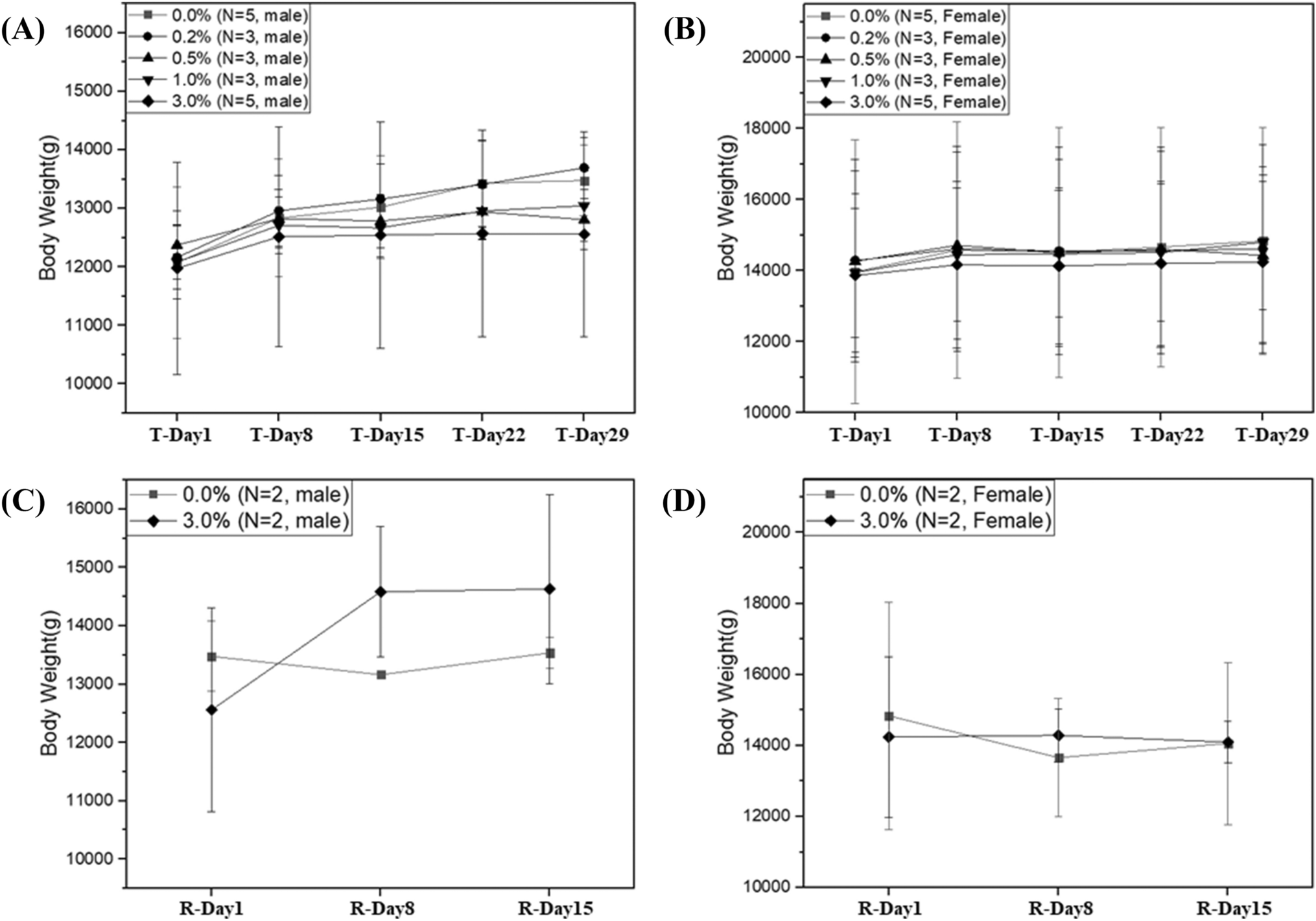
F344-Il2rg/Rag2em1Iexas rats were bred and maintained at the animal facility of Seoul National University Hospital (SNUH). The founding colony was originally established with animals purchased from the National Bio Resource Project-Rat at the University of Tokyo, Japan. Animals were acclimatized for at least 5 days in the experimental room prior to experimentation and housed under a 12/12-h light and dark cycle, at a temperature of 22 ± 2℃, and 40 − 60% humidity, with access to food (Teklad Certified Irradiated Global 18% Protein Rodent Diet 2918C, Envigo, USA) and sterilized water freely. To ensure statistical reliability while adhering to the 3R principles, the sample size was determined to be 6 animals per group. Rats were injected 5.0 × 10⁶ cells/kg of Luc-NALM-6 cells via the tail vein. Post-injection, animals were monitored for clinical signs daily, and body weight was measured twice a week. The observation period was set to 5 weeks, representing the maximal feasible duration determined by the mortality of the untreated disease control group in preliminary experiments, while being sufficient to cover acute to sub-acute toxicity. Animals were euthanized upon a decrease in body weight of more than 20% or the onset of hind limb paralysis due to leukemic burden under deep anesthesia with isoflurane. No animals or data points were excluded from the analysis in any experimental group.
Tumor cell lines
Luciferase-expressing NALM-6 cells were kindly provided by Dr. Kyongho Choi (Seoul National University, College of Medicine). These cells were originally generated by transducing parental NALM-6 cells (ATCC) with a lentiviral vector encoding firefly luciferase and cultured in RPMI-1640 media (Welgene, Republic of Korea) supplemented with 10% fetal bovine serum (FBS, Gibco, USA) and 1% penicillin/streptomycin (Pen-strep, Gibco, USA.). Cell viability was assessed using trypan blue and an automated cell counter (Luna II, Logos Biosystems, Republic of Korea).
CD19 CAR-T reference material and characterization
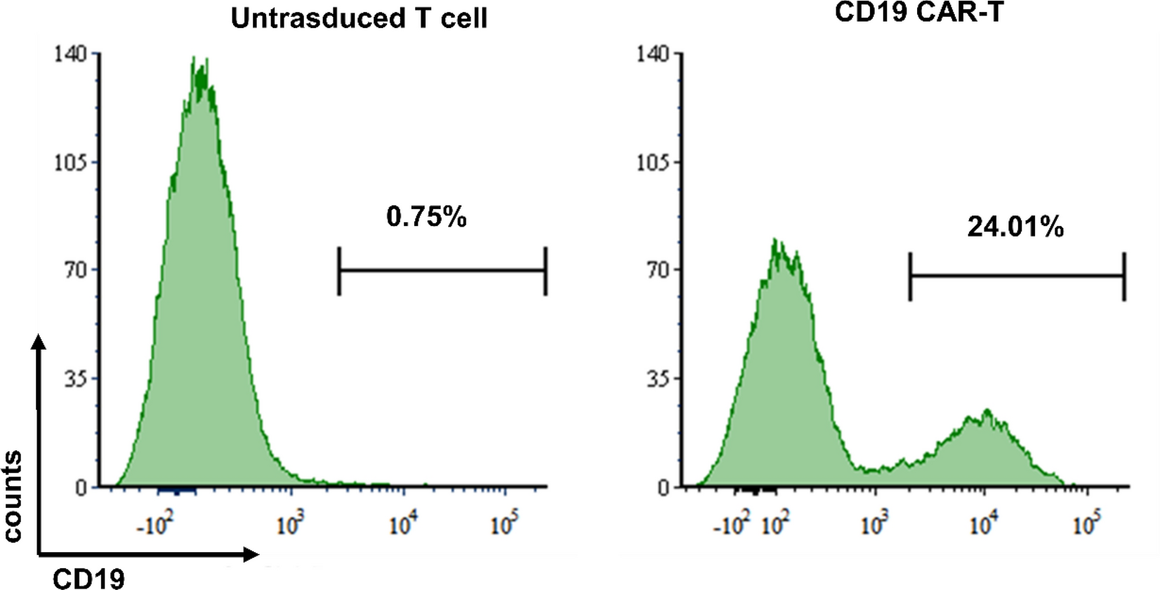
The CD19 CAR-T cells used as a reference standard for this platform validation study were produced in accordance with previously published methods [12]. All procedures involving human participants were approved by the Institutional Review Board (IRB) of Seoul National University Hospital (SNUH-IRB; 1606–033-768). Briefly, peripheral blood mononuclear cells (PBMCs) from healthy volunteers were transduced with a lentiviral vector (LTG1563; Lentigen) encoding a CAR construct (FMC63 scFv, 4-1BB costimulatory domain, CD3-zeta signaling domain). T cells were activated, transduced, and expanded using the automated CliniMACS Prodigy system (Miltenyi Biotec) as described previously [12]. The transduction efficiency of the final CAR-T cell product was confirmed by flow cytometry using a biotin-labeled anti-CD19 antibody and a PE-conjugated anti-biotin antibody (FACS-LSR II; BD) prior to injection.
Efficacy assessment in the B-ALL rat model
To validate the B-ALL xenograft rat model's capacity for integrated assessment, a study was conducted using five experimental groups. All rats were randomly divided into five groups based on their body weights (6 per sex per group, total 60 rats). Naive control group received 10 ml/kg of saline without a tumor xenograft, while the other four groups were injected with 5.0 × 10⁶ cells/kg of NALM6 via the tail vein. Three days later, these groups received intravenous injections of either saline (saline control), untransduced T cells (mock T control), or 1.0 (low-dose) or 2.0 × 10⁸ cells/kg (high-dose) of CD19 CAR-T cells (Table 1). The model's ability to track efficacy was evaluated using an in vivo imaging system (Lumina II, PerkinElmer, USA) on days 7, 14, 18, 21, 25, 28, 32, and 35 after injection. Rats were anesthetized with 2–3% isoflurane gas and imaged 20 min after receiving 150 mg/kg D-Luciferin (Promega, USA).
Table 1 Experimental design of efficacy evaluation in B-ALL xenograft ratsFive-week toxicity assessment in the B-ALL rat model
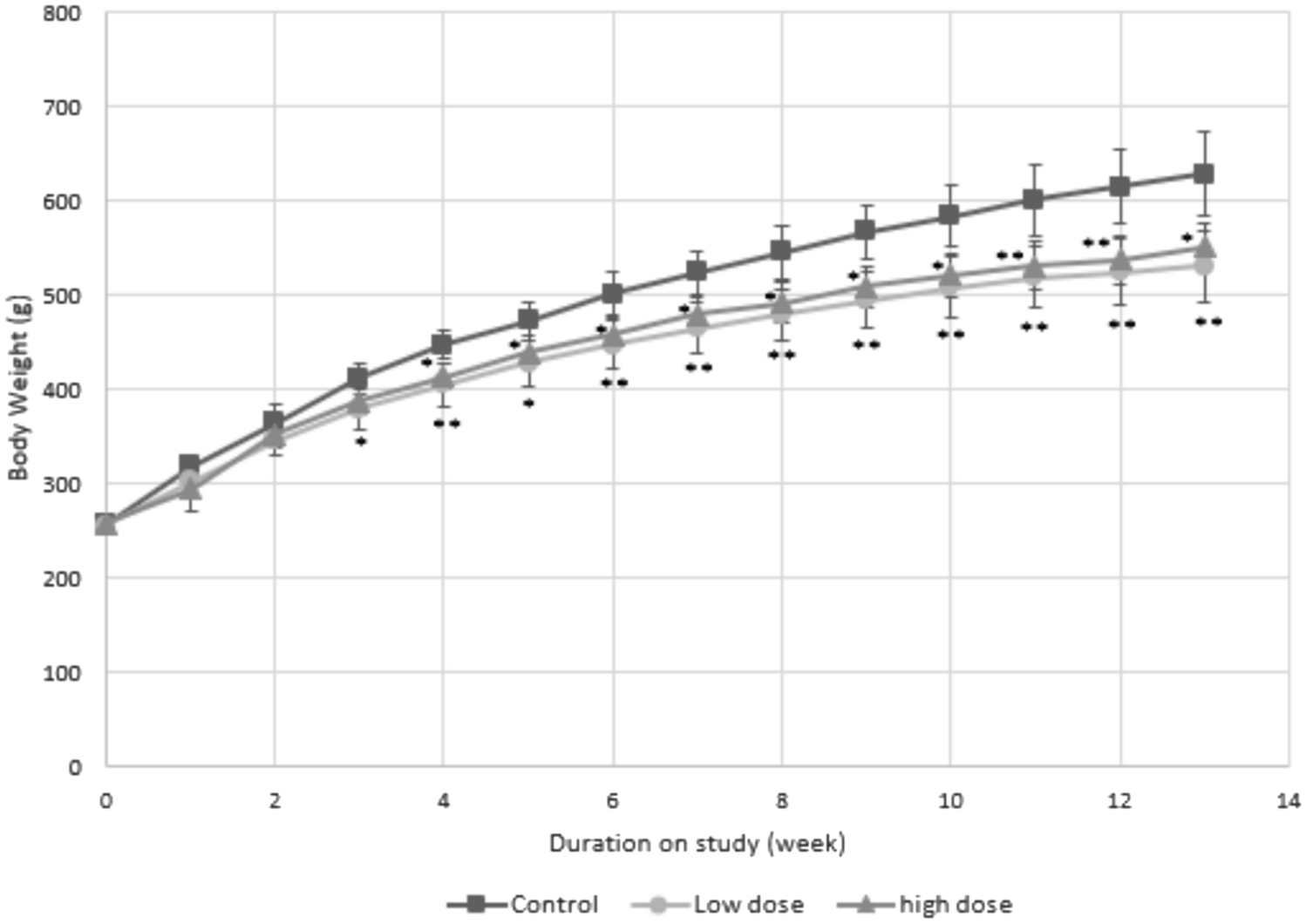
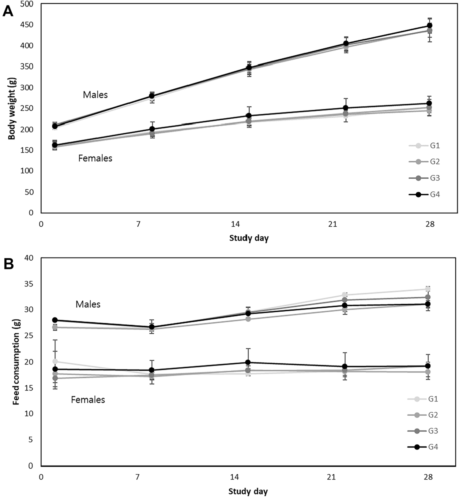
During the experiment, body weights, food, and water consumption were measured twice a week, and clinical signs were observed daily. Rats were euthanized under 2–3% isoflurane 5 weeks after CAR-T cell injection. Whole blood was collected from the vena cava for hematologic analysis (ADVIA 2120i, Siemens Healthcare, Tarrytown, NY, USA). Hematology parameters measured included white blood cell (WBC), red blood cell (RBC), platelet (PLT), hematocrit (HCT), reticulocytes (RETIC), hemoglobin (HGB), mean corpuscular hemoglobin (MCH), mean corpuscular volume (MCV), mean corpuscular hemoglobin concentration (MCHC) and differential WBC. To assess serum chemistry changes during the study, serum was analyzed using an automatic chemistry analyzer (Hitachi 7070, Hitachi, Tokyo, Japan). Serum biochemistry parameters included blood urea nitrogen (BUN), low density lipoprotein (LDL), high density lipoprotein (HDL), total cholesterol (TC), total protein (TP), albumin, total bilirubin (TB), alkaline phosphatase (ALP), aspartate transaminase (AST), alanine transaminase (ALT), creatinine, triglyceride, glucose, potassium, chlorine, sodium, calcium, and phosphorus.
Five weeks after CAR-T cell injection, animals were euthanized and organs were macroscopically examined. Liver, spleen, kidney, brain, lung, heart, adrenal glands, and testes were weighed and fixed in 10% neutral buffered formalin, while the eyes and testes were preserved in Davidson's and Bouin’s solution, respectively. The fixed organs were embedded in paraffin wax, sectioned into 4–6 μm thick pieces, and stained with hematoxylin and eosin (H&E). Histopathological analyses were conducted on the liver, spleen, pancreas, kidney, brain, lung, heart, adrenal glands, gastrointestinal tract, femur, sternum, nasal cavity, ovary, and testes. All slides were subsequently examined by a board-certified toxicologic pathologist who was blinded to group assignments.
Pharmacokinetic (PK) analysis in the B-ALL rat model
To validate the platform's suitability for kinetic analysis via serial sampling, the persistence of CD19 CAR-T cells was monitored in peripheral blood of leukemia xenograft rats, blood was collected from tail veins weekly after CD19 CAR-T cell injection (n = 3 / sex / group). Whole blood was collected in K2EDTA containing tubes (BD Microtainer, USA), from which 200 μL of the buffy coat was isolated and stored at − 80℃ in a deep freezer until DNA extraction. DNA was extracted using DNeasy Blood & Tissue Kits (QIAGEN, Germany) as per the manufacturer’s instructions, and its concentration was determined using a microplate reader (Epoch, BioTek, USA). CD19 CAR specific primers and probes were provided by Bosung Scientific Co. (Seoul, Republic of Korea) with the following sequences: Probe (FAM-ACT TGG AAC AAG AGG ACA TCG CCA-QSY), forward primer (5’-AAA CTG CTG ATC TAC CAT AC-3’), and reverse primer (5’-TCC TTG TTG ACA GAA GTA AG-3’). All reactions were performed using a ViiA7 Real-Time PCR System (Applied Biosystems, USA).
Ethical statement
All experiments received approval from the Institutional Animal Care and Use Committee at Seoul National University Hospital (SNUH-IACUC; 20–0170) and this study was conducted as a non-GLP study. The animals were housed in a facility accredited by AAALAC International (#001169), adhering to the 8th edition of the Guide for the Care and Use of Laboratory Animals, the National Research Council, and the ARRIVE guidelines. Information regarding animal group allocation was accessible solely to the researcher overseeing the randomization process. During the experiments, researchers administering injections and recording data were blinded to group assignments, and those assessing outcomes were similarly kept unaware of the group allocations to minimize bias. The group allocation was disclosed only after the analysis of data was concluded.
Statistical analysis
Data are presented as mean ± standard deviation (SD). Statistical analyses were conducted using one-way ANOVA, followed by Duncan's multiple range test with SPSS software version 22.0 (SPSS Inc., Chicago, IL, USA). P-values less than 0.05 were considered statistically significant.

Comments (0)