
Remember me

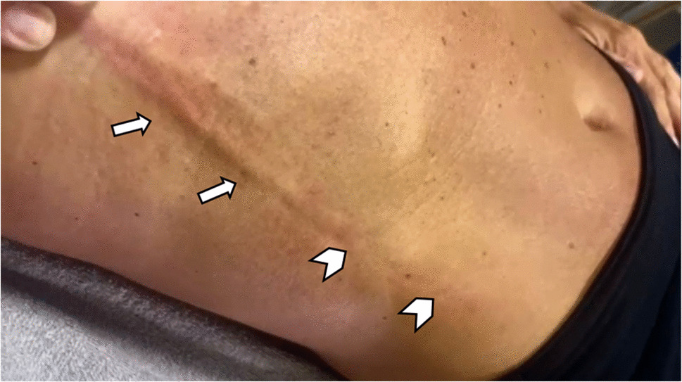
Mondor disease (MD) in a 59-year-old yoga teacher developed gradually after intense physical activity, accompanied by mild discomfort and localized tenderness on physical examination, without systemic symptoms or known risk factors for venous thrombosis. Photograph of the patient with the right arm abducted shows a tender, cord-like subcutaneous induration along the thoracoepigastric vein with skin retraction (Fig. 1). High-frequency ultrasound (L3–22 MHz) demonstrates a non-compressible, hypoechoic tubular structure corresponding to a thrombosed thoracoepigastric vein, with absence of color Doppler flow on longitudinal and transverse images, confirming superficial venous thrombosis (Fig. 2). There was no clinical evidence of deep vein involvement or lymphatic obstruction.
Fig. 1
Clinical findings in Mondor disease. Photograph of the patient with the right arm raised overhead in abduction shows a cord-like induration along the thoracoepigastric vein. There is a linear, palpable, and tender subcutaneous cord extending along the anterolateral aspect of the thoracoabdominal wall (arrows). Notice the adjacent skin retraction (chevrons), which follows the course of the thrombosed vein and appears as a shallow groove
Fig. 2
Ultrasonographic findings in Mondor disease. Longitudinal B-mode ultrasound images (A, B) obtained with a high-frequency linear probe (L3-22 MHz) demonstrate a non-compressible, hypoechoic, tubular structure (arrows), corresponding to a thrombosed thoracoepigastric vein at the anterolateral aspect of the right thoracoabdominal wall. Longitudinal (C) and transverse (D) Doppler ultrasound images acquired at 48 Hz show the absence of color flow within the affected vein (arrows), confirming the presence of thrombus. Adjacent ribs, intercostal muscles, and underlying lung were unremarkable, and the overlying skin demonstrated normal thickness and echogenicity, underscoring the importance of evaluating surrounding soft tissues beyond the area of clinical concern
Patient was managed conservatively with nonsteroidal anti-inflammatory drugs, and warm compresses were recommended, with gradual improvement of symptoms, consistent with the self-limiting course of MD. On clinical follow-up, no additional imaging examinations were performed, as the patient experienced progressive improvement of symptoms, and a residual area of skin retraction persisted but became less evident over time.
MD is a rare, self-limiting superficial thrombophlebitis of the anterior chest wall veins. It typically presents as a tender, cord-like lesion and is often associated with vigorous physical activity, trauma, or breast-related procedures. It primarily affects the thoracoepigastric, lateral thoracic, or superior epigastric veins, leading to a palpable, tender, cord-like structure beneath the skin. The painful linear induration often becomes more noticeable with arm movement or when the skin is stretched, as seen in this patient, distinguishing it from other causes of chest wall pain [1, 2]. It results from venous thrombosis causing partial or complete occlusion of the affected vessel, followed by fibromuscular hyperplasia and surrounding subcutaneous fibrosis. Adhesion of the thrombosed vein to the overlying skin may then produce retraction and the characteristic cord-like groove [1].
Ultrasound typically shows a tubular, non-compressible superficial vein with a beaded appearance and absent color Doppler flow. Surrounding soft tissues are usually unremarkable, helping distinguish it from inflammatory or neoplastic causes [4,5,6,7,8]. In earlier or resolving stages, partial compressibility or residual intraluminal flow may be observed, and mild surrounding edema or hyperemia can be present. A targeted ultrasound examination of the symptomatic region is generally sufficient, with extension to a comprehensive deep venous thrombosis study reserved for cases with atypical imaging features or clinical suspicion of deeper venous involvement, such as diffuse limb swelling, pain, tenderness, and occasionally erythema [9].
MD may mimic lymphangitis, mastitis, and other causes of superficial thrombophlebitis, while inflammatory breast cancer is an uncommon clinical mimic [3]. Lymphangitis may produce linear subcutaneous changes but lacks an intravascular thrombus, and catheter- or hypercoagulability-related thrombophlebitis typically involves deeper venous territories [10]. MD, while classically described in the anterior chest wall, may also involve other superficial venous territories, including axillary and penile variants, and should be recognized by musculoskeletal radiologists when evaluating superficial soft-tissue abnormalities [3, 4, 8].
In patients with a classic presentation, clinical diagnosis is often sufficient, allowing reassurance and conservative management without further testing. Imaging is reserved for atypical, persistent, or progressive cases, or when alternative diagnoses are suspected. Treatment is usually conservative, focusing on pain relief with anti-inflammatory medications, warm compresses, and reassurance [1, 4].
In summary, MD is a rare yet self-limiting condition that presents with a distinctive cord-like lesion due to superficial thrombophlebitis of the chest wall veins [2, 3]. This case highlights the importance of clinical recognition and imaging, particularly ultrasound, in distinguishing MD from more serious conditions.
Comments (0)