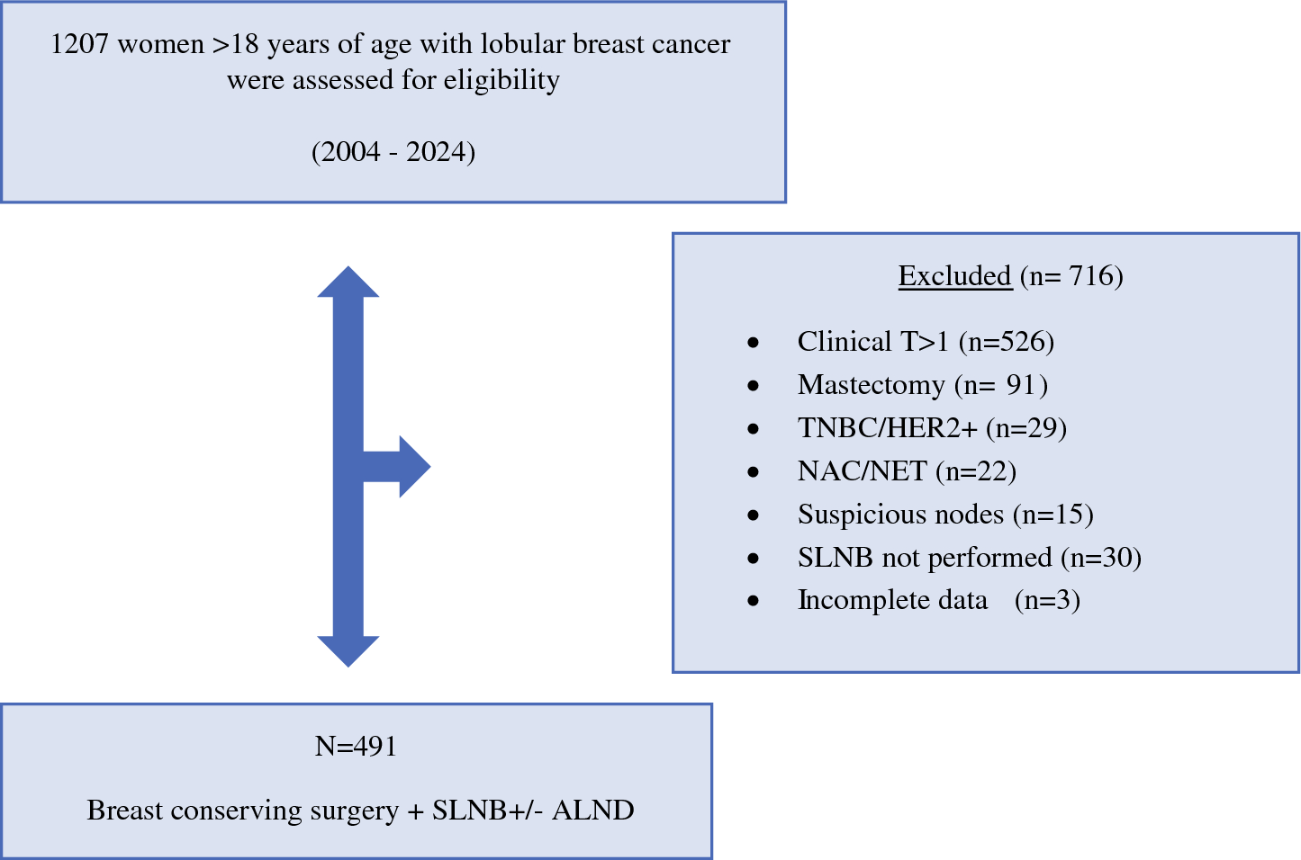
In our series, 99 (20.2 %) of 491 patients with early-stage, ER-positive/HER2-negative ILC and clinically node-negative axilla were found to have pathologic nodal metastases (pN1mi–pN3) at definitive surgery. Despite careful preoperative assessment with both ultrasound and MRI, approximately one in five patients presented with occult nodal metastases, underscoring the limited sensitivity of current imaging methods in accurately identifying nodal involvement in ILC.
The intrinsic challenges of axillary imaging in ILC complicate the safe omission of SLNB. Schumacher et al.16 reported that in 349 stages I to III ILC patients, 38 % of those with advanced nodal disease (pN2–pN3) were misclassified as cN0 on imaging despite MRI showing the highest sensitivity (65 %) followed by ultrasound (42 %) and mammography (10 %). These findings reinforce that imaging alone is insufficient for axillary staging in ILC and that SLNB remains essential, particularly for guiding adjuvant therapy.
Emerging methods, such as [18F]FDG PET/MRI, have shown promise in detecting nodal metastases in early breast cancer.17 However, lobular carcinomas represented a minority in these studies, and the infiltrative often hypometabolic nature of ILC may limit the accuracy of fluorodeoxyglucose (FDG)-based imaging.
The unique behavior of ILC in the axilla is further supported by Adachi et al.18 who demonstrated a higher frequency of non-sentinel lymph node metastases in ILC than in IDC when the sentinel node was positive. This suggests that nodal disease in ILC is often diffuse and unpredictable, indicating the need for precise surgical assessment even in cN0 patients. Consistently, Mukhtar et al.19 reported that accurate preoperative axillary staging in ILC remains difficult due to the tumor’s subtle and diffuse infiltration pattern, which may escape both clinical and imaging detection.
In older patients, the omission of SLNB in selected ILC cases may be safe. Carleton et al.20 found no significant difference in locoregional recurrence-free survival among women ≥70 years old with early-stage, ER-positive/HER2-negative, cN0 breast cancer, including those with ILC, whether SLNB was performed or omitted.
In our cohort, menopausal status was not associated with axillary nodal involvement (p = 0.96). In addition, a subgroup analysis of patients age ≥70 years showed that 18 % harbored nodal metastases, a proportion that did not differ meaningfully from the overall rate observed in the study population.
Collectively, these findings indicate that neither menopausal status nor chronological age alone reliably predicts nodal involvement in invasive lobular carcinoma and therefore should not be used in isolation to guide axillary surgical decision-making Among the variables analyzed, tumor size emerged as the strongest predictor of nodal burden. Larger tumors were significantly more common in node-positive groups, particularly among those with four or more positive lymph nodes. This finding, although somewhat expected, reinforces the well-established notion that greater tumor size is directly associated with increased axillary spread, even in luminal A-like tumors.21
As expected, after the cohort was restricted to patients with clinical T1 disease, a measurable rate of pathologic upstaging was observed. On final histopathologic examination, 69 patients (14.0 %) were upstaged to pT2, underscoring the intrinsic limitations of preoperative tumor size assessment in invasive lobular carcinoma. This finding was not unexpected because ILC is characterized by a diffuse growth pattern and ill-defined tumor margins, which may lead to underestimation of true tumor extent on imaging and clinical evaluation.22
Histologic subtype did not demonstrate a statistically significant association with axillary nodal status in the analysis. However, pleomorphic and mixed variants showed a numerically higher prevalence of nodal involvement than classic invasive lobular carcinoma. Although classic ILC was more frequently observed among node-negative patients, these more aggressive variants tended to be overrepresented in higher nodal categories. Although this trend did not reach statistical significance, it suggests potential biologic heterogeneity within the ILC spectrum that may warrant consideration when axillary management strategies are evaluated.23
Recent trials have promoted selective de-escalation of axillary surgery in early-stage breast cancer. However, the 2025 St. Gallen International Consensus highlighted that these trials included only a small proportion of ILC patients, and a majority of panelists (60 vs 40 %) recommended against omitting SLNB in this histologic subtype.24 National guidelines differ: the 2025 AGO Breast Commission integrates SOUND and INSEMA data, but does not distinguish between ductal and lobular cancers, recommending SLNB omission based on low-risk criteria regardless of histology.25
In the SOUND and INSEMA trials, which enrolled predominantly IDC, sentinel lymph node positivity rates were 13.7 % and 15 %, respectively, whereas in our pure ILC population, the rate reached 20.2 % (Table 3). However, the current study aimed to underscore that ILC constitutes a biologically distinct entity, characterized by unique pathophysiologic features that limit the applicability of axillary de-escalation strategies traditionally developed for IDC. Even in small, luminal-type tumors, lobular histology appears to retain a non-negligible risk of axillary metastasis, which may not be adequately predicted by conventional staging tools.26
Table 3 Lymph node positivity rates of SLNB omission trials and the current studyIn addition, it should be considered that the INSEMA trial included a small proportion of patients with clinical T2 tumors, whereas in our analysis, similar to the SOUND trial, only tumors clinically assessed as ≤ 2 cm were included, based on preoperative ultrasound and MRI. Despite this strict selection of clinically small tumors, a pathologic upstaging rate of 14 % was observed, further highlighting the intrinsic challenges of accurately estimating tumor extent in invasive lobular carcinoma.
Importantly, 3 % of the patients harbored four or more positive nodes, a nodal burden that would significantly influence adjuvant treatment decisions, including the indication for chemotherapy or regional nodal irradiation. These data suggest that omitting SLNB in this subgroup may result in understaging and potential undertreatment.27,28
Limitations of this study included its retrospective, single-center design which may have introduced selection bias and may limit generalizability. The long inclusion period may have encompassed evolving imaging, surgical, and pathologic practices. Finally, only patients undergoing breast-conserving surgery were included, potentially underrepresenting the full spectrum of early-stage ILC.

Comments (0)