Endocrine carcinoma with amphicrine carcinoma component at esophagogastric junction
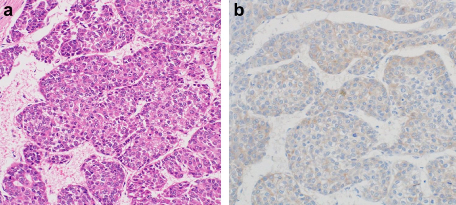
Amphicrine carcinoma is a rare malignancy exhibiting both endocrine and exocrine differentiation within the same tumor cells. We report a case of a 70-year-old male who underwent screening upper endoscopy, which revealed a 15 mm depressed lesion with a reddish elevated margin at the esophagogastric junction. Narrow-band imaging with magnification demonstrated an irregular, densely packed vascular pattern, suggesting malignancy. Endoscopic ultrasound indicated third-layer disruption, raising suspicion of submucosal invasion. Endoscopic resection was performed after informed consent. Histopathological analysis confirmed submucosal invasion and identified three tumor components: (1) an endocrine carcinoma component with a solid-nest structure, (2) a glandular component, and (3) a goblet cell-like signet ring cell carcinoma component. Immunohistochemistry revealed synaptophysin, INSM1, and CD56 positivity in all components, while MUC2 was expressed in the signet ring cell carcinoma component and partially in the others. The tumor exhibited a high Ki-67 labeling index, indicating aggressive proliferation. Based on these findings, the lesion was diagnosed as an endocrine carcinoma with an amphicrine carcinoma component. Additional surgery confirmed no residual tumor or lymph node metastasis. Given the rarity of amphicrine carcinoma and the limited understanding of its clinical behavior, further research is necessary to determine its prognosis and optimal management strategies.

Comments (0)