In this study, the effects of the endogenous cannabinoids AEA and ODA, as well as the PPAR-γ receptor inhibitor GW9662, were evaluated on different functional markers in two GB cell lines, C6 and RG2. Both AEA and ODA induced anti-proliferative effects in C6 and RG2 cells, while they did not affect viability in primary astrocytes, used as non-tumor controls as GB is known to be tumors derived from the malignant transformation of astrocytes. These findings indicate that the mechanisms activated by AEA and ODA for anti-proliferative effects on GB cells are distinct from those inherent to primary cells, suggesting that these differential mechanisms might be accounted for the differences in cannabinoid receptor levels, or by mutated key proteins in glioblastoma cell lines. Moreover, the fact that AEA and ODA decreased cell viability in GB lines, but not in primary astrocytes, suggest tumor selectivity, thus highlighting their therapeutic potential.
Several receptors linked to the activity of the endocannabinoid system are altered in GB cells, including CB1, GPR55, TRPV1, and PPAR-γ [28,29,30,31], as well as enzymes like fatty acid amide hydrolase (FAAH), which is responsible for the degradation of AEA and ODA [32]. PPAR-γ is a key player in mitochondrial function in neurological diseases, as well as an important physiological mediator of the beneficial effects of cannabinoids; however, its role in GB tumors remains poorly understood. Here, we evaluated the role of the PPAR-γ antagonist GW9662 in two GB cell lines, as PPAR-γ activation is known to enhance cell death in GB tumors using temozolomide (the main chemotherapeutic used in the treatment in GB therapy) by regulating the activity of metabolic enzymes [33]. Therefore, to determine whether PPAR-γ is involved in the effects mediated by AEA and ODA as an additional mechanism to those already described previously, we used the antagonist GW9662, hypothesizing that it would reverse their antiproliferative effects. Furthermore, because endocannabinoids act through multiple receptors and trigger numerous signaling pathways, the mechanisms associated with their anti-proliferative activity in different types of tumor cells have not been fully described, and their study could be key to understanding their different and sometimes dual actions.
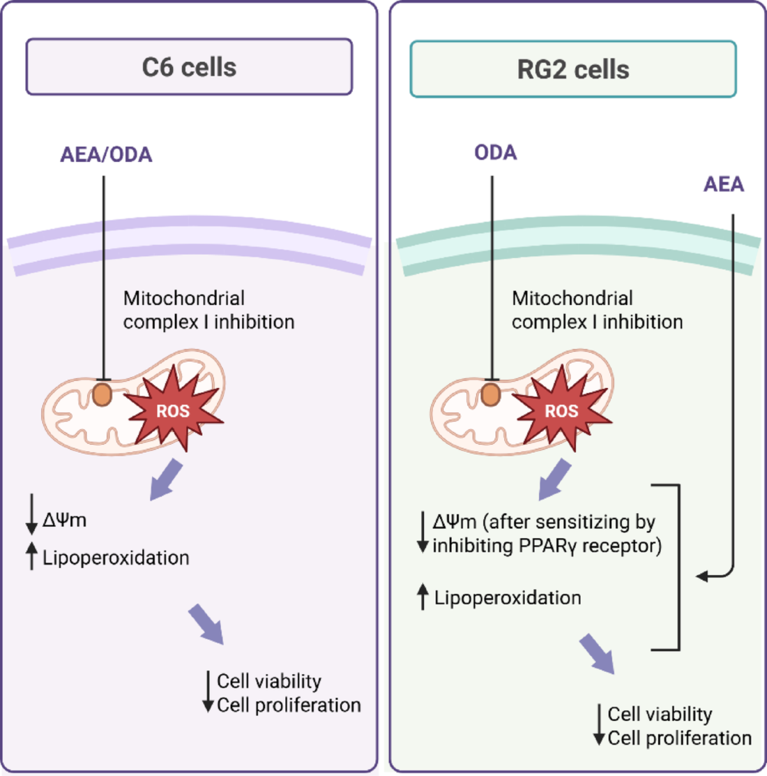
We observed differential roles of PPAR-γ in the tumor cell lines used. While the lack of effects of GW9662 on cannabinoid-induced cell death in C6 cells suggests that the cytotoxic effects of AEA and ODA could be mostly mediated by alternative pathways such as CB1/CB2 receptors, TRPV1 channels, or direct modulation of mitochondrial function, in RG2 cells PPAR-γ may play a protective role in this cell line. These effects could be inherent to the metabolic and genotyping differences between the two GB cell lines. In this regard, PPAR-γ inhibition by GW9662 reduces stem cell-like properties in GB and increases sensitization to antitumor treatments [34]. Moreover, in GB cells, when PPAR-γ is inhibited, cells become more vulnerable due to deficiency in mitochondrial ATP supply, which is the main source of ATP required for proliferative maintenance. This vulnerability was evident in RG2 cells, where the endocannabinoid-induced loss of cell viability was enhanced when PPAR-γ was inhibited with GW9662, unlike in C6 cells, which exhibited less accelerated proliferation [35]. Again, genotypic and metabolic differences might underline these differential responses between the two cell lines.
A particularly relevant finding was the increase in ΔΨm observed in RG2 cells only after pretreatment with GW9662. This paradoxical effect could indicate a compensatory mechanism where PPAR-γ inhibition transiently restores mitochondrial membrane potential, possibly through the upregulation of respiratory chain complexes or other mitochondrial bioenergetic processes. Such an adaptive response in RG2 cells, but not in C6 cells, underscores the differential role of PPAR-γ in regulating mitochondrial bioenergetics between these two cell lines. RG2 cells, which possess an inherently high invasive capacity, compared to the greater angiogenic capacity of C6 cells [36], may be more critically dependent on PPAR-γ regulation to maintain mitochondrial homeostasis under stress conditions.
AEA and ODA increased lipid peroxidation in C6 and RG2 cells, as part of their anti-proliferative mechanisms. These data also suggest that eCBs promote changes in mitochondrial metabolism, either by increasing OXPHOS activity or by increasing mitochondrial mass via cannabinoid receptor activation [37]; however, it remains necessary to determine whether lipid peroxidation results from increased mitochondrial ROS production.
Metabolic differences between GB cell lines have been documented in previous reports. While RG2 tumor cells possess a higher level of choline pathway substrates, such as phosphocholine and glycerophosphocholine, C6 cells contain higher levels of mitochondrial pathway substrates, such as creatine and taurine, as well as higher levels of glutathione, the main endogenous antioxidant [36]. Precisely, this last feature corroborates that redox activity is higher in C6 cells than in RG2 cells, rendering them more sensitive to mitochondrial and redox alterations.
Two other major differences noted in the responses of these cell lines to eCBs include changes in ΔΨm and in the activity of mitochondrial Complex I. As expected, AEA and ODA significantly decreased the ΔΨm in C6 cells, while only ODA decreased this parameter in RG2 cells. We hypothesize that the effects induced by eCBs in C6 cells described herein were tightly related to the altered activity of mitochondrial Complex I, in contrast to RG2 cells. These differences might be related to the fact that C6 cells have a greater dependence on oxidative metabolism compared to RG2 cells, as suggested by the substrates found within the tumor [36]. Therefore, AEA and ODA may affect mitochondrial function to a greater extent in C6 cells. Furthermore, OXPHOS is an essential generator of ATP in GB cells [38]. Likewise, the mitochondrial respiratory chain plays a fundamental role in the regeneration of NAD+ and FAD, which are necessary to sustain a functional tricarboxylic acid (TCA) cycle, thus favoring vital biosynthetic processes such as the generation of cytosolic acetyl-CoA and the production of aspartate [39]. Thus, alterations in the components of the respiratory chain could compromise cellular homeostasis and even increase the sensitivity of tumor cells to anticancer drugs [40, 41]. Mitochondrial Complex I has been reported as a promoter of metastasis in murine models and in patients with some metabolically altered types of cancer [42]. In addition, downregulation of Complex I activity may stimulate Complex II to increase its activity, and when altered [43], it would promote an increase in ROS generation, exacerbating oxidative damage, as observed mostly in C6 cells.
Here, we observed that AEA and ODA can inhibit Complex I in GB cells, highlighting an additional mechanism to those previously attributed to the antitumor effects of eCBs. It has been reported that cannabidiol (CBD) can induce apoptosis in GB cells by altering mitochondrial Complex I activity, while reducing oxygen consumption, an effect that was reversed by pharmacological inhibition of the Voltage Anion Channel VDAC1 [44]. This ion channel functions as a gatekeeper for the entry and exit of mitochondrial metabolites, which in turn could associate the engagement of NADH reductase with the induction of apoptosis via cytochrome c release into the cytosol [45]. This event is also related to the already reported inhibition of Complex I by a clinically applied inhibitor, which blocks proliferation and induces apoptosis in OXPHOS-dependent solid tumor models of brain cancer and acute myeloid leukemia (AML) [46, 47].
The activity of Complex I contributes approximately 40% of the proton-motive force; thus, it is essential for the generation of the membrane potential required for mitochondrial ATP synthesis. In addition to its energetic contribution, it also supports cell proliferation by maintaining the cellular pool of the NAD+ used as an electron acceptor in different metabolic pathways, while also maintaining the NAD+/NADH ratio necessary for the production of aspartate involved in the synthesis of nucleic acids and macromolecules that sustain the high cellular proliferation of tumor cells [48, 49]. Furthermore, the continued production of pyruvate from glycolysis requires NAD+; thus, the use of glucose-derived pyruvate as a biosynthetic intermediate requires an exogenous source of NAD+ regeneration to maintain redox balance.
The differential effects evoked by AEA compared to ODA on ΔΨm and Complex I activity across both cell lines reflect mechanisms of action potentially different triggered by each eCB. In fact, even though both AEA and ODA seem to involve PPARγ modulation, they do that in a different manner, as evidenced by the effects of the antagonist GW9662. Then, how to explain these differences? Unlike AEA, which has high affinity for specific receptors, ODA is known for its effects more linked to the modulation of gap junctions. Disruption of gap junctions by oleamide is known to alter mitochondrial function by inducing Ca2+ and cytochrome C release [50]. Furthermore, it should be noted that while AEA is rapidly degraded by the enzyme FAAH, ODA, being also a substrate for this enzyme, often acts as a competitive inhibitor or alternative substrate that saturates FAAH, allowing its intracellular levels to remain elevated for longer periods and act on several targets, including mitochondria [51]. Taken together, our novel findings advance the understanding of mechanisms responsible for cannabinoid-induced effects on glioblastoma cells associated with the regulation of OXPHOS activity.
Under certain circumstances, cannabinoids reduce the expression of OXPHOS genes [52], while increasing oxidative stress by altering the activity of NADH-ubiquinone oxidoreductase and other mitochondrial complexes (II, III, and IV) [53]. This effect cannot be ruled out for AEA and ODA, as these eCBs may act by inhibiting other mitochondrial complexes, thereby inducing an uncoupled respiratory chain and promoting the activation of cell death pathways.
The decrease in ΔΨm observed following AEA and ODA treatments can be directly attributed to the inhibition of NADH-ubiquinone oxidoreductase. In this regard, we found that inhibition of PPAR-γ receptor with GW9662 reverted the AEA-induced decrease in ΔΨm in C6 cells, indicating that AEA is acting through this receptor to regulate mitochondrial functions. Notably, neither AEA nor ODA decreased membrane potential in RG2 cells, thus confirming that C6 and RG2 tumor cell lines utilize differential genotypic and metabolic mechanisms that ultimately determine active signals for anti-proliferative pathways. Finally, further studies are needed to determine whether the mitochondrial regulation exerted by these eCBs is also mediated by mitochondrial biogenesis and function.

Comments (0)