
Remember me
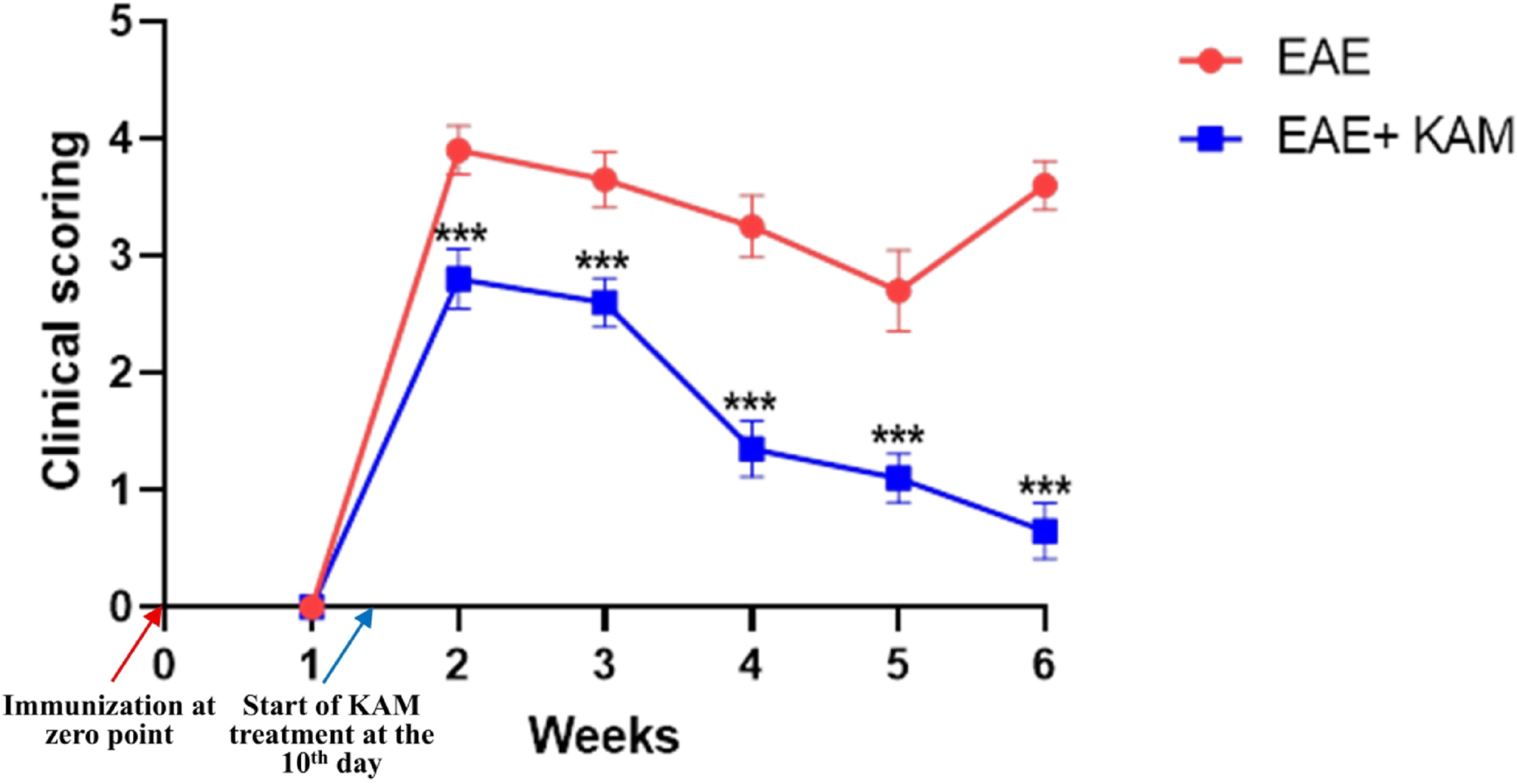
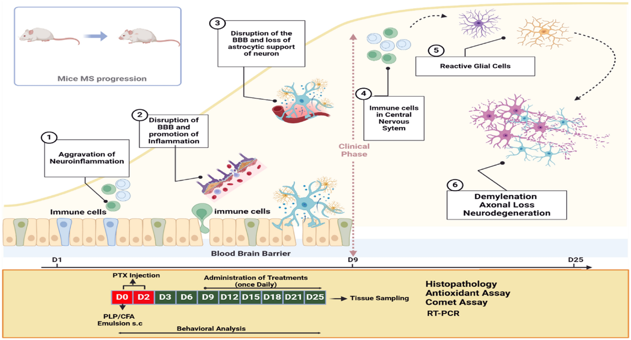
To investigate the neuroprotective effect of KAM on MS pathogenesis, (8–10)-week-old female C57BL/6 mice were immunized with MOG35–55 peptide to establish the EAE model (a valid model of MS) and then treated with KAM. Clinical scoring was used to assess neurological dysfunction across different groups. As shown in Table 3; Fig. 1, the untreated immunized EAE group exhibited a peak clinical score at the beginning of the second week, reaching 3.9 ± 0.21. This was followed by a gradual decrease (remission) to a mean of 2.7 ± 0.35 by the 5th week, with a subsequent relapse by the 6th week to a mean score of 3.6 ± 0.31. In contrast, the EAE + KAM group showed that KAM treatment (50 mg/kg) significantly reduced the peak clinical disability score (maximum score: 2.8 ± 0.26), accelerated remission, and prevented relapse in the 6th week, with a considerably lower score of 0.65 ± 0.24. These results indicated that KAM effectively alleviated the severity of clinical symptoms in EAE mice and halted disease progression.
Table 3 Weekly-based clinical scores recorded post-immunization in EAE and EAE + KAM groupsFig. 1
Progress of the clinical disability score in experimental autoimmune encephalomyelitis (EAE group; red line) vs. EAE treated by kaempferol (EAE + KAM group; blue line) to detect difference at each weekly recording points. Values were presented as mean ± SD (n = 10). ***p < 0.001, by Mann-Whitney U test
KAM Ameliorated Histopathological Alterations in the Brain and Spinal Cord of EAE MiceH&E-stained sections from the control groups of the cerebral cortex showed the normal structure of cerebral cortex neurons, like pyramidal cells with their pyramidal shape and apical dendrite, granule cells with its pale nuclei and prominent nucleoli, separated by compact eosinophilic neuropil containing blood capillaries (Fig. 2a). The EAE group examination depicted severe focal disruption and marked inflammatory cellular infiltrations within the molecular layer, other layers, and surrounding the dilated blood capillaries forming perivascular cuff with separated meninges (Fig. 2b&c) and depicted an extremely significant increase in the mean number of inflammatory infiltrate/mm2 as compared to the control group (F (3,56) = 283, P < 0.0001, Fig. 2e). It is noteworthy that administration of KAM alleviated most of the previous changes except for the presence of some inflammatory infiltrates and a significant decrease in the number of inflammatory infiltrates as compared to the EAE group (Fig. 2d&e).
Fig. 2
Effect of KAM (50 mg/kg) treatment on brain (cerebral cortex) histopathological alteration in the EAE model of MS. a Control of the cerebral cortex showing normal pyramidal cells (angled arrows) with their apical dendrite, granule cells with pale nuclei and prominent nucleoli (arrows) separated by eosinophilic neuropil. (b & c) EAE group with marked inflammatory cellular infiltration forming perivascular cuffs within the layers (thick arrows). Also, separated overlying meninges are present (arrowhead). d The KAM-treated group shows improvement with few inflammatory infiltrates (thick arrows). e Comparison between the studied groups as regards the mean number of inflammatory cells/mm² in cerebral cortex sections. (Magnification: X400, H&E, scale bar: 50 µ). NC: normal control; KAM: kaempferol; EAE: experimental autoimmune encephalomyelitis
Examination of spinal cord sections from control groups revealed neuronal cell bodies with prominent nuclei within the grey matter and myelinated axons within the white matter (Fig. 3a). EAE group showed neuronal cell bodies with dark dense nuclei within the grey matter, a marked loss of myelinated axons within the white matter in some sections (Fig. 3b) and focal others with inflammatory cellular infiltration within the white matter and around blood capillaries in separated meninges (Fig. 3c) and depicted an extremely significant increase in the mean number of inflammatory infiltrates/mm2 as compared to the control group (F (3,56) = 167, P < 0.0001, Fig. 3e). KAM treated group depicted marked improvement and restoration of myelinated axons within the white matter and a significant decrease in the number of inflammatory infiltrates as compared to the EAE group (Fig. 3d&e).
Fig. 3
Effect of KAM (50 mg/kg) treatment on the spinal cord histopathological alteration in the EAE model of MS. a Control of the spinal cord shows neuronal cell bodies within the grey matter (curved arrow) and myelinated axons within the white matter (arrows). b EAE group with irregular neuronal cell bodies with dark dense nuclei (curved arrows) within the grey matter and white matter with marked loss of myelinated axons (arrows). c EAE group shows focal inflammatory cellular infiltration within the white matter (angled arrows) and around blood capillaries within the separated meninges (arrowhead). d The KAM-treated group shows restoration of the myelinated axons within the white matter (arrows) and normal neuronal cell bodies within the grey matter. e Comparison between the studied groups as regards the mean number of inflammatory cells/mm² in spinal cord sections. (Magnification: X400, H&E, scale bar: 50 µ). NC: normal control; KAM: kaempferol; EAE: experimental autoimmune encephalomyelitis
KAM Mitigated TEM Abnormalities in the Brain and Spinal Cord of EAE MiceTEM examination of ultrathin sections from the cerebral cortex of control groups revealed pyramidal neurons with euchromatic nuclei and surrounded by an intact neuropil composed of unmyelinated and myelinated axons (Fig. 4a). EAE group examination showed severely disturbed architecture with abnormal neurons with irregular nuclei surrounded by marked vacuolation of the neuropil, abnormal unmyelinated axons contained mitochondria with destroyed cristae. Myelinated axons within the neuropil appeared with irregularly split myelin and contained mitochondria with destroyed cristae (Fig. 4b&c). Interestingly, neurons within the KAM-treated group appeared with euchromatic nuclei, surrounded by an intact neuropil of myelinated axons and few appeared containing mitochondria with cristolysis (Fig. 4d).
Fig. 4
Effect of KAM (50 mg/kg) treatment on brain (cerebral cortex) TEM changes in the EAE model of MS. a control cerebral cortex showing pyramidal neuron with euchromatic nucleus (N) and surrounded by neuropil composed of both unmyelinated (curved arrow) and myelinated axons (arrows). b EAE group with marked deterioration, as most of the neurons appear irregular with irregular nuclei (N), surrounded by disturbed unmyelinated axons containing mitochondria with destroyed cristae (curved arrows). c EAE group shows myelinated axons with disturbed, split thin myelin (arrows) and mitochondria with destroyed cristae within the myelinated (arrowhead) and the unmyelinated (curved arrow) axons. d The KAM-treated group shows neuron with euchromatic nucleus (N) and is surrounded by intact neuropil of myelinated axons (arrows) with few with mitochondria with cristolysis (arrowhead). (Magnification: X2000, scale bar: 2 µ, TEM). NC: normal control; KAM: kaempferol; EAE: experimental autoimmune encephalomyelitis
Ultrathin sections examination from the spinal cord of control groups depicted crowded myelinated axons within the white matter containing normal mitochondria (Fig. 5a). The EAE group showed marked disturbance of the architecture with loss of myelinated axons or the presence of thin, split myelin or even discontinuous myelin and mitochondria with destroyed cristae (Fig. 5b&c). KAM administration improves myelination and the presence of myelinated axons (Fig. 5d).
Fig. 5
Effect of KAM (50 mg/kg) treatment on the spinal cord TEM changes in the EAE model of MS. a The control group showing white matter of the spinal cord with crowded myelinated axons (arrows) containing normal mitochondria (arrowheads). (b & c) EAE group with severe deterioration of myelinated axons, which appear with either thin discontinuous myelin (arrows) or even loss of myelinated axons (stars). Neurons appear with an euchromatic nucleus (N), a prominent nucleolus (n), vacuolated cytoplasm (v), and are surrounded by vacuolated neuropil (stars). d The KAM-treated group shows improvement in the presence of myelinated axons (arrows), except some of them are still surrounded by split myelin (thick arrows). (Magnification: X2000, scale bar: 2 µ, TEM). NC: normal control; KAM: kaempferol; EAE: experimental autoimmune encephalomyelitis
KAM Effectively Attenuated Ferroptosis and Oxidative Damage in the Brain and Spinal Cord of EAE MiceRecent studies have revealed that ferroptosis, a nonapoptotic type of regulated cell death initiated by iron accumulation and lipid peroxidation-mediated stress, accelerates oligodendrocyte loss, demyelination, and neurodegeneration, significantly contributing to MS pathogenesis [7]. To explore whether the neuroprotective impact of KAM was related to the suppression of ferroptosis, we evaluated key ferroptosis markers, including Fe²⁺, MDA, Fpn1, SLC7A11, GSH, and GPX4. Our findings revealed a drastic elevation in the level of intracellular Fe2+ in the brain and spinal cord of MOG35–55-immunized EAE mice, which was effectively reduced by KAM treatment (F (3, 36) = 54.4, P < 0.0001, Fig. 6A and F (3, 36) = 54.6, P < 0.0001, Fig. 6A−, respectively). Similarly, the brain and cord tissue levels of MDA (F (3, 36) = 442, P < 0.0001, Fig. 6B and F (3, 36) = 442, P < 0.0001, Fig. 6B−, respectively) were greatly elevated in EAE mice compared to the NC group, but these levels were obviously mitigated by KAM (50 mg/kg) administration in the EAE + KAM group. While the EAE group displayed a notable decline in the mRNA levels of the Fpn1-encoding gene (SLC40A1) in the brain and spinal cord tissues from untreated MOG35–55-immunized mice, meanwhile KAM (50 mg/kg) treatment considerably enhanced SLC40A1 relative expression in the CNS from the EAE + KAM group compared to the EAE-untreated group (F (3, 36) = 162, P < 0.0001, Fig. 6C and F (3, 36) = 273, P < 0.0001, Fig. 6C−). These findings suggest that KAM may inhibit iron overload by promoting Fpn1-mediated iron efflux.
Fig. 6
Effect of KAM (50 mg/kg) treatment on the brain and spinal cord Fe+ 2 level (A and A−, respectively); MDA level B and B−, respectively); and SLC40A1 (ferroportin1-encoding gene) mRNA expression (C and C−, respectively) in an EAE model of MS. Values were presented as mean ± SD (n = 10). *p < 0.05; **p < 0.01; and ***p < 0.001, by one-way ANOVA followed by Tukey’s post hoc test. Fe+ 2: ferrous iron; MDA: malondialdehyde; SLC40A1: solute carrier family 40 member 1; NC: normal control; KAM: Kaempferol; EAE: experimental autoimmune encephalomyelitis
The levels of SLC7A11 in the brain and spinal cord of EAE mice were remarkably lower compared to those of the NC and KAM groups, whereas their levels were substantially increased in the EAE group treated with KAM (50 mg/kg) compared to those of the EAE group (F = 199, P < 0.0001, Fig. 7A and F = 172, P < 0.0001, Fig. 7A−). Moreover, the content of antioxidant marker GSH (F = 142, P < 0.0001, Fig. 7B and F = 121, P < 0.0001, Fig. 7B−) and the level of GPX4 (F = 515, P < 0.0001, Fig. 7C and F = 960, P < 0.0001, Fig. 7C−) were obviously reduced in the brain and spinal cord of MOG35–55-immunized EAE mice, but these reductions were effectively reversed by KAM treatment in the EAE + KAM group versus the EAE-only group. Taken together, these results indicate that KAM inhibits neuronal ferroptosis in the EAE mouse model of MS by restoring the SLC7A11/GSH/GPX4 axis.
Fig. 7
Effect of KAM (50 mg/kg) treatment on the brain and spinal cord SLC7A11 level (A and A−, respectively); GSH level (B and B−, respectively); and GPX4 level (C and C−, respectively) in an EAE model of MS. Values were presented as mean ± SD (n = 10). *p < 0.05; **p < 0.01; and ***p < 0.001, by one-way ANOVA followed by Tukey’s post hoc test. SLC7A1: solute carrier family 7 member A1; GSH: reduced glutathione; GPX4: glutathione peroxidase 4; NC: normal control; KAM: Kaempferol; EAE: experimental autoimmune encephalomyelitis
KAM Significantly Suppressed Neuroinflammation in the Brain and Spinal Cord of EAE MiceDysregulation of pro-inflammatory mediators (cytokines and chemokines) contributes significantly to the development of immune-mediated neuroinflammation, demyelination, and neurodegeneration in the CNS, as well as in the context of MS [25]. Therefore, we assessed the levels of IL-17 and CCL-19 in the brains and spinal cords of all tested groups using the ELISA technique. It was found that the brain and spinal cord levels of proinflammatory cytokine IL-17 (F = 3154, P < 0.0001, Fig. 8A and F = 3031, P < 0.0001, Fig. 8A−, respectively) and chemokine CCL-19 (F = 1720, P < 0.0001, Fig. 8B and F = 1249, P < 0.0001, Fig. 8B−, respectively) in EAE mice were remarkably increased compared to those in the NC and KAM groups. It was noteworthy that their levels were considerably reduced in the EAE + KAM group compared to those in the EAE group. This highlighted the immunomodulatory and anti-inflammatory potential of KAM, which may contribute to its neuroprotection.
Fig. 8
Effect of KAM (50 mg/kg) treatment on the brain and spinal cord IL-17 (A and A−, respectively) and CCL-19 (B and B−, respectively) levels in an EAE model of MS. Values were presented as mean ± SD (n = 10). ***p < 0.001, by one-way ANOVA followed by Tukey’s post hoc test. IL-17: interleukin-17; CCL-19: C-C motif chemokine ligand-19; NC: normal control; KAM: Kaempferol; EAE: experimental autoimmune encephalomyelitis
KAM Notably Upregulated CNTF Through Activation of the cAMP/CREB Signaling Pathway in the Brain and Spinal Cord of EAE MicecAMP activation is pivotal in promoting neuroplasticity, neurotransmission, neuronal survival, and myelination, while suppressing neuroinflammation and immune activation [13]. Indeed, cAMP activation regulates various biomolecules such as CNTF, a neurotrophic cytokine identified as a survival factor for neurons that exhibits effective promyelinating properties [14]. Thus, we clarified whether the cAMP/CREB/CNTF axis may be a potential underlying molecular mechanism of KAM’s neuroprotective activity against EAE. Protein levels of cAMP and its downstream transcription factor, p-CREB, were unequivocally reduced by EAE induction. Controversially, the i.p. injection of KAM (50 mg/kg) counteracted this suppressive effect, as manifested by a noticeable elevation of the brain and cords cAMP (F = 69, P < 0.0001, Fig. 9A and F = 142, P < 0.0001, Fig. 9A−, respectively) and p-CREB (F = 109, P < 0.0001, Fig. 9B and F = 195, P < 0.0001, Fig. 9B−, respectively) levels in the EAE + KAM group compared to the MOG35–55-immunized group. These effects coincided with alterations in CNTF relative expression, which was substantially downregulated in the brains and cords of EAE mice versus control groups. Compared with the EAE group, KAM treatment (50 mg/kg) considerably increased CNTF mRNA levels in the CNS tissue from KAM-treated EAE mice (F = 922, P < 0.0001, Fig. 9C and F = 1253, P < 0.0001, Fig. 9C−, respectively). Collectively, these results demonstrate that KAM promotes the activation of the cAMP/CREB axis and upregulates CNTF.
Fig. 9
Effect of KAM (50 mg/kg) treatment on the brain and spinal cord cAMP level (A and A−, respectively); p-CREB level (B and B−, respectively); and CNTF mRNA expression (C and C−, respectively) in an EAE model of MS. Values were presented as mean ± SD (n = 10). ***p < 0.001, by one-way ANOVA followed by Tukey’s post hoc test. p-CREB: phosphorylated-cAMP response element binding protein; CNTF: ciliary neurotrophic factor. NC: normal control; KAM: Kaempferol; EAE: experimental autoimmune encephalomyelitis
KAM Effectively Enhanced miR-367–3p Expression in the Brain and Spinal Cord of EAE MiceAccording to recently published studies, miR-367–3p upregulation negatively regulates microglia activation, inflammatory response, and ferroptosis in various CNS disorders such as ischemic stroke [16] and MS [18]. In this regard, upregulation of miR-367-3p may constitute a promising therapeutic candidate for MS. Consequently, we planned to examine its gene expression in all studied mouse groups. Brain and spinal cord miR-367–3p gene expression levels were remarkably downregulated in the MOG35–55-immunized EAE group compared to the NC group (F = 289, P < 0.0001, Fig. 10A and F = 542, P < 0.0001, Fig. 10A−, respectively). In contrast, its mRNA levels were effectively upregulated upon KAM treatment (50 mg/kg) in the KAM-treated EAE mice compared to EAE-untreated mice. These findings provide a novel molecular mechanism for KAM’s neuroprotective potential.
Fig. 10
Effect of KAM (50 mg/kg) treatment on the brain and spinal cord miR-367–3p mRNA expression (A and A−, respectively) in an EAE model of MS. Values were presented as mean ± SD (n = 10). ***p < 0.001, by one-way ANOVA followed by Tukey’s post hoc test. NC: normal control; KAM: Kaempferol; EAE: experimental autoimmune encephalomyelitis
KAM Promoted Myelination in the Brain and Spinal Cord of EAE MiceExamination of cerebral cortex sections stained immunohistochemically with MBP antibody showed strong positive immunoreaction within the molecular layer (Fig. 11a). While the EAE-induced group showed a weak reaction in the remaining myelinated axons within the molecular layer (Fig. 11b) and an extremely significant decrease in the mean area percentage of MBP as compared to the control group (F (3,36) = 74.4, P < 0.0001, Fig. 11d). Noteworthy, the KAM-treated group depicted a moderate positive reaction for MBP and a significant increase in the mean area percentage of MBP as compared to the EAE group, which indicated it enhanced myelination (Fig. 11c&d).
Fig. 11
Effect of KAM (50 mg/kg) treatment on brain (cerebral cortex) MBP immunohistochemistry in the EAE model of MS. a control cerebral cortex showing strong widespread positive immunoreaction of MBP in myelinated axons within the molecular layer (arrows). b EAE group with limited weak positive immunoreaction of MBP in myelinated axons within the molecular layer (arrow). c The KAM-treated group had a moderate positive immunoreaction of MBP in myelinated axons within the molecular layer (arrow). e comparison between the studied groups as regards the mean area percentage of MBP in cerebral cortex sections. (Magnification: X400, scale bar: 50 µ). MBP: myelin basic protein; NC: normal control; KAM: kaempferol; EAE: experimental autoimmune encephalomyelitis
Sections from the spinal cord showed strong positive immunoreaction to MBP within the white matter (Fig. 12a). The EAE group showed weak immunoreaction in the remaining myelin within the white matter (Fig. 12b) and an extremely significant decrease in the mean area percentage of MBP as compared to the control group (F (3,36) = 138, P < 0.0001, Fig. 12d). KAM treatment depicted a moderate immunoreaction to MBP and a significant increase in the mean area percentage of MBP as compared to the EAE group as an indicator of its effect on the improvement of myelination (Fig. 12c&d).
Fig. 12
Effect of KAM (50 mg/kg) treatment on the spinal cord MBP immunohistochemistry in the EAE model of MS. a The control spinal cord shows strong widespread positive immunoreaction of MBP in myelinated axons within the white matter (arrows). b EAE group with limited weak positive immunoreaction of MBP in the remnants of myelinated axons within the white matter (arrow). c The KAM-treated group had a moderate positive immunoreaction of MBP in myelinated axons within the white matter (arrow). d Comparison between the studied groups as regards the mean area percentage of MBP in spinal cord sections. (Magnification: X400, scale bar: 50 µ). MBP: myelin basic protein; NC: normal control; KAM: kaempferol; EAE: experimental autoimmune encephalomyelitis
Comments (0)