
Remember me
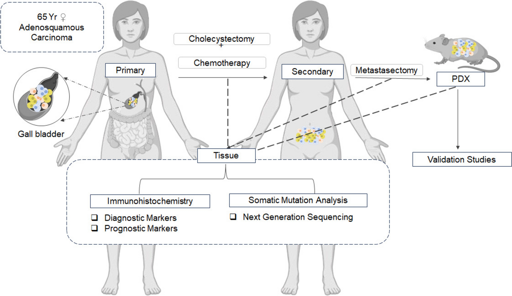
Gallbladder cancer has a poor prognosis due to its aggressive nature and late detection. Adenosquamous subtypes are rare and poorly characterized at the molecular level. Patient-derived xenograft (PDX) models preserve tumor heterogeneity and support evolutionary studies. Longitudinal specimens—primary, metastatic, and PDX—from the same patient, combined with immunohistochemistry (IHC), allow investigation of tumor progression and cellular origin.
MethodologyA 65-year-old woman with gallbladder cancer underwent cholecystectomy, followed by omental metastasectomy upon relapse. A single-cell suspension from the metastatic lesion was injected into NOD-SCID mice to generate a PDX model. IHC assessed marker expression in primary, metastatic, and PDX tumors. A targeted next-generation sequencing somatic panel analyzed clonal evolution.
ResultsP63 confirmed the adenosquamous subtype. The PDX model retained histopathological and marker features of the primary and metastatic tumors. Diagnostic (CK7, CK17, Muc1, Muc5AC) and prognostic markers (EpCAM, CRP, S100P, AQP1, Podoplanin) were preserved across all tumors. Genomic analysis identified KRAS (G12V) mutation as the main oncogenic driver and LRP1B (Q48R) mutation as a putative tumor suppressor retained in all tumors. The secondary tumor acquired PIK3CA (E65D) and LRP1B (G37437) mutations. The PDX gained mutations in chromatin remodeling genes ARID2, ARID1A, and BAP1. Clonal trajectory analysis identified four subclones, indicating branched and linear evolution patterns.
ConclusionWe successfully established a PDX model of gallbladder adenosquamous carcinoma, with KRAS (G12V) identified as a potential initiating mutation, and the probable tumor-initiating cell is derived from an EpCAM-positive putative cancer stem cell. Sequential analysis revealed the emergence of resistant clones and adaptive selection of chromatin remodelers in the PDX model.
Graphical Abstract
Comments (0)