A predominantly female sample (93.81%) was identified, with a mean age of approximately 54 years, as expected for RA patients in Brazil. This finding is corroborated by Sacilotto et al. [17], who reported a sample composed of 90% women with a mean age of 56.7 years. Although studies from other countries report slightly different samples, most also indicate a female predominance greater than 70% and a mean age between 50 and 60 years [18,19,20]. The mean duration of diagnosis (12.11 years) remained within the range observed in recent studies, demonstrating compatibility of the sample with the contemporary profile of RA.
In the present study, more than 50% of patients tested positive for RF and more than 30% for anti-CCP. The presence of these markers is associated with a more aggressive disease profile, which can hinder and prolong disease control. Rocha, Baldo, and Andrade [21] highlighted that patients positive for RF tend to have less effective clinical responses to TNF-α inhibitors but may respond better to B-cell depletion therapies, such as rituximab. Additionally, the presence of anti-CCP can interfere with the effectiveness of DMARDs, often requiring the early initiation of combination therapy [22]. This profile has the potential to lead to more challenging clinical management, as demonstrated in the present study’s sample.
Regarding pharmacological treatment, patients were undergoing appropriate therapy in accordance with recommendations from the main rheumatology societies, including Brazil’s Clinical Protocol and Therapeutic Guidelines for RA [23, 24]. More than 75% of the sample were on DMARDs, either as monotherapy or in combination. However, despite appropriate therapy, most patients continued to report high pain levels and high disease activity, based on NRS and CDAI scores. The persistence of pain despite adequate treatment reinforces the importance of investigating other pain mechanisms in this patient group [25]. One contributing factor often described in the literature is the coexistence of FM in RA patients [22], present in an average of 21% of cases, ranging from 4.9% to 52.4% according to the meta-analysis by Duffield et al. [26].
The mechanism behind the development of nociplastic pain in RA is not yet fully understood. Clauw [22] suggests that CS in RA patients results from the chronic inflammation characteristic of the disease, lowering the pain threshold and amplifying pain perception even in the absence of noxious stimuli. This may be exacerbated by dysfunction in the descending inhibitory pathways, which normally modulate and reduce pain, leading to CS associated with the primary disease [22]. In turn, Meert et al. [27] identified that CS may result from two distinct mechanisms: “top-down” (from the brain to the periphery) or “bottom-up” (from the periphery to the brain). In RA, the inflammatory stimulus could result in prolonged hyperactivity of dorsal horn neurons, contributing to an exaggerated pain response and leading to predominantly “bottom-up” CS. Both authors propose that top-down and bottom-up mechanisms may coexist and overlap [22, 27].
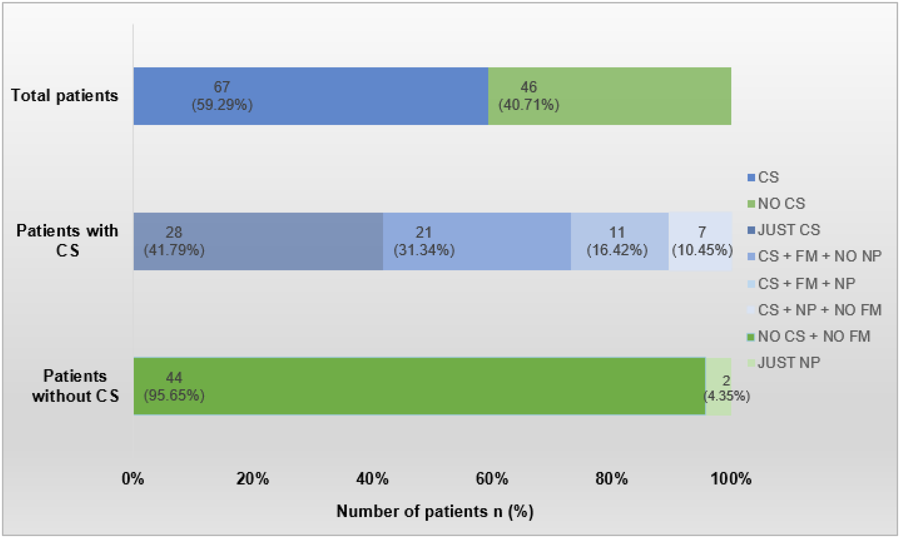
The prevalence of CS among RA patients in our sample was nearly 60%, a higher percentage than even the elevated rates reported by Mesci et al. [28] 48.3% and Guler, Celik, and Ayhan [29] 41.10%. Adami et al. [11] and Salaffi et al. [31] reported lower rates of CS in this group, 29% and 36.5%, respectively, reinforcing the association between RA and CS and the need to better understand this phenomenon and its impact on the clinical profile. Results from our study showed that just over 30% of patients presented with CS without FM, demonstrating that RA can evolve with CS-related pain in a significant portion of the sample, even without meeting FM criteria.
Considering the neurophysiology of pain, anxiety and depression may contribute to the development of a more primary form of nociplastic pain represented by FM or a secondary presentation, as seen in CS without FM [26], or even a mixed CS profile (“top-down and bottom-up”) in RA [27]. From this perspective, the high prevalence may be justified. Furthermore, the presence of CS may explain discrepancies between physician and patient assessments regarding clinical improvement, pain levels, and disease perception, as well as the frequent occurrence of residual pain in RA patients [25, 30,31,32].
The study by Mesci et al. [28] found a positive association between pain levels and disease activity patients with CS tend to have higher levels of pain and higher CDAI values which corroborates the results of our study. In the presence of CS, greater pain reactivity is expected. The study by Adami et al. [11], which evaluated the association between CSI, disease activity, and functional disability, also identified a relationship between pain intensity and disease activity via CSI scores. It found that the association between CSI scores and disease activity was mainly mediated by persistent inflammation and was not the result of faulty global assessment or subjective pain intensity reported on the NRS. The authors concluded that CS in RA significantly impacts functional disability.
The study by Saitou et al. [20] also found an association between CSI and disease activity using different disease activity metrics (CDAI, Simplified Disease Activity Index – SDAI, and DAS-28. However, Guler et al. [29] did not identify any relationship between CSI and pain level or disease activity (DAS-28). The use of a single metric that incorporates an inflammatory marker may have been a confounding factor, as CS can occur even in the absence of inflammation—especially when nociplastic pain is primary.
Sarzi-Puttini et al. [25] found that patients with residual pain are frequently considered to have treatment failure, leading to medication overuse or increased polypharmacy, as well as increased treatment costs and adverse effects. Since pain amplification caused by RA may be responsible for a portion of residual pain symptoms, caution is necessary regarding the excessive use of analgesics, corticosteroids, NSAIDs, and DMARDs.
The results of this study showed that patients with CS predominantly reported severe pain and moderate and/or high disease activity, even when under appropriate pharmacological treatment. If residual pain and disease activity are being driven by an undiagnosed CS, the use of anti-inflammatory agents and DMARDs alone may not sufficiently control pain or induce remission and/or low disease activity. In this regard, Clauw [22] suggests the inclusion of interventions that directly modulate the CNS, as a complement to traditional inflammation-focused approaches.
In this context, the use of tricyclic antidepressants and SNRIs is recommended to restore the function of descending inhibitory pathways, along with medications that modulate calcium and/or sodium influx, which may be effective in reducing neuronal hyperexcitability [25, 27, 31]. Salaffi et al. [33] demonstrated that Janus Kinase (JAK) inhibitors appear to have a positive effect on pain-related variables, particularly central sensitization and pain catastrophizing, which may originate from extra-synovial mechanisms.
The aforementioned studies did not find any association between disease duration and CS, which aligns with our study. However, this is a surprising result, given that longer exposure to nociceptive damage would theoretically have the potential to trigger CS, as suggested in the literature. Likewise, no correlation was found between autoantibody markers (RF and anti-CCP), despite their association with more severe disease presentations. Smoking and physical activity also did not appear to influence the additional pain mechanisms identified, although this result may have been affected by the low frequency of these variables in the sample. Other studies did not evaluate smoking status or physical activity levels.
The presence of NP in chronic inflammatory diseases has gained increasing attention in recent years, due to its potential impact on quality of life and its interference with treatment efficacy. Just over 17% of this study’s sample presented with probable NP, according to the PD-Q. These values are consistent with those reported in the literature, which range between 12.5% and 40%, depending on the instrument used [34,35,36,37,38].
Di Carlo, Smerilli, and Salaffi [39] argued that NP in RA results from a complex combination of factors. It may arise due to compressive mechanisms triggered when chronic joint inflammation alters local or even neuronal morphology, leading to nerve compression by adjacent structures. This is seen in some presentations of carpal tunnel syndrome when triggered by RA.
Another possibility is that NP may also develop due to the activation of glial cells in the central nervous system (CNS), particularly in the spinal cord. These cells, when activated by pro-inflammatory cytokines released during RA such as IL-1β, IL-6, and TNF-alpha—produce mediators that amplify NP through neuronal damage and continued activation of glial cells. This neurosensitization process can occur from the early stages of RA, suggesting that NP is not merely a consequence of prolonged inflammation but may also be an early feature of the disease [39].
The presence of synovial inflammation can lead to perineuritis, an inflammation of peripheral nerves, as observed in the palmar digital nerves of RA patients. This theory of peripheral nerve damage is supported by the study by Pereira, Lourenço, and Assis [37], who assessed the presence of neuropathies in RA patients and found that 48.5% had some form of peripheral neuropathy (either axonal or demyelinating), confirmed by electroneuromyography. Sousa et al. [40] identified axonal damage caused by cytokines, antibodies, or cytotoxic cells as likely causes of NP in rheumatologic disorders.
The findings of this study indicated a positive correlation and predictive value between NP (as defined by probable PD-Q), disease activity, and pain intensity. According to these results, patients with probable NP tend to report higher pain levels and greater disease activity scores, similarly to what is observed in CS. Similar findings were reported by Koop et al. [35], who found that RA patients with NP had higher pain levels, higher DAS-28 scores, poorer quality of life, and a higher likelihood of disability. In turn, Garip et al. [34] identified an association between NP and pain intensity but did not find a relationship between NP and the number of painful joints. Conversely, Radwan and Borai [38] found an association between pain intensity and the number of painful joints, and the latter also identified a relationship between disease duration and NP levels unlike the findings in this study.
PD-Q was a predictor of CS, with a proportion of patients exhibiting overlapping NP and CS. Similar results were observed in other studies [19, 20]. Some authors argue that NP may be a symptom of CS, that tools designed to identify NP may be useful for identifying CS, or that the co-occurrence of NP and CS may be explained by shared mechanisms of pain development. However, this relationship is not yet well-established in the literature, and further studies are needed to identify and analyze these associations, their impacts, and when NP functions as a symptom of CS and/or as a distinct primary or secondary pain mechanism [19, 20].
The differences found among studies investigating NP in RA may be influenced by racial variations and sample size, since these are generally small studies, as well as differences in pain management strategies used in different populations, the inclusion or exclusion of patients with comorbidities that may cause NP, such as diabetes, and the use of different assessment tools (e.g., PD-Q vs. DN4), as suggested in several studies [20, 35, 38].
The early identification and treatment of mechanisms associated with NP development are essential for effective pain management in RA patients and for achieving disease remission, just as in the presence of CS in RA patients. Effective pain management requires strategies that address both central modulation and peripheral inflammatory mechanisms. A comprehensive therapeutic approach including both pharmacological and non-pharmacological components, and the use of different drug classes to control nociception, CS, and NP is necessary to mitigate the harmful potential of the various CP mechanisms associated with RA.
The results of this study reinforce that pain reported by patients with RA may, in many cases, be dissociated from the objective inflammatory activity of the disease, being instead related to CP mechanisms. This dissociation is particularly relevant in the clinical setting, considering that widely used tools to guide therapeutic decisions such as the CDAI and DAS-28 include subjective components directly influenced by the patient’s pain perception, such as global assessment and tender joint count. Thus, the presence of CS and/or NP may result in artificially elevated scores, even in the absence of active inflammation, raising important concerns about the accuracy of these indices in reflecting the true inflammatory activity of the disease [9, 41].
This distortion has relevant clinical implications, especially in the context of the treat-to-target strategy, which is widely recommended and adopted in modern rheumatology practice to achieve remission or low disease activity [41]. As this strategy relies directly on clinical scores to guide therapeutic decisions, the influence of non-inflammatory pain mechanisms may lead to unnecessary intensification of immunosuppressive treatment, increasing the risk of adverse effects and healthcare costs. Therefore, it is crucial to consider these mechanisms when interpreting RA activity scores, incorporating specific tools to detect CS and NP, as well as promoting the development of new assessment instruments that more comprehensively evaluate disease activity in RA, including the presence of these other CP mechanisms. This integrated approach may support more individualized, safe, and effective clinical decisions, ensuring adequate pain control without compromising the accuracy of inflammatory disease activity assessment.
Considering its relevance in the context of RA, CP treatment cannot be disregarded. In the present sample, the low use of antidepressants and gabapentinoids may reflect the underdiagnosis of CS and NP, as well as other contributing factors such as financial constraints, limited access to specialized care, tolerability issues, and concerns about side effects. Although treatment was briefly mentioned, a detailed discussion of all therapies used or not used was beyond the scope and methodological design of this study. Given the breadth, complexity, and amount of data required for a thorough analysis of pharmacological management and its implications for RA, CS, and NP, we believe this topic deserves dedicated future investigations.
This study presented some limitations that should be recognized. The limited timeframe available for data collection and study implementation restricted the sample size. In addition, the scarcity of previous studies on this topic limited the possibility of in-depth comparisons and made it difficult to contextualize findings within the existing body of knowledge. Other limitations inherent to studies of this nature include self-reporting bias, participants’ difficulties in completing or bringing requested tests to consultations, and methodological limitations related to the study design, which may impact the generalizability of the results. Despite these constraints, the findings contribute significantly to the understanding of the topic and may serve as a foundation for future investigations.
In contrast, an important strength of this study is the careful exclusion of patient groups that could act as major confounders in the interpretation of pain mechanisms, such as individuals with diabetes, psychiatric disorders, or significant neurological conditions. By applying these criteria, the final sample was composed of patients less influenced by overlapping conditions that themselves may generate or amplify CP. This methodological choice allowed for a clearer characterization of CS and NP specifically within the context of RA, reducing bias and increasing the internal validity of the findings.


Comments (0)