July in focus in HCB
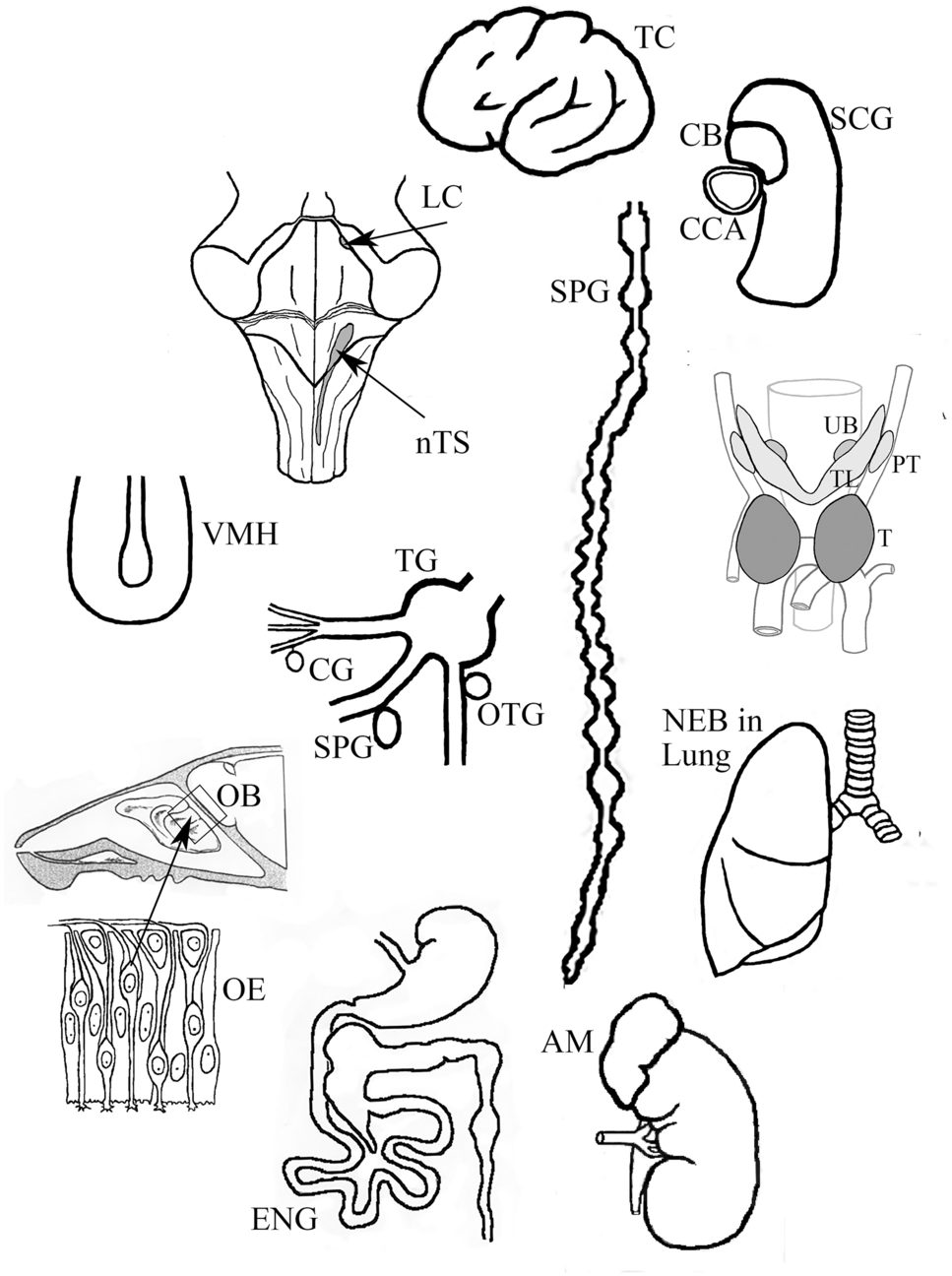
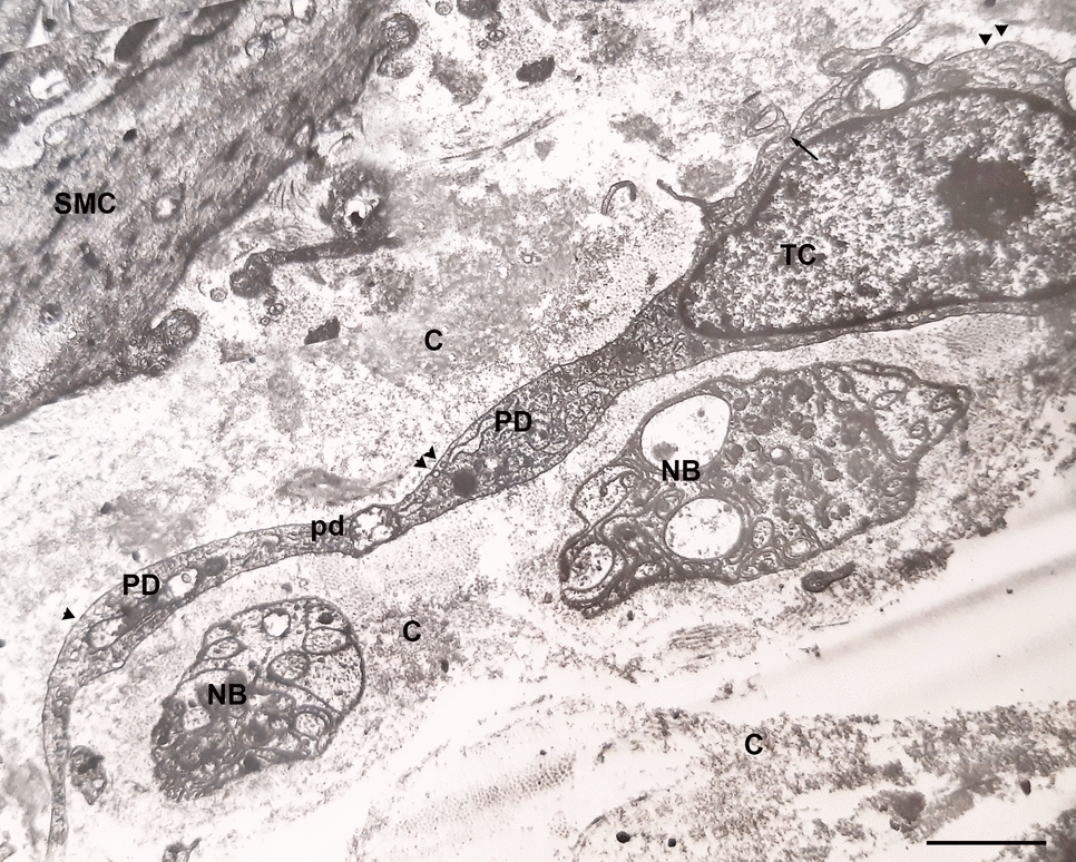
The inhibitor of growth protein family (ING) consists of at least five family members (ING1–5), with a range of proposed activities, including epigenetic regulation, tumor suppression, hematopoietic stem cell homeostasis, cancer cell stemness, cell migration, tumor cell invasion and metastasis, and innate immunity regulation (see Dantas et al. 2019; Jacquet and Binda 2021, for reviews). All the ING family proteins possess a nuclear localization signal including a nuclear targeting sequence, but some immunocytochemical and immunohistochemical studies have identified these proteins in a cytoplasmic location as well. As we opined in the “June in focus in HCB” editorial (Taatjes and Roth 2025), the veracity of immunocytochemical and immunohistochemical staining results is absolutely dependent upon the validated specificity of the primary antibody for its intended antigen target. Indeed, the validation of antibody specificity becomes even more complicated when investigating a family of proteins with very homologous configurations, such as those belonging to the ING family. It is of course quite possible that an antibody raised against one family member protein may cross-react with other family member proteins sharing epitope sequence homology. Over many years, Karl Riabowol’s group has generated antibodies against specific ING proteins to assess their presence in a variety of cells and tissues (Boland et al. 2000; Nabbi et al. 2015; Suzuki et al. 2011). They have now continued this line of work by generating two monoclonal antibodies raised against ING4, validated their specificity for this ING protein, and characterized their intracellular localization in a cultured cell line and variety of mouse and human tissues (Dantas et al. 2025). The ING4 antigen was produced in bacteria, and mouse monoclonal antibodies were generated via hybridoma. From their clones, two monoclonal antibodies (designated 4F4 and 6C6) were found by enzyme-linked immunosorbent assay (ELISA) to recognize both mouse and human ING4. By Western blotting, both antibodies were able to detect ING4 protein in both overexpressing cells and the endogenous level in non-transfected cells, and importantly, the signal was knocked down by treatment of cells with ING4 small interfering RNA (siRNA). They then showed by Western blotting that the two antibodies specifically recognized ING4 protein, and did not cross-react with ING1, ING2, ING3, or ING5. Having diligently validated the antibodies to specifically recognize ING4 using two of the validation pillars recommended by the International Working Group for Antibody Validation (“genetic” via siRNA editing and “orthogonal” via Western blotting; see Uhlen et al. 2016), they then performed immunocyto- and immunohistochemical staining. By immunofluorescence analysis on HEK293 cells, they found (1) high levels of ING4 expression in cells overexpressing ING4, and lower levels of staining for endogenous protein; and (2) while most of the fluorescence signal was found in the nucleus, some staining was also observed in the cytoplasm of overexpressing cells. Finally, by immunohistochemical staining on mouse and human tissue sections, they found a small subset of cells associated with secretory activity in various tissues to be positively stained. Interestingly, the staining pattern in these cells was cytoplasmic, with no nuclear staining evident. The different staining patterns observed in the cultured cell line versus the embedded tissue sections are certainly of interest. Parameters potentially affecting immunostaining results include initial fixation, sample processing, and immunostaining protocols, to name a few. With the very diligent validation of the antibodies for ING4 protein, it will be of interest to perform further immunostaining experiments to analyze the intracellular compartmental localization of ING4 in a variety of cells and tissues, including tumor samples, comparing the potential effects of the abovementioned parameters on immunostaining localization results.


Comments (0)