Traditional surgical methods for accessing posterior tongue and oropharyngeal tumors present several drawbacks and postoperative complications, potentially leading to non-unions and compromised masticatory functions, and resulting in aesthetic concerns [3,4,5]. A modified pull-through approach was proposed to overcome these limitations [1]. However, the self-retentive wedge osteotomy creates an iatrogenic weak point in the mandibular symphysis that increases the risk of pathologic fractures [17]. Biomechanically, the symphysis region has to absorb or compensate for significant bending, torsion, and shear stresses [18]. Yet, a wedge osteotomy potentially leads to an increased susceptibility of the symphysis to fatigue crack propagation [19]. Therefore, the osteotomy design plays a key role in promoting mechanical stability and reducing the risk of pathologic fractures. In this study, we evaluated variations of the previously proposed wedge osteotomy design [1] to identify features associated with reduced fracture risk.
Four wedge osteotomy designs (W1–4) for a modified pull-through approach in the mandibular symphysis were compared by assessing mechanical strain distributions during right-sided unilateral clenching in a maximum bite force condition. The cortical bone at the right canine was designated as the region of interest for quantifying the mechanical environment, as the intersection of the osteotomy planes here, the presence of the long canine roots, and the resulting thinning of the overlying cortical layer may create weak points prone to crack initiation. A maximum principal strain failure criterion [14] was chosen to identify areas experiencing peak tensile and shear loading, which are the primary failure mechanisms for cortical bone tissues [19]. Maximum principal strain and maximum shear strain in the ROIs were quantified and compared to their respective tensile and shear yield strain values [15]. The results suggest that osteotomy designs with less acute angles (e.g., W4) are biomechanically more advantageous since they lower tensile and shear strain concentrations at the right canine region, due to a smoother transition between the osteotomy planes.
Reported values of yield strain in tension and shear are 0.45% ± 0.05% and 0.57% ± 0.03% [15]. In this study, in the ROIs, W1 consistently exhibited the highest strain values εpeak = 0.20% and γpeak = 0.14%. On the other hand, W4 consistently showed the lowest values εpeak = 0.08% and γpeak = 0.07%, respectively, 62% and 51% lower than W1. Overall, the strain values in this study were always below the reported yield thresholds, suggesting that none of the osteotomy configurations are at immediate risk of failure under the simulated physiological loading conditions. However, the stress and strain concentrations in W1 at the intersection of the straight bone cuts represent a critical point for damage accumulation and fatigue failure. W4 showed the most favorable mechanical response thanks to the larger angle (96°) between osteotomy planes, compared to the 63° of W1. The strain concentration reduction was also noticeable in W2 and W3, which had angles of 92° and 86°, respectively. The osteotomy angle plays, therefore, a fundamental role in the distribution of strain within the bone. Our results well align with Bujtar et al. [20] who showed that a beveled, rather than a right-angled, osteotomy cut helps reduce the stress at the osteotomy cut. This principle has also been previously applied in structural engineering. For example, Mattheck showed how a smoother transition in a fillet improved the fatigue Life of a part by up to 40 times [21].
The wedge osteotomy designs proposed in this study represent a progressive refinement of the original W1 design [1]. The rationale underlying these modifications was to introduce smoother curvatures to help lower the cutting point in the canine region, which also provides a greater distance between the osteotomy and the dental roots. This aimed to preserve a greater symphyseal cross-sectional area, while still allowing surgical access to the posterior oral cavity. Although the arch-shaped osteotomies W3 and W4 helped reduce the strain in the middle of the symphysis compared to W2, the main reason for lower strains in the canine region was the less acute osteotomy angle in W2 and W4. Thus, this seems to be the most critical parameter when designing the wedge osteotomy. The performance of the wedge osteotomy and the associated weakening of the mandible at the symphysis should be carefully evaluated, especially if stabilizing osteosynthesis material is omitted. It must be noted that such a surgical approach still coincides with a decrease in stability and a risk of fracture. This is why the edentulous highly atrophic mandible is generally considered a contraindication for this approach [1]. However, preoperative biomechanical studies may potentially permit the application of the modified pull-through approach in individual cases presenting with an edentulous jaw with sufficient bone height.
Since the form of the osteotomy needs to be individualized, no single shape can be considered a universally optimal solution. With this in mind, a compromise between the biomechanically optimized shape and surgical feasibility must be identified based on virtual planning and biomechanical assessment for each individual situation. Overall, a patient-specific modification of the shape of the original osteotomy of the modified pull-through technique (W1) [1] is recommended. Reducing the segment size and thus the functional gap generally helps reduce the risk of fracture.
While this study provides valuable insights into the biomechanical behavior of different osteotomy designs, several limitations must be acknowledged.
A key limitation is that only four wedge designs were tested in a single patient-specific model, representing a preliminary analysis based on surgically feasible configurations. While this approach allowed for a controlled comparison and identification of design principles, such as avoiding acute angles and preferring rounded transitions, generalization to broader anatomical and surgical conditions (e.g., edentulous mandibles, mandibular atrophy, variable bone densities) remains limited. Future work should incorporate parametric variation or computational optimization techniques to identify potentially better-performing configurations to develop patient-specific osteotomy planning tools.
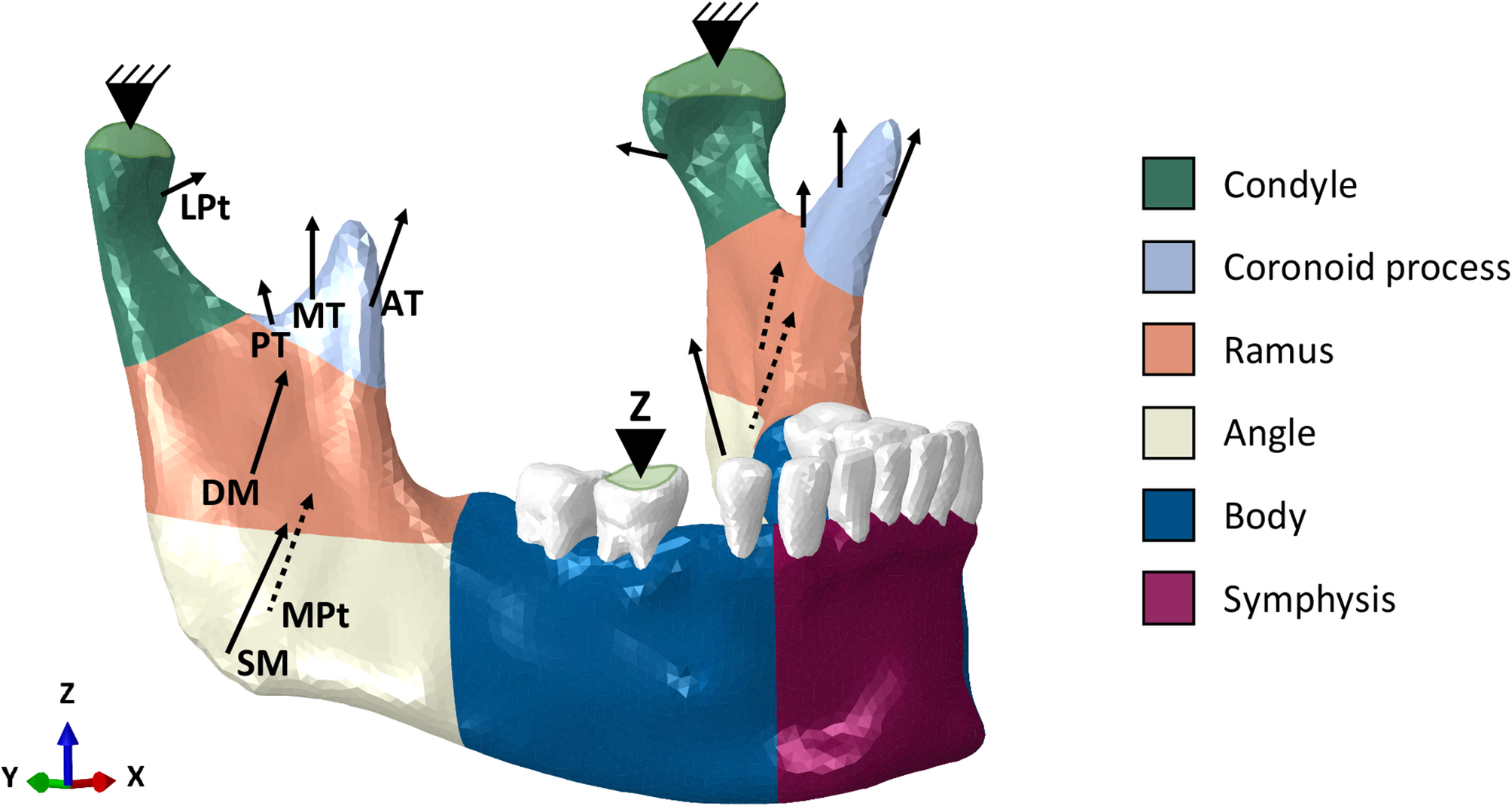
The chin wedge was omitted from the model to simulate a worst-case post-operative scenario, in which no osseous union is present and the wedge provides no mechanical support. As the healing progresses, the mineralization of the interface tissue between the mandible and the wedge increases the load-bearing contribution of the wedge, thereby lowering strain concentrations at the osteotomy junctions. By focusing on an immediate post-operative condition, the analysis allowed testing the most critical mechanical scenario in all osteotomy cases. The material properties used in the finite element model were assumed homogeneous and elastic, whereas bone exhibits heterogeneous properties. This is, however, a reasonable simplification considering that the study focused on the comparison of the performance of different osteotomy designs. Additionally, only static biting was included in the analysis, while in vivo conditions involve dynamic and cyclic loading. However, since all models use the same material properties and loading and boundary conditions, the relative differences in stress and strain remain valid. Furthermore, only unilateral clenching was tested, but evidence shows that this represents the most mechanically solicited scenario [9, 22] and clinically the most Likely to occur post-surgery. All designs were moreover tested in a maximum bite force condition as the worst-case scenario, with a bite force at the occlusion equal to 330 N. Previous studies on mandibular fractures and reconstruction have reported maximum bite forces of approximately 100 N during early recovery [23, 24]. However, unlike in those cases, in the modified pull-through approach, the mandibular continuity is preserved, which may allow for greater functional loading. Moreover, involuntary high bite forces (i.e., bruxism) remain clinically plausible and pose additional risk in the immediate post-operative phase. Therefore, in this study, the upper-bound load was used to ensure a conservative assessment of bone failure risk, allowing a coherent comparison across the different cases. As other FEA analyses have shown, a closed mouth position can decrease the fracture risk in the symphysis area and provide the maximum passive traction to the muscle, forcing the bony segment into its socket [22]. Therefore, a postoperative intermaxillary fixation with elastics might help to avoid unfavorable forces and outcomes. Future studies should also consider incorporating patient-specific bite force measurements to improve the clinical validation of finite element predictions.




Comments (0)