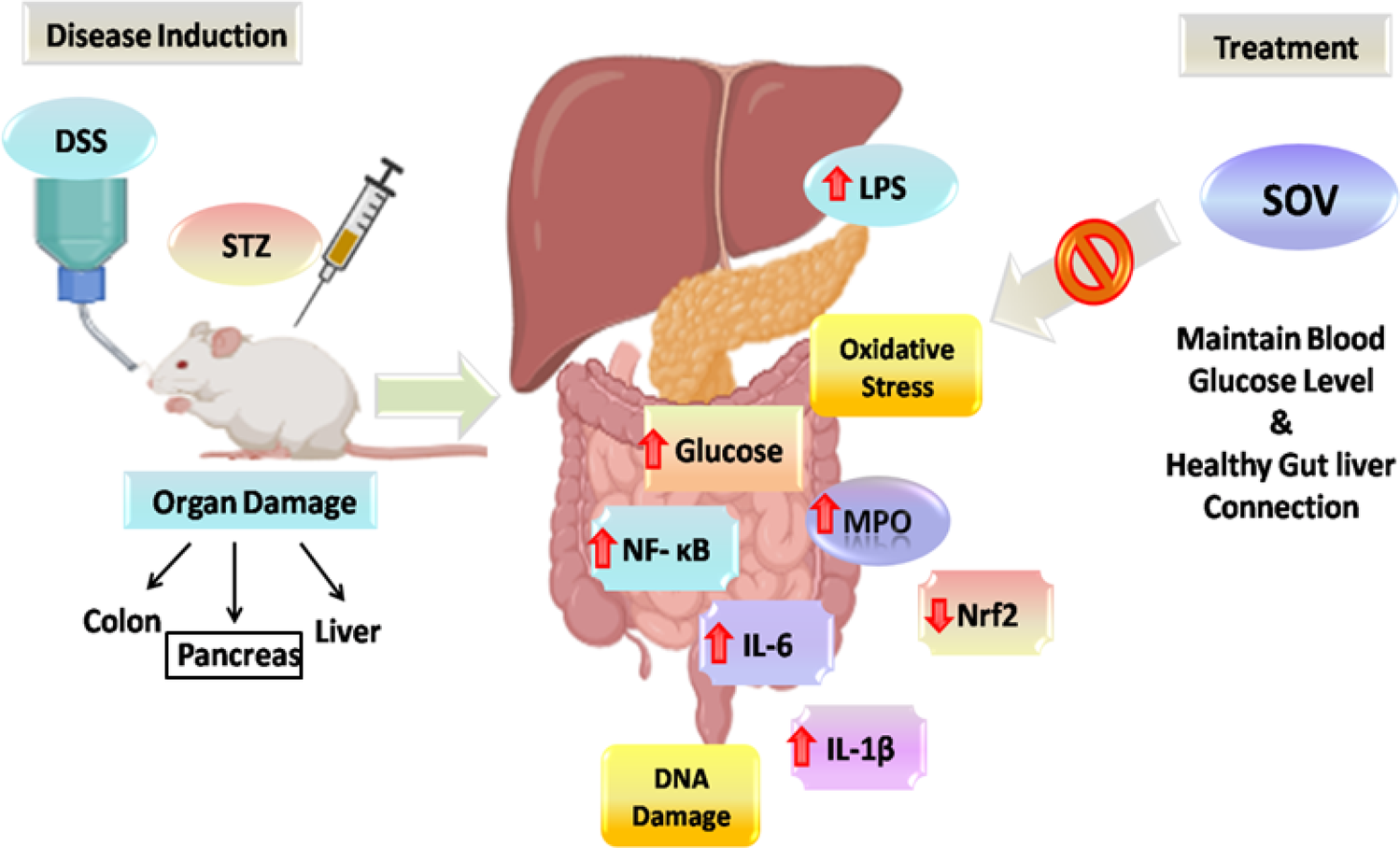
Animal model
Fifty rats were purchased from Shulaibao Biotech (Wuhan, China) and kept at constant temperature (23–25 ° C) with controlled light/dark cycles (12 h/12 h) and bred and housed in the Affiliated Shanghai East Hospital, Tongji University School of Medicine. We monitored the health and behavior of rats every day, including weight, food intake, water intake, fur and skin condition, as well as the occurrence of any abnormal activities. Injection of sodium pentobarbital (40 mg/kg) intraperitoneally was used to anesthetize rats prior to the experiment. No rats died during feeding and experiment. All rats were sacrificed by exposure to carbon dioxide at a concentration of 60% of the container volume per minute after the experiment. We established the UUO model by blocking the unilateral ureter. Sprague–Dawley (SD) male rats, weighing 180 to 200 g, were randomly classified into the sham, UUO and UUO + KAR-10, -20 and − 40 IU/kg groups (n = 10). Following UUO surgery, the renal obstruction modeling was maintained for 14 days post-operation, consistent with established protocols for observing renal fibrosis progression in rodent models. After disinfection, we incised the left abdomen, exposed the kidneys, separated the ureter, interrupted the suture by a 4 − 0 operation at the end of the kidney, cut from the middle of the two ligations and reset the organs, sutured the incision, and disinfected the skin. The rats in the sham group were treated with UUO except for ligation and cutting of the ureteral. The UUO rats were treated with low, medium and high doses of KAR. Throughout the 14-day modeling period, all animals were monitored daily for surgical integrity (e.g., wound healing, ligation stability) and physiological stability (body weight, food intake, activity level). The study was approved by the Ethics Committee of the Shanghai East Hospital, Tongji University School of Medicine.
Hematoxylin and eosin (HE) staining analysis
We fixed the left kidney in 4% formaldehyde for several days and embedded it with paraffin. The samples were dewaxed in xylene and dehydrated with ethanol and HE staining was performed to analyze renal dysfunction. Images were taken at 400× magnification using a microscope.
Masson and prostate-specific antigen staining analysis
The renal tissues were stained with prostate specific antigen (PAS) for the detection of glycogen and Masson was used to analyze the degree of fibrosis. Images were taken at 400× magnification using a microscope.
Serological analysis
Serum levels of urea nitrogen (BUN) and serum creatinine (SCr) were measured using kits purchased from Beyotime Biotechnology (Shanghai, China).
Cell culture
We purchased HK-2 cells (RRID: CSTR:19375.09.3101HUMGNHu47) from the Cell Bank of the Shanghai Life Science Institution. HK-2 cells were cultured in DMEM/F12 (Thermo Fisher Scientific) and treated with 10% fetal bovine serum and 100 U/ml penicillin–streptomycin and incubated in a cell incubator at 37 ° C with 5% CO2. Then the effect of KAR was studied in the HK-2 cell by being exposed for 24 h to normoxia (21% O2) or hypoxia (O2 < 0.1%). Cells were stimulated with or without KAR-50 µM for 24 h and treated with 5 µM ML385 for 24 h. Cells were collected for subsequent analysis. Transforming growth factor-β (TGF-β) is a type of multifunctional cell factor that can regulate cell growth, differentiation, remodeling of the ECM, endothelial mesenchymal transition, and induce cell fibrosis. Therefore, we established a cellular fibrosis model by treating HK-2 cells with TGF-β. The provided sequence represents small interfering RNA (siRNA) molecules, including Nrf2-siRNA and si-NC. These sequences are, respectively, composed of the following nucleotides: 5′-UAAUUGUCAACUACUGUCAGUU-3′ and 5′-GACGAAAGCAGTTCGTCTA-3′.
Quantitative real-time polymerase chain reaction (RT-qPCR)
The total RNA of the samples was extracted using a Trizol reagent. The reverse transcription of RNA into cDNA was performed using the Bio-Rad iScript cDNA synthesis kit. Differential mRNA expression was determined using a CFX ConnectTM real-time system (BIO-RAD, United States), following the instructions provided by the manufacturer (Applied Biosystems). RT-qPCR results were analyzed using the 2 − ΔΔCt method, with GAPDH used as the standardized control. The primers used were as follows. TGF-β1: forward 5’-GGCCAGATCCTGTCCAAGC-3’ reverse 5’-GTGGGTTTCCACCATTAGCAC-3’, FN: forward 5’- CGGTGGCTGTCAGTCAAAG-3’ reverse 5’- AAACCTCGGCTTCCTCCATAA-3’, SMAD3: forward 5’-TGGACGCAGGTTCTCCAAAC-3’ reverse 5’- CCGGCTCGCAGTAGGTAAC-3’, SNAI1: forward 5’- TCGGAAGCCTAACTACAGCGA-3’ reverse 5’- AGATGAGCATTGGCAGCGAG-3’, Kim1: forward 5’-TGGCAGATTCTGTAGCTGGTT-3’ reverse 5’- AGAGAACATGAGCCTCTATTCCA-3’, GAPDH.
Western blot
RIPA lysis buffer (Solarbio, Beijing, China) was utilized to extract total protein. The BCA protein concentration determination kit (Beyotime, Wuhan, China) was used to determine the protein concentration. The proteins in different samples were separated by SDS-PAGE and transferred to the polyvinylidene fluoride (PVDF) membrane. The membrane was sealed with 5% BSA at room temperature (RT, 25 ° C) for 1 h. It was then incubated overnight at 4 ° C with different primary antibodies (1:500 ~ 1,000). Subsequently, once the unbound primary antibodies were removed from the membrane, the corresponding HRP-conjugated antibodys (1:5,000 ~ 8,000) were incubated at RT for 1 h. The film was eventually processed with the ECL detection kit (Beyotime, Wuhan, China) for analysis of the grayscale values of the bands was done using the image processing software Image J. The following primary antibodies were used: GAPDH (Santa Cruz, CA), fibronectin (FN) (1:1000, Proteintech, China), α-SMA (1:1000, Proteintech, China), Snail1 (1:1000, Proteintech, China), Kim1 (1:1000, Proteintech, China), small mother against decapentaplegic (Smad)3 (1:1000, Proteintech, China), p-Smad3 (1:1000, Abcam, USA), TGF-β1 (1:500, Bioworlde, China), Nfr-2 (1:1000, Abcam, USA), (1:1000, CST, China), HO-1 (1:1000, CST, China), Hif-1α (1:1000, Proteintech, China) and Keap1 (1:1000, CST, China).
Immunofluorescence (IF) staining analysis
After fixation with 4% paraformaldehyde, samples were permeabilized with Triton-X 100 (0.1%) and blocked with an immunostaining blocking solution (3% H2O2). Cells after treatment with 5% BSA for 30 min were then incubated at 4 ° C with α-SMA (1:500, Proteintech, China), differentiation cluster (CD) 206 (1:500, Proteintech, China), F4/80 (1:200, CST, China), TGF-β1 (1:100, Bioworlde, China) and Nrf-2 (1:200, Abcam, USA) for 12 h. The corresponding secondary antibodies were incubated at 37 ° C for 1 h. We then used 4’,6-diamidino-2-phenylindole to stain the nucleus. We obtained IF images using a laser scanning microscope.
RNA interference
We seeded HK-2 cells in 6-well plates and small interference RNA (siRNA) targeting the Nrf-2 gene (siNrf-2), which were transfected with the siNrf-2 plasmid using RNA transfection reagent. The specific operation is as follows: inoculate cells one day before transfection, and the cell density for inoculation is around 30%-50%. Subsequently, for each cell well, dilute 8.4 ng of siRNA (0.6 pmoles) in 100 µL serum-free medium (OPTI-MEM medium) and mix well. Immediately add 2 µL transfection reagent (Lipofectamine 2000) to 100 µL siRNA, vortex for 10 s to thoroughly mix. Incubate at room temperature for 10 min to form siRNA-PEI cationic nucleic acid transfection complexes. During the formation of the complexes, remove the cell growth medium and add 500 µL of fresh preheated complete medium to each well. Directly add 100 µL of siRNA-PEI cationic nucleic acid transfection complexes to the cells, gently shake the culture plate to mix. The final volume is 600 µL, with a siRNA concentration of 1 nM. Incubate at 37 °C, 5% CO2 in a cell culture incubator for 48 h, and then detect the expression level of the target protein.
Statistical analysis
We use the Statistical Package for Social Sciences software version 26.0 to analyze the results. Multiple comparisons were performed using a one-way analysis of variance. Comparison between the two groups was made using Student’s t-test. Data in all figures with histograms were represented using the mean ± standard deviation. P-values < 0.05 indicated statistical significance.


Comments (0)