
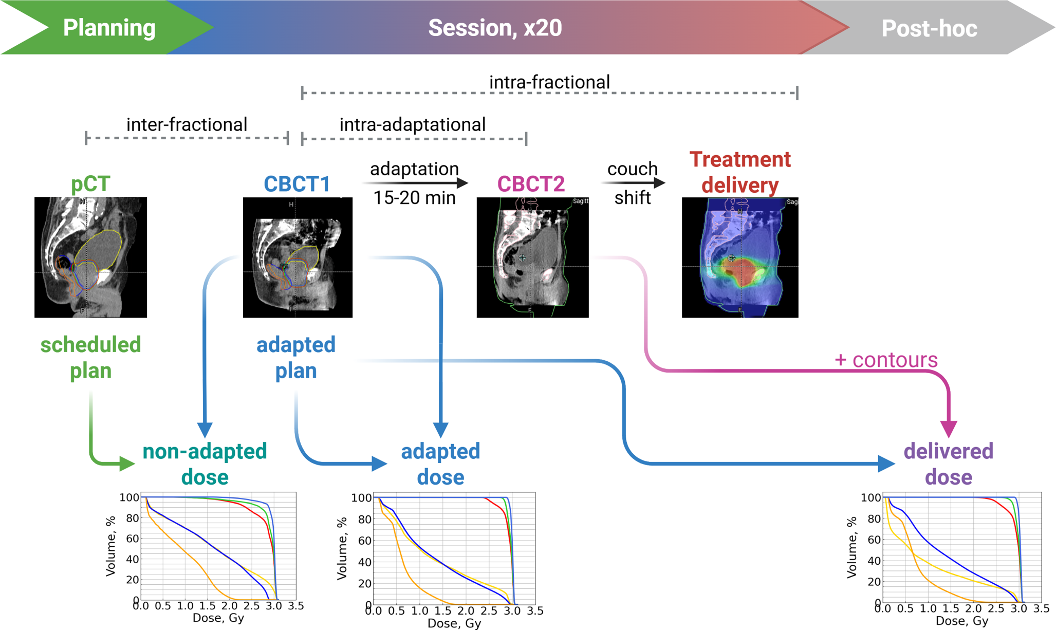
Remember me
The prostate contour volume remained largely unchanged during the short interval between CBCT1 and CBCT2 (approximately 15–20 min, see Fig. 1 in the Supplement), with the mean volume difference between the two scans per patient ranging from 0.01 to 0.73 ml (Fig. 3).
Seminal vesiclesSimilarly, no significant changes in the seminal vesicle volumes are anticipated between CBCT1 and CBCT2; the observed mean differences ranged from 0.12 to 2.8 ml across individual patients (Fig. 3).
Organs at riskChanges in bladder and rectum volumes have physiological reasons, additionally to variations in contouring. For the bladder, we saw a systematic increase in volume between CBCT1 and CBCT2 (Fig. 3), which is plausible since bladder filling continued during the treatment session. The mean difference in bladder volume between CBCT2 and CBCT1 across patients ranged from 19.5 to 130.9 ml (the increase was statistically significant for each patient, with \(p<0.001\) according to the Wilcoxon test), and the mean filling rate ranged from 1.14 to 5.92 ml/min.
For the rectum, no such systematic effect could be discerned (Fig. 3), with mean volume differences between CBCT1 and CBCT2 staying rather small and nonsignificant for most patients: between −3.6 and 6.3 ml, with \(p > 0.1\) (except for patient 7 with an always full rectum, who exhibited a significant decrease in rectum volume, with mean values of 8.9 ml and \(p=0.003\)). However, in addition to some variability in manual contouring, we observed changes in shape due to peristalsis and movement of air pockets (see below).
Dosimetric impactExamples of two patientsExamples of the metric distributions for single patients (3 and 4) are depicted in Fig. 4. We comment in some detail on these plots, as they provide a representative and intuitive view of the most relevant effects.
Fig. 4
Metric distributions for patient 3 (a) and for patient 4 (b). The y‑axes present the metric values, while the x‑axes show the combination of a plan (“sch” or “adp”) and a CBCT (“cbct1” or “cbct2”). Bold lines represent the metric value with the scheduled plan on the pCT. Dotted lines correspond to the objective for each metric. Each box extends from the first quartile (Q1) to the third quartile (Q3), with a line indicating the median. Dots represent outliers (data points lying outside the interval \([Q1-1.5IQR, Q3+1.5IQR]\), where IQR denotes the interquartile range). The metric name and its objective are provided on top of each plot
For both these patients, it is evident that the scheduled plan will result in poorer dose coverage when evaluated on CBCT1 (which is, in part, due to the contouring method for the prostate, as described above; see also Fig. 3 for the prostate volumes). Regardless of the origin of this discrepancy, it will be considered a “real underdosage” in the adaptive workflow, and the aim of the adaptation will be to correct this. Consequently, the reoptimized, adapted plan (adp@cbct1) will again provide perfect target coverage, as defined in our treatment intents, i.e., 100% of the PTV/SIB1/SIB2 volume covered by 95% of the prescribed dose to this volume. In a way, this would correspond to a scaling of the plans in a planning study, and the adapted plan (on CBCT1) will always strive for this target coverage. In comparison with the ideal adapted plan (on CBCT1), the delivered dose (adp@cbct2) falls well short of this coverage, while remaining better than the scheduled plan. This effect is quite strong, as the loss in coverage between adp@cbct1 and adp@cbct2 cannot be explained by a change in prostate and target volume contouring. Since both adp@cbct1 and adp@cbct2 were contoured based on CBCT images only, these target volumes should be consistent with one another and no marked deviation in dose should ensue.
Regarding the OAR doses, the dose–volume metrics reflect the general changes in organ volumes as outlined above. However, these two patients represented different cases. Patient 3 had non-ideal bladder filling on the planning CT (Fig. 3). The bladder showed a small change between CBCT1 and CBCT2; thus, the metrics could not improve due to geometrical effects. Although all the bladder metrics stayed below the limits, significant degradation was observed with adp@cbct2 in comparison to adp@cbct1. Somewhat better performance of the scheduled plan on CBCT1 might be caused by a smaller prostate on the pCT: the scheduled plan had a smaller high-dose region (an example of such a session is shown in Fig. 2 in the Supplement) that expanded into the bladder (and the rectum).
For the rectum, no relevant change in volume or in dose metrics can be seen between CBCT1 and CBCT2 for patient 3 (although the differences for the rectum metrics were statistically significant, all the metrics remained well under the limits). There was slightly more dose to the PRW on CBCT2 because the plan was not optimized on the CBCT2 anatomy but rather on the CBCT1 anatomy.
In contrast to patient 3, patient 4 had high bladder filling on the planning CT (Fig. 3), but rather low filling on the CBCTs. This explains the very poor performance of the scheduled plan applied to the CBCT1 contours. Since the bladder volume on CBCT2 is larger than on CBCT1, the dose metrics will appear improved on CBCT2 due to geometry, even with a “less-adapted” plan.
Also for patient 4, no relevant change in rectum volume was observed between CBCT1 and CBCT2. The dose metrics showed, however, a notable deterioration with the adapted plan on CBCT2. Partially, this can be attributed to the fact that the plan was not optimized on the CBCT2 anatomy. Additionally, this very patient exhibited substantial changes in rectum shape between CBCT1 and CBCT2 (one session is depicted in Fig. 2), which impacted both the rectum and the PRW metrics.
Adapted vs. delivered dose distributionsThis comparison addresses the question of how the anatomical changes between CBCT1 and CBCT2 influence plan accuracy.
Statistically significant differences between adapted and delivered dose distributions were observed for most patients in the target volume metrics: PTV/SIB1/SIB2 V95% and SIB1 D95% yielded better results with the adapted plan on CBCT1 (see Fig. 5 as well as Fig. 4 in the Supplement). In contrast, no significant differences were noted for PTV/SIB2 D95% in most cases, with few exceptions. When significant differences were observed for PTV D95% and SIB2 D95%, adp@cbct1 demonstrated better results. The only instance where a target volume metric was significantly better with adp@cbct2 occurred for SIB2 D95% for patient 6; however, the median difference for this case was only 0.25% (here and below, we refer to absolute changes, e.g., percentage points).
Fig. 5
Distributions of absolute metric differences: “adapted–delivered” dose. Each subplot represents one metric, and each box corresponds to a single patient. Each box extends from the first quartile (Q1) to the third quartile (Q3), with a line indicating the median. Dots represent outliers (data points lying outside the interval \([Q1-1.5IQR, Q3+1.5IQR]\), where IQR denotes the interquartile range). Significant differences (\(p < 0.05\)) are marked with an asterisk
Considering the entire cohort, all target volume metrics significantly degraded with the delivered dose compared to the adapted one, as evidenced by positive medians of the difference distributions (Table 2 and Fig. 6). However, PTV and SIB2 metrics stayed within the alternative constraints for most sessions with the delivered dose.
Table 2 Percentage of sessions with an unmet alternative constraint for a specific metric for each dose distribution: non-adapted, adapted, and delivered as well as median and range of metric difference distribution with the corresponding p-values (obtained from the Wilcoxon signed-rank test for paired data with a two-sided alternative hypothesis) for each pair of dose distributions (adapted–delivered and delivered–non-adapted) across the entire cohortFig. 6
Metric distributions for all 155 fractions. Medians are shown with thick lines. For each violin plot, maximum and minimum values as well as 5% and 95% quantiles are shown
There are two main factors affecting the bladder metrics: (1) the adapted plan is calculated based on CBCT1 (meaning that adp@cbct1 is expected to be better than adp@cbct2), but (2) the bladder continues to fill between CBCT1 and CBCT2 (which means that adp@cbct2 is expected to be better than adp@cbct1). For some patients, one factor predominated, while for others, the other factor played a more significant role. This may explain the opposing results observed in some patients (Fig. 5). If the bladder did not fill much between the two CBCT scans, bladder metrics could be worse with adp@cbct2. Patient 8 could serve as example here: he exhibited a very small bladder volume increase (19.5 ml on average), and bladder V48Gy degraded with adp@cbct2, while bladder V60Gy and V40Gy changed nonsignificantly. Considering the entire cohort, only the V48Gy and V40Gy metrics exhibited significant improvement with the delivered dose (Table 2).
The rectum may also change its shape between CBCT1 and CBCT2, but no consistent trend is expected regarding whether the rectum volume increases or decreases. Consequently, no definitive behavior of rectum metrics can be predicted based solely on rectum volume. However, as the adapted plan is based on CBCT1, it is expected that adp@cbct1 would perform better or, at least, not worse. This was confirmed for 7/8 patients (Fig. 5). For the entire cohort, both V48Gy and V24Gy were significantly worse with adp@cbct2 (Table 2), but they still met objectives for most sessions.
As mentioned above, V37Gy for the PRW is often zero. Therefore, we focused on V30Gy, using V37Gy as a supplementary measure. For four patients, V30Gy exhibited a statistically significant difference, showing for one patient a small improvement with adp@cbct2, while for 3 others, a degradation (Fig. 4 in the Supplement). For the entire cohort, V30Gy was significantly better with adp@cbct1 (Table 2). V37Gy yielded a significant difference only in two patients. Frequent zero values of V37Gy led to a counterintuitive result: for the entire cohort, the Wilcoxon test revealed a significant difference between these two dose distributions (\(p = 0.003\)), but the median difference was exactly 0. The mean difference was −1.0% though, which means that adp@cbct1 outperformed adp@cbct2.
Delivered (adaptive) vs. non-adapted dose distributionThis comparison addresses the clinical question of whether the adaptation remains beneficial in the face of the intra-adaptational anatomical changes.
It is important to note that the cohort is relatively small (8 patients), and we cannot cover all possible relationships between organ volumes on pCT and CBCTs evenly. These relationships strongly influence results of the “non-adapted vs. delivered” comparison (discussed below). Among the cohort, 3/8 patients had \(V_\mathrm < \mathrm(V_\mathrm)\), also 3/8 patients had \(V_\mathrm < \mathrm(V_\mathrm)\), and only 1 patient had both the prostate and the bladder volumes smaller on the pCT, while 3 patients had both volumes bigger on the pCTs.
Two sets of anatomical changes play a role in this comparison. First, the changes between pCT and CBCT1 affect the scheduled plan on CBCT1, worsening it compared to the same plan on the pCT (which almost certainly meets all the objectives). On the other hand, the changes occurring between CBCT1 and CBCT2 could worsen adp@cbct2 compared to adp@cbct1. The key comparison is the following: which of these two sets of changes has a stronger influence? Intuitively, the changes between CBCT1 and CBCT2 should be smaller than those between pCT and CBCT1. Therefore, we expect adp@cbct2 to be better/not worse than sch@cbct1.
This expectation was quantitatively proven for all the target volume metrics for each patient individually (see Fig. 7 as well as Fig. 5 in the Supplement). Furthermore, when considering the entire cohort, all target volume metrics significantly increased by 1–4% with the delivered dose distribution (Table 2 and Fig. 6).
Fig. 7
Distributions of absolute metric differences: “delivered–non-adapted” dose. Each subplot represents one metric, and each box corresponds to a single patient. Each box extends from the first quartile (Q1) to the third quartile (Q3), with a line indicating the median. Dots represent outliers (data points lying outside the interval \([Q1-1.5IQR, Q3+1.5IQR]\), where IQR denotes the interquartile range). Significant differences (\(p < 0.05\)) are marked with an asterisk
Considering individual patients, the OAR metrics showed inconsistent results. Only for the middle-dose bladder metrics did the majority of patients exhibit improvement with the delivered dose (see Fig. 7 as well as Fig. 5 in the Supplement). Moreover, for the entire cohort, bladder V48Gy and V40Gy as well as rectum V24Gy were lower with adp@cbct2 (Table 2). At the same time, bladder V60Gy and both PRW metrics yielded no statistically significant differences. Rectum V48Gy, however, was better with the non-adapted dose.
Unmet constraintsAs discussed above, the adapted dose is expected to yield better outcomes than the delivered dose. Although this was confirmed for most metrics, a key question is how often is this difference large enough to result in an unmet objective with the delivered dose? Table 2 presents the percentages of sessions with unmet constraint for all three dose distributions. We observed an increase in constraint failures between the adapted and the delivered dose for all metrics (except for bladder V48Gy and V40Gy). The increase varied from 0.6 to 19% (percentage points) among the different metrics. A high increase of 10–19% was observed for the PRW and SIB1 metrics, while for all other metrics, the increase did not exceed 4.5%. A high number of sessions with a PRW V30Gy constraint failure (even with the adapted dose) could be explained by a low priority of this metric during plan calculationFootnote 2.
Additionally, we assessed the number of sessions in which a goal for a given metric (an alternative goal, when it exists) was satisfied with adp@cbct1 but not with adp@cbct2. These session counts are summarized in Table 1 in the Supplement.
There were 15 sessions in which only both SIB1 goals were not achieved with the delivered dose (being achieved with the adapted one). The possible reason for this is a steep gradient in the dose in the craniocaudal direction, which is additionally “reinforced” by the definition of the SIB1 used in CHHiP [10]: SIB1 and PTV contours are identical almost along the entire contour, except for seminal vesicle area and posteriorly. But the isodose lines for SIB1 and PTV differ everywhere: the 95% prescribed-dose isoline for PTV includes more area than the one for SIB1. In the case when the prostate contour would be slightly shifted downwards on CBCT2 with respect to CBCT1 (as depicted in Fig. 3 in the Supplement), the probability of the SIB1 goals being unmet is the highest among all target volumes.
Comments (0)