Albrektsson T, Wennerberg A. On osseointegration in relation to implant surfaces. Clin Implant Dent Relat Res. 2019;21 Suppl 1:4-7. https://doi.org/10.1111/cid.12742. Epub 2019 Feb 28. PMID: 30816639.
Heydecke G, Boudrias P, Awad MA, De Albuquerque RF, Lund JP, Feine JS. Within-subject comparisons of maxillary fixed and removable implant prostheses: Patient satisfaction and choice of prosthesis. Clin Oral Implants Res. 2003 Feb;14(1):125-30. https://doi.org/10.1034/j.1600-0501.2003.140117.x. PMID: 12562375.
Trulsson U, Engstrand P, Berggren U, Nannmark U, Brånemark PI. Edentulousness and oral rehabilitation: experiences from the patients’ perspective. Eur J Oral Sci. 2002 Dec;110(6):417-24. https://doi.org/10.1034/j.1600-0722.2002.21394.x. PMID: 12507214.
Pinotti FE, de Oliveira GJPL, Aroni MAT, Marcantonio RAC, Marcantonio E Jr. Analysis of osseointegration of implants with hydrophilic surfaces in grafted areas: A Preclinical study. Clin Oral Implants Res. 2018 Oct;29(10):963-972. https://doi.org/10.1111/clr.13361. Epub 2018 Sep 20. PMID: 30238514.
French D, Grandin HM, Ofec R. Retrospective cohort study of 4,591 dental implants: Analysis of risk indicators for bone loss and prevalence of peri-implant mucositis and peri-implantitis. J Periodontol. 2019 Jul; 90(7):691–700. doi: 10.1002/JPER.18-0236. Epub 2019 Feb 6. PMID: 30644101; PMCID: PMC6849729.
Oliveira VXR, Pinotti FE, Marcantonio RAC, Marcantonio E Jr, Oliveira GJPL. Comparison of osseointegration in areas grafted with different osteoconductive biomaterials. Preclinical study. Braz Dent J. 2022 Jan-Feb;33(1):105–111. doi: 10.1590/0103-6440202204378. PMID: 35262548; PMCID: PMC9645139.
Frisch E, Wild V, Ratka-Krüger P, Vach K, Sennhenn-Kirchner S. Long-term results of implants and implant-supported prostheses under systematic supportive implant therapy: A retrospective 25-year study. Clin Implant Dent Relat Res. 2020 Dec;22(6):689-696. https://doi.org/10.1111/cid.12944. Epub 2020 Sep 23. PMID: 32969180.
Schuster AJ, de Abreu JLB, Pola NM, Witek L, Coelho PG, Faot F. Histomorphometric analysis of implant osseointegration using hydrophilic implants in diabetic rats. Clin Oral Investig. 2021 Oct;25(10):5867-5878. https://doi.org/10.1007/s00784-021-03892-x. Epub 2021 Mar 25. PMID: 33765194.
Leocádio ACS, Júnior MS, Oliveira GJPL, Pinto GDCS, Faeda RS, Padovan LEM, Júnior ÉM. Evaluation of Implants with Different Macrostructures in Type I Bone-Pre-Clinical Study in Rabbits. Materials (Basel). 2020 Mar 26;13(7):1521. https://doi.org/10.3390/ma13071521. PMID: 32224982; PMCID: PMC7178163.
Spin-Neto R, Stavropoulos A, Coletti FL, Faeda RS, Pereira LA, Marcantonio E Jr. Graft incorporation and implant osseointegration following the use of autologous and fresh-frozen allogeneic block bone grafts for lateral ridge augmentation. Clin Oral Implants Res. 2014 Feb;25(2):226-33. https://doi.org/10.1111/clr.12107 . Epub 2013 Jan 25. PMID: 23346871.
Gonçalves VP, Pinotti FE, Spolidorio LC, Junior EM, de Oliveira GJPL, Steffens JP. Supraphysiologic Testosterone Administration Impairs Late Osseointegration in Male Rats. Int J Oral Maxillofac Implants. 2024 Feb 27;39(1):50-56. https://doi.org/10.11607/jomi.10263. PMID: 38416000.
Al Hezaimi K, Naghshbandi J, Nooh N, Schupbach P, Nevins M. Buccal Bone Remodeling Around Immediate Implants in STZ-Induced Diabetic Dogs: A Histologic and Microcomputed Tomographic Analysis. Int J Periodontics Restorative Dent. 2021 Sep-Oct;41(5):683–690. https://doi.org/10.11607/prd.4589. PMID: 34547070.
Gondim PHCC, Limirio PHJO, Rocha FS, Batista JD, Dechichi P, Travençolo BAN, Backes AR. Automatic Segmentation of Bone Canals in Histological Images. J Digit Imaging. 2021 Jun;34(3):678–690. https://doi.org/10.1007/s10278-021-00454-1. Epub 2021 May 4. PMID: 33948761; PMCID: PMC8329125.
Rodrigues LF, Backes AR, Travençolo BAN, de Oliveira GMB. Optimizing a Deep Residual Neural Network with Genetic Algorithm for Acute Lymphoblastic Leukemia Classification. J Digit Imaging. 2022 Jun; 35(3):623–637. doi: 10.1007/s10278-022-00600-3. Epub 2022 Feb 23. PMID: 35199257; PMCID: PMC9156643.
Hosseini MS, Bejnordi BE, Trinh VQ, Chan L, Hasan D, Li X, Yang S, Kim T, Zhang H, Wu T, Chinniah K, Maghsoudlou S, Zhang R, Zhu J, Khaki S, Buin A, Chaji F, Salehi A, Nguyen BN, Samaras D, Plataniotis KN. Computational pathology: A survey review and the way forward. J Pathol Inform. 2024;15:100357. https://doi.org/10.1016/j.jpi.2023.100357. PMID: 38420608; PMCID: PMC10900832.
Andrade AR, Vogado LHS, Veras RMS, Silva RRV, Araujo FHD, Medeiros FNS. Recent computational methods for white blood cell nuclei segmentation: A comparative study. Comput Methods Programs Biomed. 2019 May;173:1-14. https://doi.org/10.1016/j.cmpb.2019.03.001. Epub 2019 Mar 6. PMID: 31046984.
Garcia-Lamont F, Lopez-Chau A, Cervantes J, Ruiz S. Nucleus segmentation of white blood cells in blood smear images by modeling the pixels’ intensities as a set of three Gaussian distributions. Med Biol Eng Comput. 2024;62(8):2371-2388. https://doi.org/10.1007/s11517-024-03065-4. Epub 2024 Apr 8. PMID: 38584206.
Martos O, Hoque MZ, Keskinarkaus A, Kemi N, Näpänkangas J, Eskuri M, Pohjanen VM, Kauppila JH, Seppänen T. Optimized detection and segmentation of nuclei in gastric cancer images using stain normalization and blurred artifact removal. Pathol Res Pract. 2023 Aug;248:154694. https://doi.org/10.1016/j.prp.2023.154694. Epub 2023 Jul 16. PMID: 37494804.
Altini N, Marvulli TM, Zito FA, Caputo M, Tommasi S, Azzariti A, Brunetti A, Prencipe B, Mattioli E, De Summa S, Bevilacqua V. The role of unpaired image-to-image translation for stain color normalization in colorectal cancer histology classification. Comput Methods Programs Biomed. 2023 Jun;234:107511. https://doi.org/10.1016/j.cmpb.2023.107511. Epub 2023 Mar 26. PMID: 37011426.
Salvi M, Michielli N, Molinari F. Stain Color Adaptive Normalization (SCAN) algorithm: Separation and standardization of histological stains in digital pathology. Comput Methods Programs Biomed. 2020 Sep;193:105506. https://doi.org/10.1016/j.cmpb.2020.105506. Epub 2020 Apr 17. PMID: 32353672.
Patil S, Albogami S, Hosmani J, Mujoo S, Kamil MA, Mansour MA, Abdul HN, Bhandi S, Ahmed SSSJ. Artificial Intelligence in the Diagnosis of Oral Diseases: Applications and Pitfalls. Diagnostics (Basel). 2022 Apr 19;12(5):1029. https://doi.org/10.3390/diagnostics12051029. PMID: 35626185; PMCID: PMC9139975.
Ilhan B, Lin K, Guneri P, Wilder-Smith P. Improving Oral Cancer Outcomes with Imaging and Artificial Intelligence. J Dent Res. 2020;99(3):241-248. https://doi.org/10.1177/0022034520902128. PMID: 32077795; PMCID: PMC7036512.
Backes, A. R., Travençolo, B. A. N., & Escarpinati, M. C. A derivative based algorithm for image thresholding. In Proceedings of the XII Workshop of Computational Vision (pp. 189–194), 2016.
Gonzalez, R. C., & Woods, R. E. . Digital Image Processing. Pearson; 3ª edição, 2000.
Dougherty, E. R. Digital image processing methods. CRC Press, 2020.
de Andrade, R.G., Durval, M.C., Ferreira, I.M. et al. Automated assessment of water holding capacity in digital images. SIViP 16, 465–472 (2022).
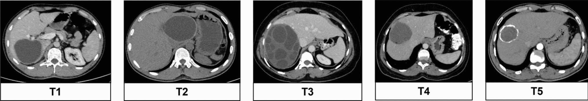
Irie MS, Spin-Neto R, Teixeira LHS, Rabelo GD, Reis NTA, Soares PBF. Effect of micro-CT acquisition parameters and individual analysis on the assessment of bone repair. Braz Oral Res. 2023 Oct 27;37:e099. https://doi.org/10.1590/1807-3107bor-2023.vol37.0099. PMID: 38055517.
Lee JH, Kim DH, Jeong SN, Choi SH. Detection and diagnosis of dental caries using a deep learning-based convolutional neural network algorithm. J Dent. 2018 Oct;77:106-111. https://doi.org/10.1016/j.jdent.2018.07.015. Epub 2018 Jul 26. PMID: 30056118.
Cantu AG, Gehrung S, Krois J, Chaurasia A, Rossi JG, Gaudin R, Elhennawy K, Schwendicke F. Detecting caries lesions of different radiographic extension on bitewings using deep learning. J Dent. 2020;100:103425. https://doi.org/10.1016/j.jdent.2020.103425 .Epub 2020 Jul 4. PMID: 32634466.
Kuwana R, Ariji Y, Fukuda M, Kise Y, Nozawa M, Kuwada C, Muramatsu C, Katsumata A, Fujita H, Ariji E. Performance of deep learning object detection technology in the detection and diagnosis of maxillary sinus lesions on panoramic radiographs. Dentomaxillofac Radiol. 2021;50(1):20200171. https://doi.org/10.1259/dmfr.20200171.
Kim Y, Lee KJ, Sunwoo L, Choi D, Nam CM, Cho J, Kim J, Bae YJ, Yoo RE, Choi BS, Jung C, Kim JH. Deep Learning in Diagnosis of Maxillary Sinusitis Using Conventional Radiography. Invest Radiol. 2019 Jan;54(1):7-15. https://doi.org/10.1097/RLI.0000000000000503. PMID: 30067607.
Nayak GS, Kamath S, Pai KM, Sarkar A, Ray S, Kurien J, D’Almeida L, Krishnanand BR, Santhosh C, Kartha VB, Mahato KK. Principal component analysis and artificial neural network analysis of oral tissue fluorescence spectra: classification of normal premalignant and malignant pathological conditions. Biopolymers. 2006;82(2):152-66. https://doi.org/10.1002/bip.20473. PMID: 16470821.

Comments (0)