Reconsideration of conversion to multiple sclerosis: one-year cerebral lesion appearance rate of Japanese aquaporin-4 antibody-negative optic neuritis patients
Purpose
To analyze the rate of new cerebral lesions’ appearance within 6–12 months in Japanese optic neuritis patients who at onset had no cerebral lesions suggestive of multiple sclerosis (MS) and were negative for aquaporin-4 (AQP4) antibodies.
Study design
Retrospective study.
Methods
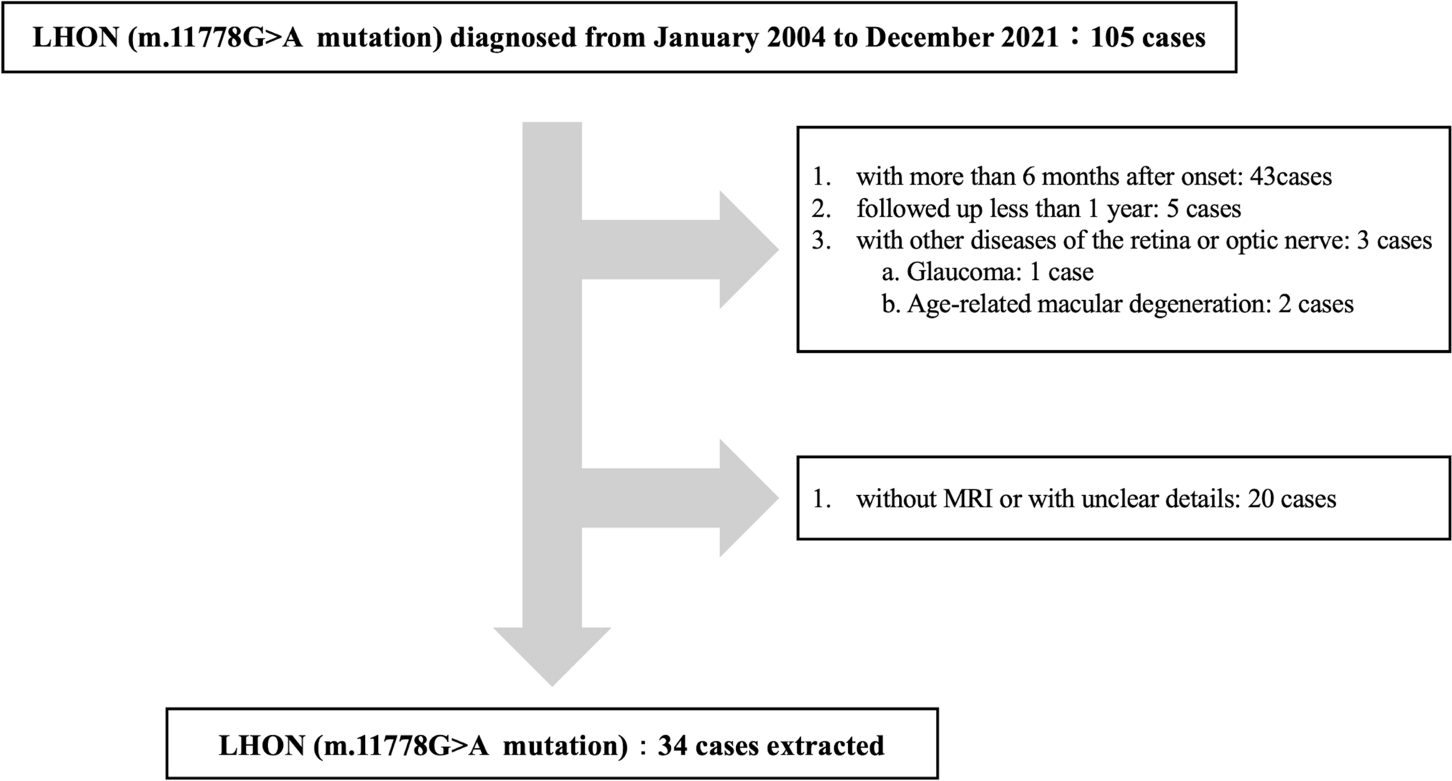
Medical records of 66 adult patients with optic neuritis were reviewed. Patients positive for AQP4, myelin oligodendrocyte glycoprotein, or antinuclear antibodies were excluded. Those without cerebral lesions on initial magnetic resonance imaging (MRI) underwent follow-up MRI within 6–12 months. Clinical characteristics and subsequent neurological diagnoses were analyzed.
Results
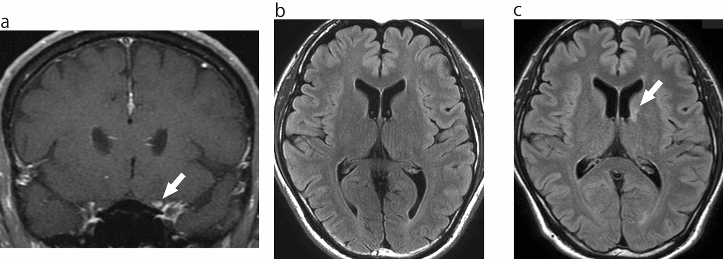
Forty-seven patients met the inclusion criteria (mean age, 41.9±16.7 years; 13 men, 34 women). Forty-two cases were unilateral, five bilateral; 19 had disc swelling, and 28 did not. The mean worst logMAR was 1.13±0.96. Two patients experienced recurrence within 1 year. Of the 27 patients without initial cerebral lesions, 20 underwent follow-up MRI; 3 (15%) developed new lesions. These three were later diagnosed as two MS and one suspected MS cases.
Conclusion
Follow-up MRI within 6–12 months revealed new cerebral lesions in 15% of patients, with 10% diagnosed with MS. This highlights the importance of follow-up imaging even in AQP4 antibody-negative optic neuritis patients without initial cerebral lesions, especially in the absence of other diseases like neuromyelitis optica spectrum disorders.

Comments (0)