Cannabidiol (CBD) exhibits diverse biological effects in the brain, including the hypothalamus. Research on CBD’s impact on hypothalamic cells revealed its potential to modulate anxiety, stress responses, and other neurobiological functions. For example, in the case of anxiolytic effects, CBD microinjected into the ventromedial hypothalamus (VMH) in rats reduced panic attack-like behaviors and unconditioned fear-induced antinociception, potentially through CB1 receptor signaling (Khan et al. 2020). CBD was also found to enhance neurogenesis and exhibit neuroprotective effects. Repeated CBD administration in chronically stressed mice increased hippocampal neurogenesis and reduced anxiety-like behaviors (Campos et al. 2013). Additionally, CBD influenced histone modifications in the hypothalamus, which can affect gene expression. A recent study revealed that systemic administration of CBD modified the levels of certain histones (e.g., H3 K9ac) in the hypothalamus, indicating potential epigenetic effects of CBD (Pastrana-Trejo et al. 2021). An important action of CBD in the hypothalamus is the modulation of the stress-induced activation of the hypothalamic–pituitary–adrenal (HPA) axis (Viudez-Martínez et al. 2018). These complex responses to CBD in the hypothalamus require a molecular basis that can be observed only in isolated and simplified models, minimizing the complexity and multicellular interactions of brain structures.
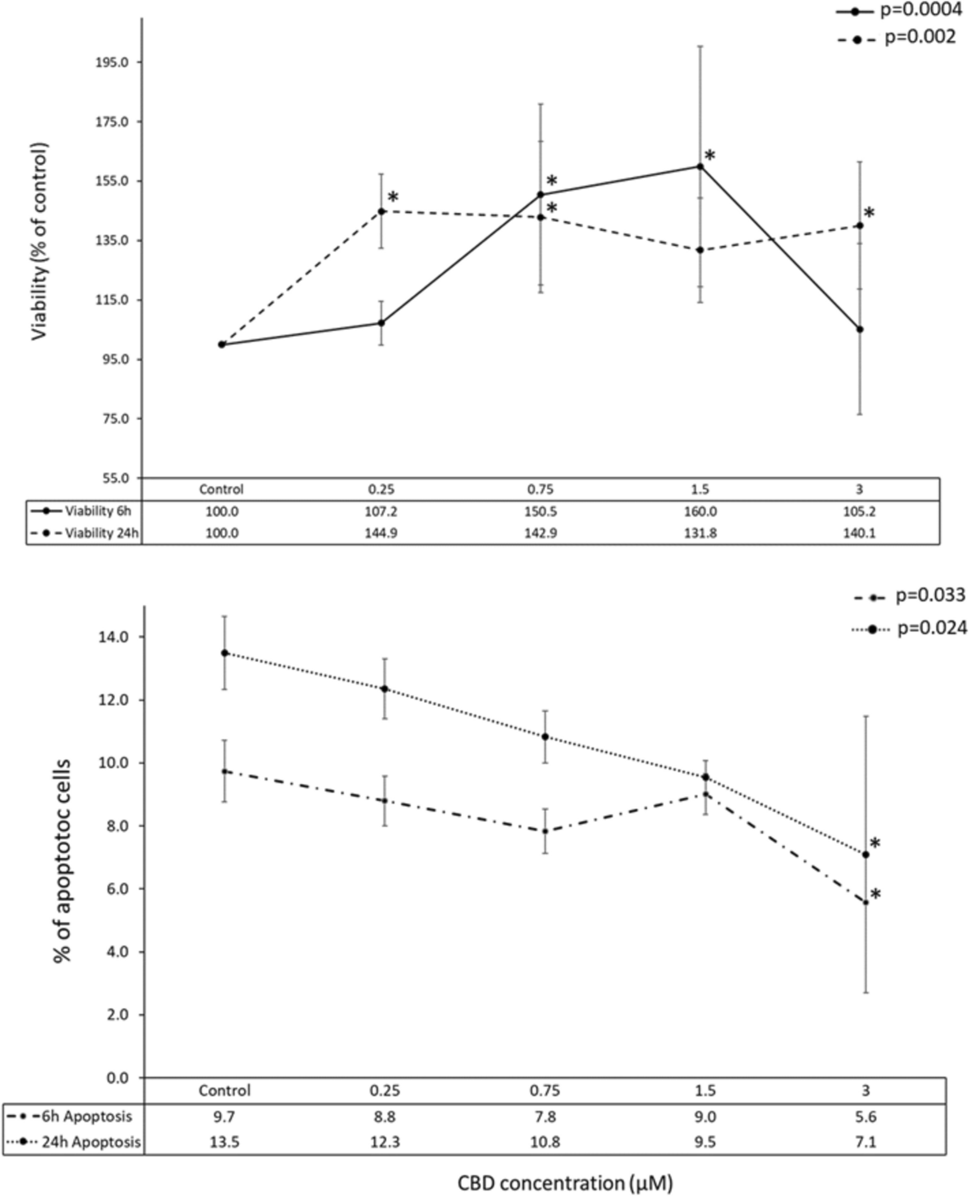
In this study, we focused on the effects of CBD on proliferation, viability, and gene expression in a cellular model comprising hypothalamic neurons. We found that CBD stimulated cell viability, especially at longer incubation times (24 h) and at lower or intermediate CBD concentrations. Our results showed a noticeable decrease in apoptotic processes intensity with increasing CBD concentrations, although statistical significance was only observed at the highest CBD concentration. These effects were accompanied by moderate changes in cellular transcriptome profiles, with a greater number of genes affected by higher CBD concentrations and shorter incubation times (indicating a stronger initial response). The transcriptomic response varied between the two time points studied, with more genes upregulated at 6 h and a higher number of downregulated genes following prolonged CBD incubation.
All these observations require in depth analysis and comparison with currently available literature data.
CBD affects hypothalamic neurons’ viability
The observation that CBD increases cell viability in in vitro cultured cells is atypical for healthy unchallenged cells, where CBD’s effect is predominantly associated with viability reduction (Pagano et al. 2020). The results obtained here contradict the findings from previously published data on hypothalamic and other neural cells (di Giacomo et al. 2020; Drummond-Main et al. 2023; Jantas et al. 2024). In most of these studies, CBD alone reduced cell viability primarily at higher concentrations, with no effect observed at lower CBD doses. However, this negative effect on cell viability was reversed when cells were challenged, e.g., with H2O2 to induce oxidative stress, where CBD treatment resulted in increased cell viability (di Giacomo et al. 2020; Jantas et al. 2024). These results indicate that CBD may exert effects that promote neuronal viability, as observed in this study on hypothalamic neurons. It remains unclear why this effect was observed here in unchallenged cells. It is possible that the CBD doses used are within a well-tolerated concertation range for mHypoA-2/12 mouse cell line, thus only the positive or neutral effect of CBD on their proliferation was observed. Alternatively, hypothalamic neural cells may react differently to CBD, similar to some other cell types such as PNT2 cells (Śledziński et al. 2023) or certain specific cancer cell lines (Omer et al. 2024), where CBD stimulates viability under basic culture conditions.
CBD affects cell apoptosis
In this study, a trend toward reduction in apoptotic process intensity (as indicated by 3/7 caspase activity assay) was observed in mHypoA-2/12 cells. This findings, similarly to the viability trend, is unusual for healthy, untreated cells. Nevertheless, reducing apoptosis levels after CBD administration is not uncommon in cells exposed to various stressors. One study demonstrated that CBD protects dopaminergic neurons from oxidative stress and apoptosis by reducing reactive oxygen species (ROS) production and maintaining mitochondrial integrity, independently of CB1 and CB2 cannabinoid receptors (Mendivil-Perez et al. 2023). Furthermore, a review on CBD’s mechanisms in regulating apoptosis and autophagy suggests that these effects may vary based on biological context, cell type, and CBD concentration (Fu et al. 2023). Another study showed that CBD could mitigate perfluorooctane sulfonic acid (PFOS)-induced cardiomyocyte apoptosis by preserving mitochondrial dynamics and metabolic energy homeostasis (Wang et al. 2023). These documented protective and anti-apoptotic effects of CBD are well-supported in the literature, reinforcing the findings of this study.
Transcriptomic evidence on apoptosis and viability regulation
The observed improvement in cell viability along with decreased apoptosis reflects neuroprotective potential and metabolic activity of CBD in hypothalamic neurons. To elucidate the underlying mechanisms at the transcriptome level, we conducted a detailed analysis of biological processes overrepresented by genes differentially expressed following various CBD treatments. Among the numerous processes affected by these treatments, those related to response to oxygen-containing compounds, cellular response to hypoxia, regulation of cell proliferation and cell cycle, regulation of programmed cell death, intrinsic apoptotic signaling pathway by p53 class mediator, DNA damage response, immune system processes, and regulation of metabolic processes were prominently altered in the 6-h treatment (Supplementary File 5; Supplementary File 7). In the 24-h treatment, processes involving regulation of cell migration, immune response, intrinsic apoptosis, and regulation of apoptosis were the most abundantly represented (Supplementary File 6; Supplementary File 9).
The analysis of these processes and the involved genes suggests that one of the important CBD actions in hypothalamic cells is the direct or indirect modulation of cellular apoptosis, particularly intrinsic apoptosis, potentially through the p53 pathway activity modulation (Supplementary File 5; Supplementary File 7). Intrinsic apoptosis involves mitochondrial proteins released in response to various stressors such as ultraviolet radiation, osmotic stress, growth factor withdrawal, chemotherapeutic agents, and natural compounds (Jan and Chaudhry 2019). Internal stimuli like high cytosolic Ca2+ concentrations, hypoxia, oxidative stress, and DNA damage also initiate mitochondrial pathway-mediated apoptosis (Carlsson et al. 2022). CBD’s activities in many of these intracellular processes have been previously documented (Ryan et al. 2009; Russo et al. 2019; Gross et al. 2021; Pereira et al. 2021). For instance, CBD has been shown to induce mitochondrial damage and cytochrome C release in cell lines derived from acute lymphoblastic leukemia of T lineage (T-ALL), and disrupt calcium homeostasis in those cells (Olivas-Aguirre et al. 2019). Additionally, CBD has been demonstrated to induce DNA damage in human-derived cell lines under conditions relevant to consumer exposure (Russo et al. 2019). While examining the key genes that were altered in our dataset and had major contributions to the regulation of apoptotic pathways, we found that the most important hub genes in 6-h treatment were Bbc3 (PUMA; which was downregulated by two lower CBD doses), and Mdm2 (that was upregulated by lowest and intermediate CBD doses). The Bbc3 (Bcl-2-binding component 3), also known as the p53 upregulated modulator of apoptosis (PUMA) is a pro-apoptotic protein, a member of the Bcl-2 protein family (Nakano and Vousden 2001). Upon activation by p53, PUMA interacts with antiapoptotic Bcl-2 family members, thereby releasing Bax and/or Bak to signal apoptosis to the mitochondria (Han et al. 2001). Subsequent mitochondrial dysfunction triggers the caspase cascade, culminating in cell death. In contrast, mouse double minute 2 homolog (Mdm2), also known as E3 ubiquitin-protein ligase, is a critical negative regulator of the p53 tumor suppressor (Mendoza et al. 2014). Mdm2 functions as both an E3 ubiquitin ligase, recognizing the N-terminal trans-activation domain (TAD) of p53, and an inhibitor of p53 transcriptional activation, thereby suppressing p53-mediated apoptosis (de Rozieres et al. 2000). Mdm2 has also been shown to promote proliferation and inhibit apoptosis in pituitary adenoma cells by directly interacting with p53 (Wang et al. 2020). Therefore, the interplay between Bbc3 (PUMA) and Mdm2 seems to be pivotal for CBD-mediated regulation of apoptosis via the p53 pathway. In the anticipated mechanism modulated by CBD, PUMA expression is inhibited directly or indirectly by CBD, thereby suppressing apoptosis, while the upregulation of Mdm2 aims to negatively regulate p53, further mitigating excessive apoptotic activity. These changes in gene expression align with the observed effects of CBD on viability and apoptosis in hypothalamus cells in this study.
The other genes identified in this study as differentially expressed, which may significantly impact apoptosis processes, proliferation regulation, and immune responses, include among others Cdkn1a (p21, Cyclin-dependent kinase inhibitor 1 A), Ndrg1 (N-myc downstream–regulated gene), and Smad3, which contributes to the maintenance of genomic integrity, cell cycle regulation, and apoptosis. The regulation of Cdkn1a and Ndrg1 by p53, and their roles in cellular stress and apoptosis, underscore their critical functions in preserving cellular integrity (Kreis et al. 2019; Schonkeren et al. 2019). Cdkn1a is a direct transcriptional target of p53. Upon cellular stress signals such as DNA damage, p53 binds to the promoter region of Cdkn1a, thereby inducing its expression. This leads to the inhibition of cyclin-dependent kinases (CDKs), resulting in cell cycle arrest at the G1 phase. Such cell cycle arrest allows for DNA damage repair before cell division proceeds, thereby preventing the propagation of mutations (Kreis et al. 2019). Smad3, a member of the SMAD family of proteins involved in TGF-β signaling, interacts with both p53 and Cdkn1a. In some contexts, Smad3 can directly regulate p21 expression, contributing to cell cycle regulation and apoptosis. Additionally, Smad3 signaling can intersect with p53 pathways to modulate cellular responses to stress and DNA damage. Smad3 involvement in TGF-β signaling adds another layer of complexity to the regulation of cell cycle, apoptosis, and stress response. Its interaction with both p53 and Cdkn1a underscores the intricate crosstalk between different signaling pathways in coordinating cellular responses to various stimuli (Wang et al. 2016a).
CBD’s ability to downregulate the expression of genes Cdkn1a, Ndrg1, Smad3, and Bbc2 can have both beneficial and potentially adverse effects. It may offer advantages such as anti-inflammatory and antitumor properties, as well as neuroprotective effects. However, there are risks, including potential disruption of cellular homeostasis, impaired cellular defense mechanisms, and unintended effects on other cellular processes. Further research would be needed to fully understand the consequences of modulating all mentioned genes and to evaluate the therapeutic potential of such interventions, particularly in the context of neurodegenerative diseases.
At 24 h of treatment, the set of genes associated with apoptosis significantly differed, suggesting the alteration of downstream elements of the intrinsic apoptosis mechanism with prolonged CBD treatments. Among the apoptosis-related genes, that were altered predominantly at 24-h treatment, there were for example Hspb1 (Heat shock protein beta-1), which product is known to inhibit apoptosis by stabilizing the actin cytoskeleton and preventing cytochrome c release from mitochondria (Park et al. 2002); Tmbim6 (Transmembrane BAX inhibitor motif containing 6), encoding an anti-apoptotic protein that inhibits mitochondrial calcium uptake and oxidative stress-induced cell death (Kim et al. 2021); Cyld (Cylindromatosis), a tumor suppressor gene that negatively regulates NF-κB signaling, promoting apoptosis by removing K63-linked polyubiquitin chains from target proteins (Fernández-Majada et al. 2016); and Dusp1 (Dual specificity phosphatase 1 also known as MAP kinase phosphatase-1), involved in the negative regulation of MAPK signaling, which can lead to the inhibition of apoptosis (Wang et al. 2016b). These and other apoptosis-related genes altered by CBD in this study suggest that CBD’s effect on intrinsic apoptosis persists over time, though later stages may involve different molecular mechanisms compared to short-term treatments.
The mechanism by which CBD affects cell viability, particularly at the transcriptome level, remains unclear. However, our findings suggest that CBD can stimulate the expression of genes associated with mitochondrial respiratory chain complex I, providing new insights into this process. In our study, the two most significantly affected genes were mt-Nd4 and mt-Nd5. These genes encode NADH-ubiquinone oxidoreductase chain proteins, which are subunits of NADH dehydrogenase (ubiquinone). This enzyme, located in the inner mitochondrial membrane, is the largest of the five complexes in the electron transport chain. NADH dehydrogenase and NAD (nicotinamide adenine dinucleotide) play a crucial role in regulating cell viability through its involvement in energy metabolism and protection against oxidative stress (Čermáková et al. 2021). Our findings align with previous studies on the brains of CBD-treated rats, where increased mitochondrial calcium accumulation enhanced the activity of calcium-sensitive dehydrogenases. This, in turn, promoted NADH availability and enhanced oxidative phosphorylation (Valvassori et al. 2013). Our results further support the broader impact of CBD on mitochondrial metabolism across various cell types (Olivas-Aguirre et al. 2019).
Modulation of stress-like neurons’ responses by CBD
CBD induces cellular responses with transcriptomic signatures resembling known stress responses, primarily evident at the 6-h treatment (initial response). Among the cellular processes overrepresented by DEGs, we identified ones associated with general stress response, cellular response to external stimuli, cellular response to chemical stimuli, response to inorganic substances, interleukin-27-mediated signaling pathway, response to hypoxia, DNA damage response signal transduction mediated by p53 resulting in cell cycle arrest, response to endoplasmic reticulum stress, response to oxidative stress, and others. While the cellular reactions to external and chemical stimuli are obvious, other processes require further analysis and explanation.
An intriguing CBD-associated cellular response is the interleukin-27-mediated signaling pathway, enriched with genes altered by the lowest CBD concentration. Interleukin-27 (IL-27) is recognized as a pivotal regulator of T cell activation and differentiation, influencing T cell responses in autoimmune conditions within the central nervous system (Iwasaki et al. 2015). Recent evidence also suggests its neuroprotective properties in the retina and brain (Nortey et al. 2022). IL-27, secreted by and interacting with infiltrating microglia, macrophages, astrocytes, and neurons, enhances neuronal survival by modulating pro- and anti-inflammatory cytokines, neuroinflammatory pathways, oxidative stress, apoptosis, autophagy, and epigenetic changes (Nortey et al. 2022). The pathway-associated genes such as Oasl1 and Oasl2 were upregulated by lower CBD doses in this research. Oasl proteins (2′−5′-oligoadenylate synthase) may indirectly affect neurodegeneration by regulating inflammatory processes and immune responses in the brain (Ghosh et al. 2019). However, their specific roles and regulatory mechanisms in the context of CBD action, and their implications for CBD consumption, warrant further investigation.
CBD’s effect on extracellular matrix
The genes altered by CBD, primarily in short-term treatments, enrich cellular components such as the extracellular region, extracellular matrix (ECM), and collagen-containing extracellular matrix. The interaction between CBD and the ECM has been previously documented, mainly in different types of fibroblastic cells. It has been shown that CBD, at lower concentrations, increases the production of metalloproteinases (MMPs), while the highest concentrations decrease both the production of MMPs and MMP-2 protein activity (Rawal et al. 2012). A previous transcriptome analysis revealed that CBD pre-treatment enriched genes and functional associations between proteins mainly related to ECM organization and cell interactions in the mouse brain (Prieto et al. 2023). In our previous transcriptomic study on human dermal fibroblasts, we found that CBD affected several genes connected with ECM formation (especially its collagen constituent), which can have serious implications for the fibrosis process (in press). In this study, several genes connected to ECM functioning were altered, the most significant being most likely Mmp3 (downregulated by all 6 h CBD treatments), Mmp13 (downregulated by all 24-h treatments and one 6-h treatment), Timp1 (downregulated by all 6-h treatments), and Col11a1 (downregulated by a single 6-h treatment).
Mmp3 (matrix metalloproteinase-3) is a matrix metalloproteinase, a proteolytic enzyme capable of degrading many ECM components, including proteoglycans, collagens (types III, IV, V, IX, and X), laminin, and fibronectin. Mmp3 plays a key role in tissue remodeling, wound healing, and pathological processes such as inflammation and cancer development (Kandhwal et al. 2022). Its activity is tightly regulated because excessive ECM degradation can lead to diseases such as arthritis and cancer (Mehner et al. 2015). Research suggests that CBD can modulate the activity of MMPs (Gęgotek et al. 2021), which leads to reduced inflammation and protection of the ECM from excessive degradation. Similarly, Mmp13 (matrix metalloproteinase-13) degrades type II collagen as well as other collagen types. It is particularly important in the remodeling of cartilage and bones (Hu and Ecker 2021). One study suggests that CBD derivate can reduce the expression Mmp13 (Jin et al. 2023) and now we further confirm this in this study.
Timp1 (tissue inhibitor of metalloproteinases-1) inhibits the activity of metalloproteinases such as Mmp3 and Mmp13, protecting the ECM from excessive degradation (Brew et al. 2000). Previous research has shown that CBD can increase the expression of Timp1, supporting the protection of the ECM against degradation and potentially benefiting the treatment of inflammation and cancer (Solinas et al. 2012, 2013).
Finally, Col11a1 (collagen type XI alpha 1 chain) is a component of type XI collagen, which supports the structural integrity of cartilage (Shi et al. 2022). In this study, we found that its transcript abundance can be modulated by CBD.
In summary, all mentioned ECM-related genes participate in the process of connective tissue remodeling, where their coordinated action enables the dynamic maintenance and reconstruction of the ECM. Mmp3 and Mmp13 are responsible for the degradation of ECM components, while Timp1 regulates their activity, protecting the ECM against excessive degradation. Col11a1, on the other hand, is important for maintaining the structure of the ECM. This complex interaction is crucial for tissue health and regeneration, and the modulation of this process by CBD can have various implications, particularly related to pathologies such as degenerative joint diseases and cancer.
Transcriptomic insights into the regulation of hypothalamic functions by CBD
In the analysis of genes altered by CBD after 24 h of treatment, we identified those enriched in the dopamine and serotonin biosynthetic processes, as well as in the dopamine receptor signaling pathway. Dopamine is crucial in the hypothalamus, influencing neuroendocrine and autonomic functions. It regulates the HPA axis, with D1 and D2 receptors activating the HPA axis in response to severe stress in rats (Belda and Armario 2009). Given these roles, we focused on genes involved in these pathways and found that Aldh2, protein of which assists in dopamine and serotonin synthesis, was upregulated by the lowest CBD dose after 24 h of treatment. What is more, a Rgs4 gene engaged in the regulation of the dopamine receptor signaling pathway was downregulated in most of the 24 h CBD treatments. Aldh2 (aldehyde dehydrogenase 2 family member) belongs to the aldehyde dehydrogenase family of proteins and is essential in the major oxidative pathway of alcohol metabolism (Adams and Rans 2013; Chen et al. 2014). This links CBD’s effect on neural cells to its presumed positive properties in therapies for alcohol use disorders (De Ternay et al. 2019), though further studies are needed to explore this aspect. Regarding the Rgs4 gene (regulator of G protein signaling 4), its downregulation observed in most 24-h CBD treatments may affect dopamine D2 and D3 receptors, which are major targets for current antipsychotic drugs (Min et al. 2012). Studies using cDNA microarrays have consistently shown decreased RGS4 expression in the prefrontal cortex of subjects with schizophrenia, suggesting implications for CBD in anxiety and mental disorder therapies (McGuire et al. 2018).

Comments (0)