
Remember me
A retrospective cross-sectional study design was applied to determine the normal configuration and alignment of the TMJ in the post-mortem setting among adult cases of non-traumatic, non-decomposed deaths reported to the coroner in our jurisdiction, which is a major urban center with a population of over 6 million people. Deaths which appear to be unnatural or unexpected are notified to the coroner and approximately 7,000 deaths are investigated each year [15].
Participant data comprised 100 deaths notified to the coroner selected consecutively from January 1st, 2023, that met the following eligibility criteria: (i) the decedent was an adult aged between 18 and 80 years; (ii) the cause of death was non-traumatic; (iii) the decedent’s body was not decomposed; (iv) a PMCT scan was performed and the image was accessible within the imaging software; (v) an autopsy and/or external examination was performed and the post-mortem examination report was available for review. Participants were excluded if: (i) the decedent had a known TMJ abnormality/injury in life; (ii) the cause of death was traumatic (as trauma, such as blows to the jaw, can cause TMJD) [16]; (iii) the body was decomposed (as the process of decomposition causes tissue breakdown resulting in disarticulation of joints including the TMJ) [14]. Additional variables included: (i) case administration (study case number; institute case number; year); (ii) demographic and case characteristics (age; sex; height; weight; BMI; decomposition of head and neck; cause of death; manner of death; suspicious circumstances); (iii) method of examination (autopsy; PMCT; additional examinations); (iv) TMJ specific data (mouth position; apparent TMJ displacement; condyle in relation fossa [right and left]). Because interrater reliability testing is recommended for heterogeneous samples greater than 30 [17] a sample size of 100 cases was chosen to increase the robustness of the interrater reliability analysis. Participants were assessed for decomposition in accordance with the decomposition scoring method established by Megyesi et al. [18]. Post-mortem photography was used to determine stages of decomposition. As different body regions can undergo decomposition at different rates [14], only the head and neck regions were assessed. Participants categorized as ‘fresh’ were included, and participants categorized as decomposed (early, advanced, skeletonization) were excluded.
A method of measuring the distance of the mandibular condyle from the articular eminence via PMCT scans was devised by a forensic radiologist and forensic pathologist using the assisted perpendicular tool (rephrased and shortened to ‘PAT’ for ease of use) provided by the imaging software, syngo.via (version VB60A software, Siemens Healthineers, Erlangen, Germany). CT technique at our institution includes a head-to-neck scan range, at 120 kVp, 420 effective mAs, 0.5 mm slice thickness, pitch of 0.6, rotation time of 0.55 s, 500 mm field of view, and reconstruction kernel of Hr68h. Data extraction was then performed independently by three researchers: an honors student who received training from a forensic radiologist over a 1-day period to learn the technique; a forensic pathologist with over a decade of PMCT experience; a highly experienced forensic radiologist. Measurements performed by the forensic radiologist were used as the gold standard.
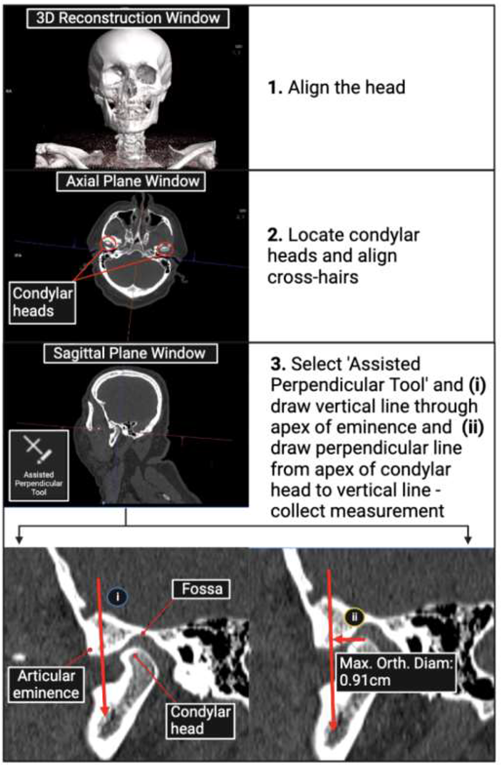
The TMJ PAT measurement methodThe TMJ PAT measurement method is described in Fig. 1. The three-dimensional (3D) reconstruction of the PMCT scan of the head and neck was firstly assessed using the 3D reconstruction window in syngo.via. Here, it was determined whether any TMJ abnormalities or pathologies could be identified, whether the TMJ appeared dislocated upon visual inspection, and the mouth position (open/partially open/closed) of the participant. Post-mortem changes, such as rigor mortis, make it difficult in some instances for the deceased person to be positioned in a straight, forward-facing position on the gantry table, as would regularly be required during clinical imaging procedures. The head of the participant was therefore aligned using the post-processing software (syngo.via) such that it was positioned in a normal anatomical orientation (forward-facing with the outer canthus of the orbits in a horizontal line with the external auditory meatus). Next, the mandibular condylar heads were located in the axial plane, and the multiplanar reconstruction crosshairs were centered over the right condyle so that it was sectioned equally into axial, sagittal and coronal planes. The sagittal plane window was then expanded and visual assessment was made to determine whether the condyle appeared to be positioned in or out of the mandibular fossa. The PAT tool was then selected and a vertical line was drawn through the apex of the eminence, followed by a perpendicular line from the apex of the condyle to the first vertical line. The ‘maximal orthogonal diameter’ measurement output was recorded, representing the right ‘TMJ PAT measurement’.
Fig. 1
Flowchart of the temporomandibular joint (TMJ) PAT (perpendicular assisted tool) measurement method devised by the research team using post-mortem computed tomography (PMCT) scans using the imaging software, syngo.via. The three-dimensional (3D) reconstructions were used to determine whether abnormal TMJ configurations could be accurately identified visually
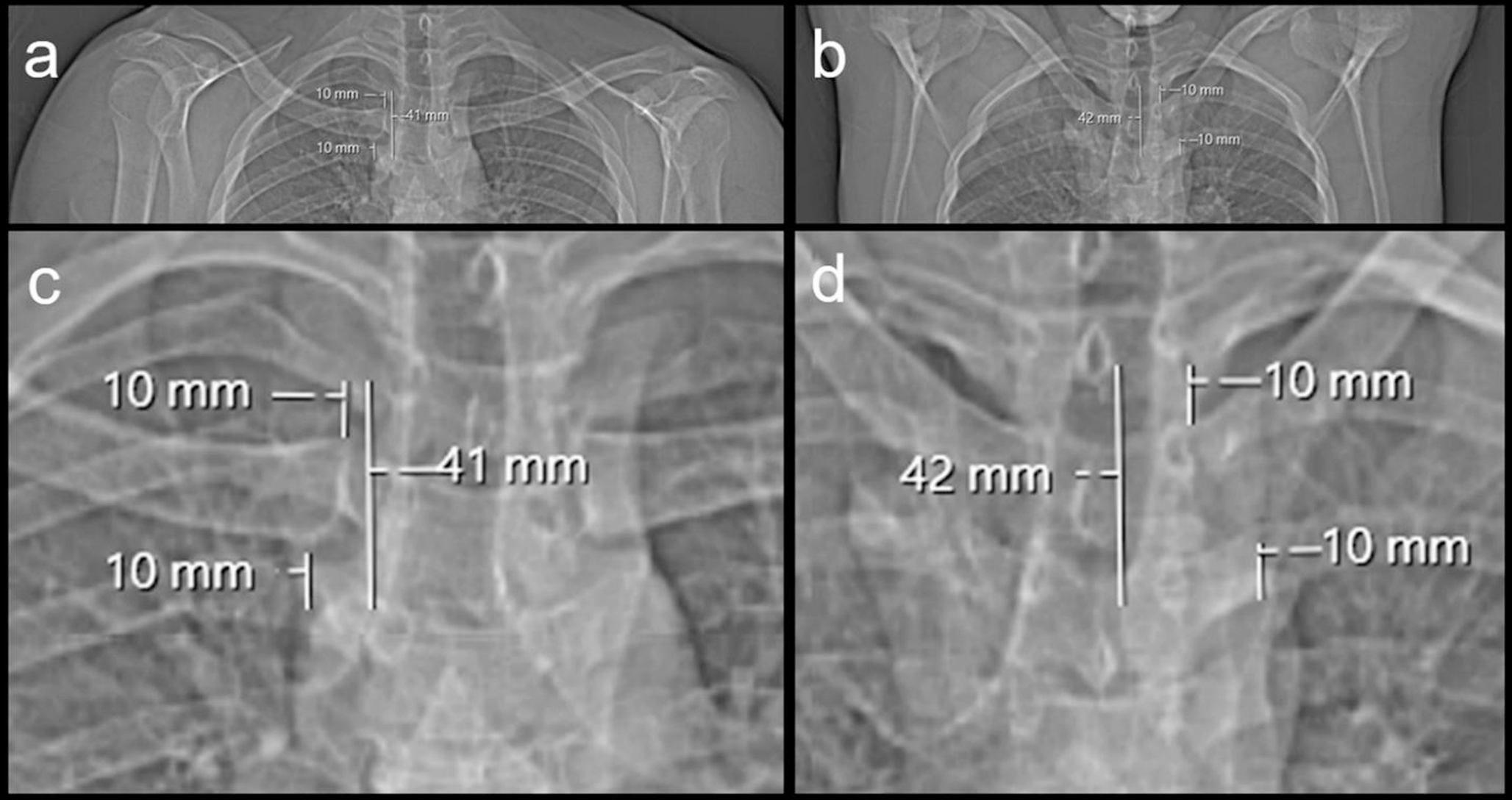
For the purpose of signifying the condyle’s position in relation to the articular eminence, the TMJ PAT measurement was assigned a ‘negative’ or ‘positive’ sign if the condyle was positioned posterior or anterior to the eminence, respectively. If the condyle was positioned directly over the eminence, the TMJ PAT measurement was zero (Fig. 2). The aforementioned steps were then repeated to measure the left TMJ. The TMJ configuration of each participant was determined using the right and left TMJ PAT measurements. A participant was deemed to have a NorTMJCon if a negative or zero TMJ PAT measurement was obtained bilaterally, or an AbTMJCon (i.e. TMJD) if a positive TMJ PAT measurement was obtained unilaterally or bilaterally.
Fig. 2
If the condyle is behind the eminence, in the fossa where it should be (green), this is a PAT negative measurement and is normal. If the condyle is at the eminence (yellow), this is a PAT zero measurement, which is normal. However, if the condyle is anterior to the eminence (red), this is PAT positive, which is abnormal and needs explanation
Established clinical criteria for the assessment of TMJD were used to form our rationale as to why a TMJ PAT measurement of zero was classified as being within the limits of a NorTMJCon in this study. Clinically, a wide-open mouth position (which may occur while yawning for example) can cause the condyle to be temporarily positioned anteriorly to the eminence. This is deemed a normal finding if the patient can close their mouth again and is not, in itself, a predictor of TMJD but demonstrates the range of TMJ hypermobility in the population. The diagnosis of TMJD is made if the mouth is unable to return to a closed position without the patient or a clinician performing a specific manipulative maneuver [12]. It is not possible to determine if a decedent can close their mouth as (a) it is not possible to assess a complaint of being unable to close the mouth and (b) assessment of jaw movement is limited by post-mortem factors such as rigor mortis (which may render the jaw immovable) and decomposition (which may cause apparent TMJ hypermobility due to muscle and ligamentous breakdown), so it could not be definitively established whether a zero TMJ PAT measurement was within the normal range of TMJ hypermobility or indicative of TMJD. A conservative approach was therefore taken to classify a zero TMJ PAT measurement as being within the range of a normal post-mortem TMJ configuration to avoid over-reporting the numbers of TMJD in the results.
Data cleaning was performed using Microsoft Excel, then data was imported into the statistical software, GraphPad Prism to perform normality testing using the Kolmogorov-Smirnov test and descriptive statistics. The Kolmogorov-Smirnov test was selected as it is the recommended normality test for larger samples (n > 50) [19]. TMJ PAT measurements were imported into Statistical Package for Social Sciences (SPSS) to perform inter-rater reliability tests. The test was repeated three times using the following data sets taken by three raters: (a) right TMJ PAT measurements; (b) left TMJ PAT measurements; (c) combined right and left TMJ PAT measurements. The test was then repeated three times using the following data sets taken by two raters: (a) right TMJ PAT measurements; (b) left TMJ PAT measurements; (c) combined right and left PAT measurements. The purpose of performing repeated tests was firstly to determine the reliability between measures taken by an experienced forensic radiologist, a forensic pathologist with experience in analyzing PMCT scans and a novice in the field, then to assess the reliability between measures taken by an experienced forensic radiologist and a novice in the field. The interrater reliability test is recommended for testing measurements performed by 3 or more raters [17] therefore the results of the test applied to two raters must be interpreted with caution.
Comments (0)