This study demonstrates the feasibility of CV-based personal identification using postmortem CT images of the maxillary sinuses, achieving an identification rate of 50% at rank 1 (the sought identity is in the first position of the consolidated list), 80% at rank 2, and 100% at rank 7 (the sought identity is within the top 7 positions of the consolidated list) across 10 identification procedures and 738 potential identities. It confirms that the method from [10] is applicable not only to antemortem data, but also to postmortem data, which can be matched with complete CT series in an antemortem CV database. However, the score is lower when comparing postmortem and antemortem images of the sought individual, making identification more challenging.
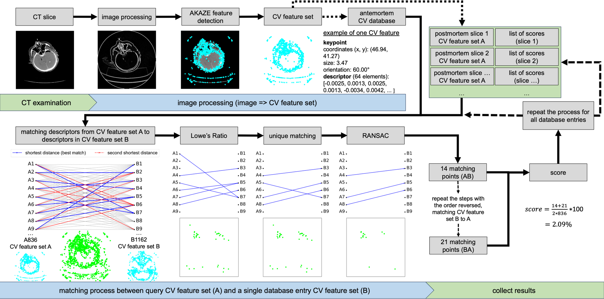
Unlike the previous study [10], which exclusively analyzed antemortem CT images, the following differences can be observed: the score for comparisons of images from the same individual (sought identity) is significantly lower for postmortem-antemortem images than for antemortem-antemortem images (1.76% vs. 6.96%), whereas the score between different individuals remains comparable (0.67% vs. 0.64%). This discrepancy is not primarily due to osseous changes after death but rather to deviations from the clinical standard in head positioning, the relative size of the head within the FOV, and injuries, which result in fewer matching points being identified. In postmortem CT imaging, the body is typically placed on the CT table without fixing the head, unlike clinical CT scans, where the head is often positioned in a standardized manner. As a result, the alignment of the head in postmortem scans can differ significantly from the antemortem reference, leading to potential misalignments in the 2D slices. In fact, even small changes in head orientation can cause half of the image to show completely different structures. In previous study [10] difficulties in identification were already observed when head alignment varied, as only 2D slices are compared. Additionally, postmortem imaging may suffer from suboptimal conditions, such as an overly large FOV in relation to the head. This can result in much lower image resolution, further hindering the identification process. In addition, the antemortem references are not always optimal, as the maxillary sinus is not always fully included in the CT series, particularly in cranial emergency examinations. CV features rely on distinctive points, primarily edges. Injuries and variations in head orientation can alter these edges, leading to a loss of matching points. However, even small matching areas can still provide enough matching points for identification. The method was simplified by storing the full antemortem CT series in the antemortem CV database, while still reliably identifying the sought individual. Additionally, the virtual autopsies were retrospectively selected, with imaging conducted as part of routine forensic practice, not specifically for this identification method. Despite these challenges, identification using the CV-based personal identification method proved to be highly promising. There is still potential to further increase reliability by evaluating only database entries with similar characteristics, such as sex, approximate age [13], and estimated body weight, as the sought identity. Additionally, postmortem search images can be optimized by removing the boundary of the FOV, for example, by setting the gray value to air, which would eliminate many false matching points between different individuals. Alternatively, one could focus only on selected areas, such as the maxillary sinuses, and hide everything else.
Previous studies [7,8,9] have demonstrated the importance of paranasal sinuses, particularly the maxillary sinuses, in forensic identification. These structures are highly distinctive due to notable individual differences in their shape, size, symmetry, and contour, which align with the criteria for reliable identifiers: uniqueness, permanence, and immutability. Evidence supporting the use of paranasal sinuses for personal identification includes methods such as visual evaluations of CT examinations by multiple radiologists with different levels of experience [7], the comparison of morphometric data obtained from paranasal sinus measurements, and advanced analysis using techniques like iterative closest point algorithms applied to 3D reconstructions of the sphenoid sinus [8]. In contrast to traditional methods, this study demonstrates the feasibility of a completely different, fully automatable approach. Our results confirm previous findings, showing that, similar to human assessment, the CV algorithm is capable of identifying and recognizing numerous individual features. This research indicates that postmortem CT slices are sufficient for personal identification, and the entire process can be automated. In addition to the maxillary sinuses, the frontal sinuses have long been recognized as valuable for personal identification due to their unique morphology [14, 15]. Since 1926, they have been used in forensic casework [16]. A major challenge in conventional postmortem radiography has been achieving the correct alignment with the X-ray beam, often requiring multiple attempts to match antemortem images [17, 18]. Postmortem CT has significantly improved this process by enabling the reconstruction of virtual radiographs from CT data [18]. This approach allows for more precise comparisons with antemortem radiographs, reduces errors related to head positioning, and enhances accuracy in forensic identification. The CV-based personal identification method presented here is also suitable for comparisons between virtual radiographs and antemortem radiographs [19]. Additionally, differences in head orientation between postmortem and antemortem CT scans can be effectively addressed in the same way by selecting oblique slices from 3D CT data.
The CV-based personal identification is fundamentally not a legally secure method for identification [13]. Its task is rather to locate suitable reference materials and/or obtain clues to the sought individual. By narrowing down the possible identity from thousands of potential identities, a legally secure and forensic identification is subsequently facilitated with appropriate reference materials by a highly qualified professional. In this study, the professional would have had to review a maximum of seven different reference materials per case to identify the sought individual - out of 738 possible identities. This was achieved despite suboptimal postmortem imaging, injuries, and only partial representation of the maxillary sinus in the CT images. Under optimal conditions, identification was very clear (see Table 4). A visual inspection of the matching points in the images could have further reduced the number of possible identities beforehand (e.g., Fig. 6b-c, far right) or by filtering the data based on sex or age [13]. Studies using panoramic radiographs [20] and thoracic CT images [21] have shown that a threshold exists, indicating when an individual has been identified with high probability, eliminating the need for a full database comparison. This threshold is, for example, the maximum number of matching points derived from millions of image comparisons between different individuals. If the sought person exceeds this threshold, their identity is highly likely to be confirmed. For comparisons between postmortem images and clinical databases, similar thresholds are expected to exist. However, further evidence and methodological optimizations are needed to establish and refine these thresholds.
The AKAZE algorithm is a traditional CV method that employs predefined mathematical techniques to extract and match features based on image intensity and gradients, without relying on training or data-driven learning. This means the process is mathematically traceable. Furthermore, the CV features are abstract numerical features that cannot be used to reconstruct the original image. However, the greatest advantage is data privacy, which can address ethical and legal concerns. CV features can be decoupled from the image and, therefore, do not contain personal data. During a matching process, it can only be determined that the postmortem CV feature set corresponds with an antemortem CV feature set. Only a pseudonymized patient ID, linked to the antemortem CV feature set, allows a conclusion about the identity it pertains to. However, decryption, for example, can only be performed by the institution where the CT examination was conducted, thereby maintaining control of the data within the originating facility. Another approach would be that large CV databases do not contain patient IDs, but only the origin (clinic, practice) of the CV feature sets. In this case, no personal data would be present in the CV database. However, in the event of a high match, data from the origin could be further examined to identify the potentially sought individual.
The limitation of this retrospective study is that it primarily aims to demonstrate the potential of the CV-based personal identification method, without addressing ethical and legal considerations, which can be explored in future research. Additionally, the small sample size limits the ability to fully assess the method’s robustness, as only a limited number of postmortem cases were available in this study. Future studies could focus on collecting additional cases to build a larger population and address this limitation. For prospective studies, the main challenge is the need for very large CV database to ensure accurate identification rates. If the identity being searched for is not in the database, non-identification reflects the limitations of the data, not a flaw in the method itself. Additionally, the study was conducted at a single location, and ethnic differences may exist. The application of additional filters, such as age, sex, and estimated body weight, or the removal of interfering elements that cause incorrect matching points, such as limiting the FOV of the CT image, could further facilitate identification. Moreover, discrepancies in head orientation between postmortem and antemortem CT scans, which posed a challenge in this study, could be addressed in future research by selecting oblique slices from 3D CT data to minimize positional differences.
In conclusion, based on the findings, it appears possible to identify an individual using postmortem CT images from a virtual autopsy in conjunction with a clinical database. The CV-based personal identification method achieved 50% correct identification at rank 1, 80% at rank 2, and 100% at rank 7 across over 738 potential identities, even without the use of additional filters. However, for optimal results, postmortem imaging should ideally adhere to clinical standards to ensure sufficient CV matching points.


Comments (0)