
Remember me
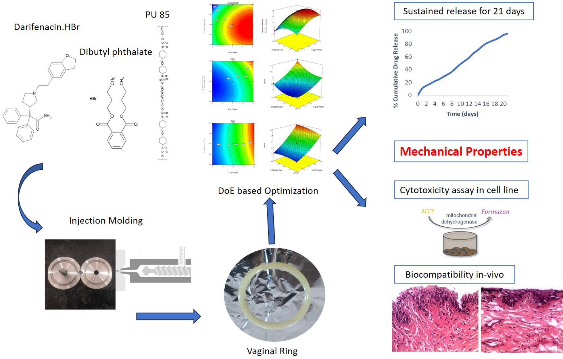


As seen in Fig. 1, IVR of different compositions was created using a stainless-steel mold. This ring-shaped mold makes it possible to create rings that are 5.5 cm in diameter and have a rim that is 0.5 cm [22]. The mold may be readily cleaned and/or washed after each sample was prepared. Three sets of molds were fabricated with same diameter and rim but with varying wall thickness comprising approx. capacities of 1.5 g, 2 g and 2.5 g polymer.
Fig. 1
Design of a ring-shaped S.S mold. a Schematic illustration of mold with location and dimensions. b Schematic of injection molding into S.S mold. c Photograph of PU 85 IVR
Analytical methodThe analysis was conducted using the HPLC system (Agilent 1100, USA), which includes an isocratic elution protocol. A reverse phase C-18 (250 X 4.6 mm, 5 µm) Phenomenex column was used for the chromatographic separation, which was carried out at 25 °C. The liquid mobile phase was made up of 50:50 v/v acetonitrile:phosphate buffer with a pH of 3.5. A flow rate of 1.0 mL/min was used to pump the degassed mobile phase. Prior to usage, it was passed through a 0.45-µm millipore nylon membrane filter. At 287 nm, the UV detection was carried out [23].
Preparation of IVRIVRs were prepared by laboratory-scale injection molding technique using in-house molds. Preliminary studies were carried out to select a suitable polymer and plasticizer for the formulation based on the mechanical properties of the IVR (data in supplementary file). PU 85 used as polymer matrix and DBP as plasticizer were melted at 180 °C in different ratios carefully avoiding bubble formation. 0.5% soya lecithin was added in the molten mass. Once the components were uniformly mixed, it was injected into the preheated mold. The mold was placed in refrigerated conditions for 1 h to get the finished product. DBr-loaded IVRs were formulated by using 30 mg of the drug, incorporated into the molten mass of PU 85 and DBP. All the batches had an outer diameter of 55 mm.
Optimization of IVR using 3 2 factorial designDoE is a usual practice in pharmaceutical product development to assess the relationship between experimental factors or conditions and desired outcomes or responses. By using Design-Expert software (version 13), a 32 factorial design was applied for optimizing IVR [24]. The critical parameters for optimization are the two independent variables, viz. the quantity of polymer (X1) and concentration of plasticizer (X2). In the present study, the tensile strength (Y1), the time required to release 50% drug release, T50% (Y2) and the time required to release 90% drug release, T90% (Y3) were the critical quality attributes governing the performance of the product and were considered as response variables [25]. Factors levels and factorial design are presented in Table 1.
Table 1 3-level 2-factor designStatistical analysisEach batch's response was noted, and then, they were statistically analyzed (p < 0.05 considered as significant) and optimized based on set constraints. The best-fitting model was chosen after the software examined several mathematical models for the experimental data. The statistical metrics, including multiple correlation coefficients (R2), adjusted multiple correlation coefficients (adjusted R2), predicted R2, and the predicted residual sum of squares (PRESS), were compared in order to determine the model selection.
Validation of experimental designTo assess the validity of the polynomial models that described the influence of the factors on the response variables, additional checkpoint experiments were conducted.
Characterization of DBr IVRDimensional analysisDigital Vernier calipers (Mitu-toyo corp.) were used for measuring wall thickness and rim diameter of the vaginal ring.
Mechanical analysisTensile strength [26] and percent compression of the IVR were measured. A texture analyzer (CT3, Brookfield Engineering Labs. Inc.) was used to measure and calculate the tensile strength (MPa) [27]. The Texture Analyser has two ring grips (one on the base and the other on the movable arm) and a 30-kg load cell for tensile testing. Before they rupture, vaginal rings were stretched at a speed of 10.0 mm/s.
$$} = F/a * b\left( + }/I} \right)$$
(1)
where F is force required to a break (N); a, b and l are width, thickness, and length of ring (m) respectively; I is the elongation of the ring (m) at the break point.
Each vaginal ring was positioned vertically in a Texture Analyser holder (CT3, Brookfield Engineering Labs. Inc.) in order to evaluate the compression properties [28]. The maximum compressive force (in N) was measured when the vaginal ring was compressed five times over a distance of 5.0 mm at a speed of 2.0 mm/s using a probe that was fastened to the movable arm. The percent compression was determined by the percent deformation recorded at compression force.
Drug contentA vaginal ring corresponding to 10 mg of drug was weighed and was cut into fine pieces, dispersed in 10 ml of methanol followed by sonication for 30 min. It was analyzed by above-mentioned HPLC method for drug content [27].
In vitro drug releaseA modified approach to drug release evaluation was developed and used for in vitro release tests. At 60 RPM and 37 °C, the rings were submerged in the dissolution beaker that contained 200 ml of the simulated vaginal fluid (SVF). The dissolution medium was changed every 24 h for 21 days. After removing 5 ml of media every 24 h, the release medium was completely replaced for 21 days. The aliquot was filtered and evaluated using the HPLC method, and the cumulative drug release was calculated [29].
Optimization and characterizationThe IVR was optimized using numerical and graphical optimization techniques employing the Design-Expert software. The optimization was done on the basis of constraints, which were set as maximizing the tensile strength (4.5–6 MPa), optimizing the T50% at target of 9–11 days and T90% at target of 19–21 days.
Selection of optimized batchThe selection of optimized batch was carried out using the desirability function from the numerical optimization and graphical optimization using the overlay plot. From the design space obtained, the batch which had the highest desirability near to 1.0 was selected as optimized batch. The optimized batch was subjected to further characterization.
Swelling studiesPlacebo (drug-free IVR prepared by the same method as mentioned earlier) and DBr IVRs were incubated for 21 days in a 25-mM sodium acetate buffer (pH 4.2) on an orbital shaker (Thermo Fisher Scientific Inc.) set to 37 °C and 80 rpm [30]. Mass measurements were taken using a digital balance (Reptech, India) to assess its swelling [31] behavior.
Infrared spectroscopy (IR)The FTIR scan of DBr and DBr IVR was assessed for compatibility by FTIR spectrophotometer (Cary 360, Agilent, India). IR spectra were recorded in the scanning range of 4000–650 cm−1 [32].
Differential scanning calorimetry (DSC)DSC of DBr and DBr-loaded IVR was done using calibrated DSC-60 Shimadzu, Japan [33]. Approximately 5 mg of the samples was taken and sealed in high-purity empty cells (alpha-alumina disks). In this technique, heat energy was applied simultaneously to reference and sample cell. At a heating rate of 10 °C per minute, measurements were taken between 20 and 250 °C.
Accelerated stability studyThe stability study was done using stability chamber (Janki Impex, India). The stability chamber had a RH and temperature of 75 ± 5% and 40 ± 2 °C, respectively. The optimized formulation was packed in an airtight container and stored for three months and evaluated by monitoring any changes in texture, tensile strength, percent compression, drug content and drug release.
Examination of vaginal tissue morphology post-DBr IVR applicationThe experimental protocol for animal studies was approved by Institutional Ethical Committee, as per the guidance of Committee for the Purpose of Control and Supervision of Experiments on Animals (registration no. BIP/IAEC/2020/17). The morphology of vaginal tissues following DBr IVR application was examined in order to look into the biological compatibility of the IVR device. After at least seven days of recuperation, oophorectomized female Sprague–Dawley rats weighing 200 ± 10 g were employed. In animals that had already been anesthetized, a DBr IVR that had been cut into a rod around 1 cm was inserted aseptically. The vaginal tissue was separated after 21 days, preserved in 10% neutral carbonated-buffered formaldehyde, embedded in paraffin, and then sliced. Following hematoxylin–eosin staining, the slices were examined under a × 400 light microscope, and observations were made [28].
In vitro cytotoxicity of DBr IVRHuman cervical cancer (HeLa) cell lines were used to assess in vitro cytotoxicity experiments. 3.7 g/L NaHCO3, 1% (v/v) nonessential amino acid solution, 10% (v/v) fetal bovine serum (FBS), 100 IU/mL penicillin, 100 µg/mL streptomycin and 1% (v/v) L-glutamine were added to DMEM medium, which was then maintained at 37ºC with 5% CO2 in the air. The cytotoxicity tests were conducted on cells with 20–40 passages, with each passage occurring every 3–4 days. The IVR samples were sterilized separately, then placed in sterile centrifuge tubes, submerged in 20 mL of DMEM media, and kept at 37ºC in a cell incubator. After being treated with 200 µL of the test sample solution, cells were seeded on 96-well tissue culture plates with a flat bottom and incubated for 30 min. Following the incubation period, the samples were collected out and 200 µL of PBS solution per well was used to wash the cells. After that, the cells were cultured for at least three hours with 100 µL of 0.5 mg/mL MTT dye. Finally, acidic isopropanol (isopropanol: 1.0 N hydrochloric acid = 25:1) was used to dissolve the formazan crystals. A microplate reader was used to measure the absorbance at 570 nm. Percentage of the untreated control was used to represent cell viability [34, 35].
Comments (0)