This study underscores the evolving landscape of GCTB management, demonstrating how a multimodal approach integrating molecular diagnostics, radiological tools, and targeted therapy can enhance patient outcomes [13,14,15,16,17,18]. H3.3 G34W immunohistochemical staining demonstrated consistent positivity across all tested tumors, reaffirming its role as a specific GCTB marker [6, 7]. In conjunction with NGS, which confirmed H3F3A mutations in most cases, these findings highlight the necessity of incorporating molecular diagnostics for both classification and potential prognostic stratification [9, 19].
The Campanacci grading system remains instrumental in stratifying tumor aggressiveness, as our results reaffirm its correlation with lesion size and recurrence rates [15]. However, its predictive capacity can be augmented through advanced imaging techniques, such as MRI-based radiomics and quantitative computed tomography (CT) analysis [2, 3, 20]. Radiomics, by extracting high-dimensional imaging features, may offer a more objective, reproducible method for assessing tumor biology beyond conventional visual grading [22]. AI-driven imaging applications hold promise in refining tumor characterization, enhancing preoperative planning, and improving recurrence risk assessment. [23]. Given the increasing feasibility of machine-learning algorithms in musculoskeletal oncology, AI-based predictive modeling holds the potential to personalize treatment strategies by integrating radiological, histological, and molecular data [20, 21, 24]. While our MRI-based radiological analysis was performed in a subset of 20 patients, these findings provide valuable insights but may require validation in larger cohorts to enhance generalizability.
Denosumab plays a pivotal role in managing high-risk GCTB, particularly in reducing surgical morbidity and facilitating joint preservation [9, 10, 18]. The classification of high-risk cases in our study aligns with prior literature emphasizing tumor size, anatomical constraints, and cortical destruction as key prognostic factors [4, 14, 15, 24]. While only 9 of 55 patients in our study received denosumab, our focus was not on its oncologic efficacy but on its role in preoperative planning and joint preservation. Larger series have established denosumab’s effect on tumor control, but our findings highlight its impact on resectability and recurrence risk in a carefully selected high-risk population [25]. In our cohort, preoperative denosumab administration led to significant tumor downsizing and bone matrix formation, enhancing joint preservation and reducing the need for radical resections. However, the risk of post-denosumab recurrence remains a critical concern, with thickened bone margins potentially obscuring the presence of residual tumor [22, 23]. To address the risk, adjuncts such as three-dimensional (3D) imaging and extended curettage should be further explored [24, 26]. Additionally, studies evaluating the optimal duration and discontinuation strategies of denosumab are needed to balance its benefits with potential recurrence risks [28, 29].
A key strength of our study is the integration of histopathological, molecular, and radiological findings to provide a more comprehensive framework for GCTB management. While retrospective in nature, our findings advocate for a paradigm shift toward a precision-based approach, leveraging both traditional classification systems and modern technological advancements [13, 17]. Previous studies with larger cohorts (> 200 cases) have focused primarily on the oncologic response to denosumab [26]. In contrast, our study integrates molecular diagnostics, radiological grading, and surgical outcomes, providing a comprehensive framework for treatment stratification. While denosumab aids surgical interventions, its impact on recurrence remains uncertain, warranting further research on optimized protocols [27]. Additionally, the study underscores the potential of AI-driven imaging analysis in refining GCTB risk stratification, an area yet to be explored in large-scale studies [28, 29].
Future research should validate radiomics-based grading models in larger cohorts, expand AI applications in tumor detection and risk stratification, and optimize denosumab protocols. [20, 25]. Integrating molecular diagnostics, advanced imaging, and AI-driven analytics could transform GCTB management, leading to more precise decision-making and improved patient outcomes [26,27,28,29]. Further multi-institutional research should aim to optimize denosumab use, including defining its ideal timing, dosage, and combination with other local therapies such as cryosurgery or radiofrequency ablation [28, 29].
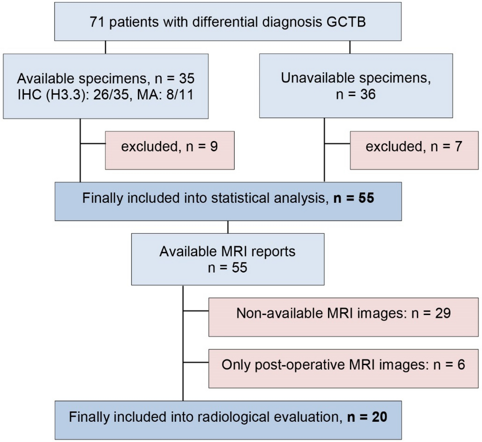
This study has several limitations. The retrospective design inherently introduces selection and information biases. Additionally, MRI-based radiological analysis was conducted on a subset of 20 patients, which may limit the generalizability of the imaging-based findings. The small sample size of patients receiving denosumab also restricts broader conclusions regarding its efficacy. Future prospective, multi-institutional studies with larger patient cohorts are needed to validate our findings and further refine treatment strategies.


Comments (0)