Patients
The study was approved by the institutional review board of the authors’ hospital (IRB nos. A-ER-112-395 and IRB20230089). This was a retrospective multicenter cohort study. Patients with acute diaphyseal humeral fractures (AO/OTA 12-A and AO/OTA 12-B fractures) that occurred within 3 weeks [6] who underwent open reduction and plate fixation between January 2017 and December 2020 in two tertiary referral hospitals were enrolled on the basis of the electronic surgical database in the authors’ hospitals. AO/OTA 12-C fractures were excluded from the study, because minimally invasive plating or nailing was preferred rather than ORPF. Moreover, patients with periprosthetic fractures, pathological fractures, revision surgeries, or incomplete medical records were excluded. A total of 112 cases of humeral shaft fracture were initially collected on the basis of electronic surgical database. After applying inclusion and exclusion criteria, 88 cases were enrolled. In this cohort, 10 cases were excluded due to nonunion and 13 cases were excluded due to loss of follow-up. Finally, 65 cases were included in the final analysis. All osteosynthesis procedures were performed using compression techniques including interfragmentary screw and/or compression plate techniques, with the aim of achieving primary bone union. Follow-up for all patients continued until bony union was achieved or nonunion was identified.
Plate osteosynthesis
Under general anesthesia, patients were positioned either supine or in lateral decubitus position, depending on the selected surgical approach, which could be either anterolateral or posterior. After aseptic draping, a standard anterolateral or posterior approach was implemented according to the operating surgeon’s preference. In cases where the posterior approach was chosen, the radial nerve was carefully dissected and isolated and protected throughout the procedure [21]. Conversely, if the anterolateral approach was selected, although the radial nerve was not routinely isolated, it was ensured that neither the plate nor the screws would compromise the integrity of the nerve [22]. The fracture sites were then exposed, and the hematoma was cleaned and irrigated. Under intraoperative fluoroscopy, reduction and provisional fixation were performed using reduction clamps and, when possible, interfragmentary screws. A conventional 4.5-mm nonlocking dynamic compression plate (DCP) or a locking 4.5-mm dynamic compression plate was applied. Stability and alignment were confirmed through fluoroscopy. The wound was copiously irrigated with normal saline and then closed in layers and dressed appropriately.
Clinical parameters
Patients’ demographic data and common comorbidities were collected, including age, gender, body mass index (BMI), presence of hypertension (HTN), diabetes mellitus (DM), alcohol and cigarette consumption, and ASA score [23]. The fracture pattern was classified according to AO/OTA classification, and the presence of open fracture was assessed using the Gustilo–Anderson classification. Surgical parameters were collected, including time between presentation and surgery, the chosen surgical approach, intraoperative blood loss, and operative duration. Implant and fixation characteristics were documented, including the total number of bicortical screws and the presence of interfragmentary screws. Postoperative complications were tracked and graded according to the Clavien–Dindo classification [24]. Grade 1 complications were defined as any deviation from the normal postoperative course without the need for pharmacological treatment or surgical, endoscopic, and radiological interventions. Grade 2 complications required pharmacological treatment with drugs other than those allowed for grade I complications. Grade 3 complications were defined as those requiring surgical, endoscopic, or radiological intervention. Grade 4 was defined as life-threatening complications. Grade 5 complications indicated patient mortality.
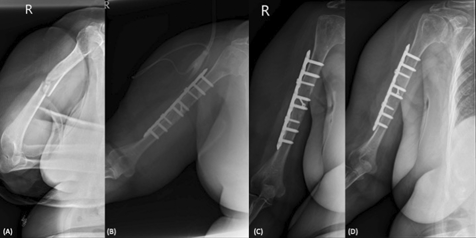
Radiographic outcomes
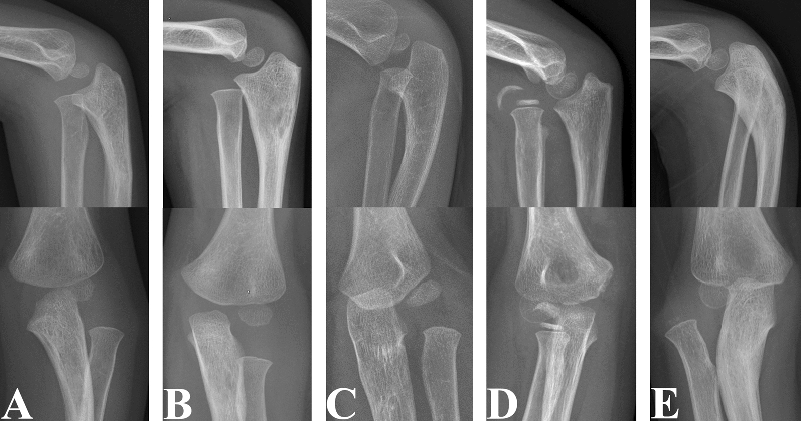
Based on follow-up plain radiographs, union time and bone healing pattern were recorded. Union was defined as the presence of cortical bridging bone formation across at least three out of four cortices, as observed on standard orthogonal radiographs and pain-free activity [6]. Timely union is defined as union achieved within 6 months postoperatively [25], while delayed union is defined as union between 6 and 12 months postoperatively [26]. If union is not achieved within 12 months, accompanied by no further fracture healing or the need for intervention, it is classified as nonunion [8, 13, 27]. For the healing pattern, primary union was defined as the absence of callus on serial radiographs, and secondary union was defined as the presence of callus formation [28]. The quality of reduction and the remaining gap/angulation immediately after the operation were also documented [16, 29]. Reduction quality was classified as excellent if the residual gap was less than 2 mm, good if the gap was between 2 and 5 mm, and poor if the residual gap exceeded 5 mm [29]. Radiographs were reviewed by two independent orthopedic surgeons who were blinded to the clinical information. Any discrepancies in the assessment of union status were resolved through consensus with a senior surgeon.
Statistical analysis
For evaluating the between-group differences, the normality assumption for all continuous variables were assessed using the Kolmogorov–Smirnov test. For the continuous data that are not normally distributed, values are presented as median with interquartile range (Q1, Q3) and analyzed using the Mann–Whitney U test. For those that are normally distributed, values are presented as mean ± standard deviation and analyzed using the Student’s t-test. For the comparison of the proportion, the data are presented with number (percentage) and analyzed with the chi-square test or the Fisher’s exact test when appropriate. To evaluate the risk factors associated with delayed union, the crude and covariate-adjusted odds ratios (ORs) and 95% confidence intervals (CIs) for the selected baseline variables and covariates were estimated using binary logistic regression. A p-value of less than 0.05 was considered statistically significant. The data were analyzed using the SPSS statistical package (version 22.0; SPSS, Chicago, IL, USA).


Comments (0)