
Remember me


Prior research shown that MMPs are crucial for angiogenesis in cancer by cleaving different proteins and producing pro-angiogenic molecules, which encourage the growth of new blood vessels. Growth factors as transforming growth factor-beta (TGF-β), FGF-2, and VEGF, which encourage the proliferation and migration of the endothelial cells—two crucial processes in angiogenesis—can become more bioavailable when MMPs are present [15]. Matrix metalloproteinase 1 (MT1-MMP), MMP-9, and MMP-2 all stimulate VEGF-mediated angiogenesis in ovarian cancer. It has been demonstrated that elevated MT1-MMP expression increases VEGF expression, which promotes tumor development and angiogenesis [2]. Moreover, MMPs can degrade forms of collagen I and IV, two extracellular matrix structural components, allowing endothelial cells to infiltrate and encouraging angiogenesis. Additionally, MMPs can generate endostatin and angiostatin, two components that restrict angiogenesis by lowering endothelial cell invasion and possibly even preventing endothelial cell growth. The intricate function that MMPs play in controlling angiogenesis in cancer underlines their importance in the development of tumors and the possibility of using MMP targeting as a therapeutic approach to prevent angiogenesis and restrict tumor expansion and metastasis [15].
Tumor microenvironment regulationMatrix metalloproteinases are essential for controlling the growth factors, cytokines and immune cells inside the tumor microenvironment. Tumor cell proliferation, survival, and angiogenesis can be enhanced by MMPs through the activation or release of TGF-β and VEGF. MMPs promote angiogenesis by altering the extracellular matrix and releasing VEGF and other pro-angiogenic substances. Furthermore, MMPs have the ability to affect immune cell invasion and activity inside the tumor microenvironment, hence influencing the tumor-fighting immune response. Targeting MMPs as a potential therapeutic method to disrupt tumor-promoting processes and increase anti-tumor immune responses is crucial because MMPs’ regulation of the tumor microenvironment promotes the growth and progression of tumor [16].
MMP in the development of ovarian cancerMMPs promote tumor development, invasion, metastasis, and angiogenesis, all of which are important factors in the advancement of ovarian cancer. Because MMPs break down extracellular matrix (ECM) components, cancer cells are able to infiltrate surrounding tissues and blood vessels. Additionally, they alter the connections between cells and the extracellular matrix, which promotes cancer cell migration and the EMT. Furthermore, by controlling the ratio of pro-angiogenic molecules like MMPs may influence tumor angiogenesis through VEGF and anti-angiogenic substances such as endostatin. The aggressive nature of cancer cells and their capacity to disseminate to distant locations are facilitated by the dysregulation of MMP activity in ovarian cancer, suggesting MMPs as possible targets for therapeutic intervention in ovarian cancer [17].
Epithelial-mesenchymal-transitionIn ovarian cancer, MMPs regulate the epithelial-mesenchymal transition, which promotes invasion and metastasis [2, 18]. MMPs play a role in triggering EMT, which is characterized by increased migratory and invasive capabilities. EMT promotes the metastasis and dispersal of cancer cells. Elevated levels of MMP, particularly MMP-2 and MMP-9, have been linked to advanced stages, greater invasiveness, and unfavorable prognoses in ovarian cancer. MMPs encourage the extracellular matrix to be remodeled, which permits cancer cells to spread to other locations and infiltrate nearby tissues. In ovarian cancer, targeting MMPs is a promising therapeutic approach to prevent tumor spread and progression while improving the effectiveness of current treatment methods [19]. One important mechanism in the development of cancer, especially ovarian cancer, is the EMT. During EMT, epithelial cells lose their distinctive characteristics and gain mesenchymal traits, which improve their motility, invasiveness, and resistance to apoptosis. The TGF-β pathway is one of the signaling pathways that promotes this transition. It does this by upregulating transcription factors like the Snail family, which repress epithelial markers like Ecadherin and promote the expression of MMPs such as MT1-MMP, MMP-1, MMP-2, and MMP7 [2]. E-cadherin, an important epithelial marker, is lost during EMT, and mesenchymal markers like vimentin and N-cadherin are often acquired in its place [20]. Empirical evidence suggests that MMP-9, in particular, promotes EMT by initiating the dissociation of the E-cadherin ectodomain [21]. Additionally, by losing additional epithelial markers and proteolyzing E-cadherin, MMP-9 can cause EMT, which in turn promotes invasion and cancer [2].
The function of TGF-β in metastasis and EMTIn many cancer types, including ovarian cancer, TGF-β is important for triggering the transition from epithelium to mesenchymal tissue and promoting metastasis [2]. The overexpression of transcription factors like the Snail family, which inhibit epithelial indicators like E-cadherin and stimulate the production of MMPs, is caused by downstream pathways that are activated by TGFβ signaling. SMAD1/2/3 signaling is activated when TGF-β connects to its receptor. SMAD4 then forms a complex with this signal and translocate into the nucleus. Snail regulatory factor family is upregulated by transcription factors that this complex binds to in the nucleus. Snail proteins promote the manufacture of MMPs and block E-cadherin, an essential cell adhesion molecule. Snail proteins promote the production of MMPs and downregulate the crucial cell adhesion molecule E-cadherin [22]. Overexpression of MMPs (MMP-1, MMP-2, MMP-7, and MT1-MMP) caused by TGF-β facilitates cell invasion and migration during EMT by degrading the extracellular matrix. Furthermore, TGF-β-induced EMT causes cancer cells to become more motile, invasive, and metastatically capable by imparting mesenchymal traits. EMT triggered by.
TGF-β causes cancer cells to become more mesenchymal, which increases their ability to migrate, invade, and spread [23]. Moreover, it has been suggested that TGF-β signaling encourages the development of a pro-invasive phenotype in cancer cells. It has been shown that pro-invasive effects resulting from TGF-β therapy alone can be mitigated by concurrent administration of broad-spectrum MMP inhibitors. This emphasizes how important MMPs are in controlling cancer cells’ ability to spread when influenced by TGF-β [24].
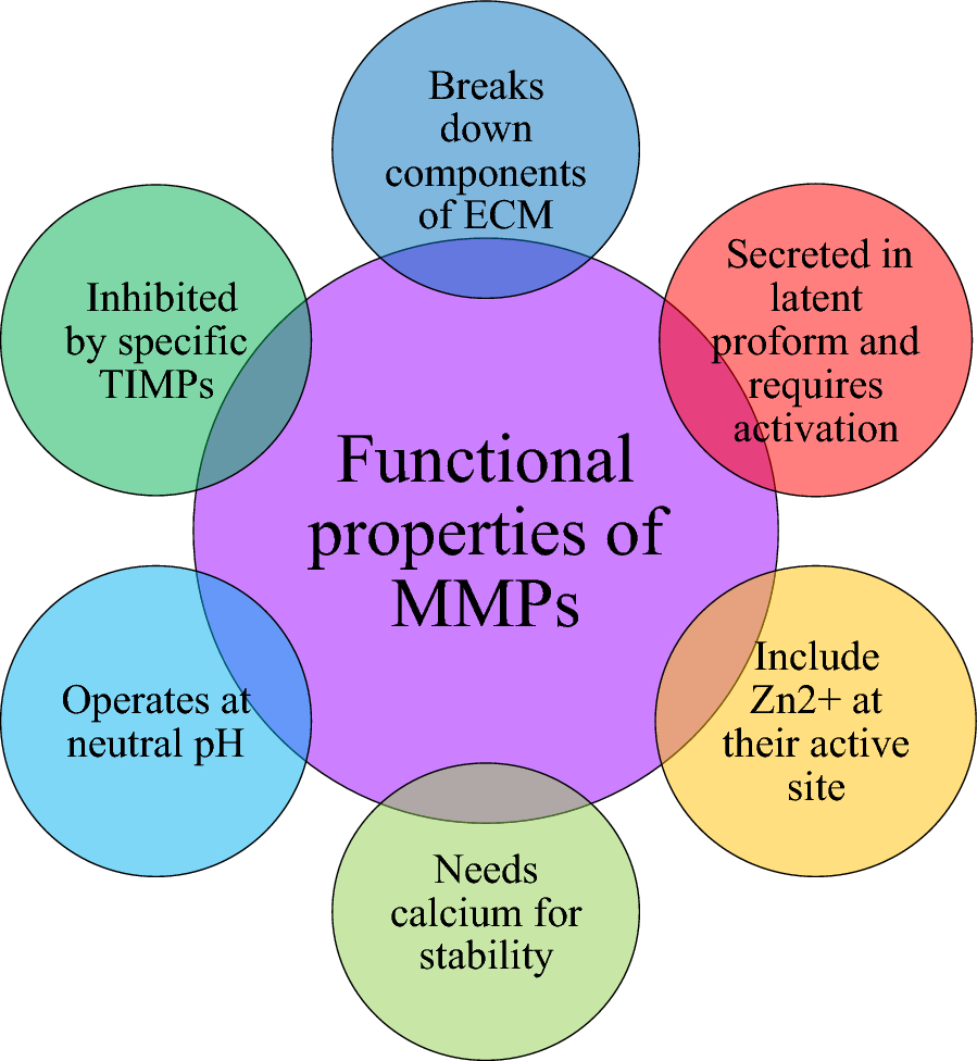
The mechanism of MMP actionA class of enzymes known as matrix metalloproteinases breaks down collagen, elastin, and proteoglycans, which are components of the extracellular matrix. They are essential to several physiological processes, such as tissue transformation, wound recovery, and embryonic growth [25]. However, a number of clinical diseases, such as cancer and arthritis, have been linked to deregulated MMP activity [26]. Here, the mechanism of action of MMP is summarized in Fig. 2.
Fig. 2
MMPs first exist as zymogens, which are inactive forms of the protein. There are several things that might activate them, such as serine proteases, other MMPs, and specific TIMP. MMPs break down extracellular matrix (ECM) constituents like collagen and fibronectin once they are activated. The migration of tumor cells to other areas of the body, known as metastasis, may be facilitated by this deterioration. By preventing their activation and function, TIMPs are essential in controlling MMP activity
MMPs are released as dormant zymogens (proenzymes) and require activation to become functional. Activation can occur through proteolytic cleavage by other MMPs, serine proteases, or specific tissue inhibitors of metalloproteinases.[27] Once activated, MMPs bind to specific ECM components or cell surface receptors through their catalytic domain [25]. MMPs cleave peptide bonds within the ECM proteins, leading to their degradation. This process allows for tissue remodeling, cell migration, and invasion. Different MMPs exhibit specificity for distinct substrates, depending on their structural features and substrate-binding domains [15]. MMP activity is tightly regulated at multiple levels to prevent excessive ECM degradation and maintain tissue homeostasis. Regulation occurs through the action of endogenous inhibitors, primarily TIMPs, which bind to active MMPs and inhibit their proteolytic activity [28]. MMPs can be localized within the extracellular space, on the cell surface, allowing for spatial control of their proteolytic activity. Overall, the balanced activity of MMPs is critical for normal physiological processes, while dysregulation can contribute to various diseases. Understanding the intricate mechanisms of MMP regulation and function is essential for developing targeted therapeutic strategies for conditions associated with abnormal MMP activity, such as cancer metastasis and tissue fibrosis [26].
Polymorphism and expression of MMP in Ovarian cancerGenetic variations in MMP genes can affect an ovarian cancer’s susceptibility and course. MMP polymorphisms and risk of ovarian cancer have been linked in a number of studies. Some of the polymorphism studies of MMP in Ovarian cancer are summarized as a table as follows [32,33,34,35,36,37].
Key studies on MMP expression in ovarian cancer is summarized as a table as follows [38,39,40,41,42,43,44,45].
S. no
Author name
Place
and
year
of the
study
Type of
the
study
No of Participants– sample size
methodology used
Description of The study
findings
1
Vos et al.,
Netherla nds,
2019
Casecontrol
study
144 patients—Caucasian population
ARMS – PCR was
done and correlated it with
Immuniohistochemi
stry
This study focused on survival outcomes by examining the relationship between clinical characteristics in patients with ovarian cancer and the polymorphisms of MMP-14 and MMP-2
+ 6767 G > A polymorphism in MMP-14 may have an impact on ovarian cancer patients’. The polymorphisms of MMP-14 and MMP-2
did not substantially correlate with survival or clinical features
2
Li et al.,
China, 2005
Casecontrol study
Sample size was not mentioned
PCR–RFLP
The study examined the association between ovarian cancer susceptibility in North China and certain SNPs in
MMP genes
Utilizing PCRRFLP, four
functional
Variations in the promoters of the MMP-1, MMP-3,
MMP-7, and MMP-
9 genes were evaluated
The study found no link between the SNPs in MMP-1, MMP-3, and MMP-9 with the risk of ovarian cancer
However, a strong correlation was found between the polymorphism of MMP-7 A/G and the likelihood of developing ovarian epithelial carcinoma, with patients having a higher frequency of the -181G variant
3
Hsiao et al.,
Taiwan, 2018
Casecontrol study
1,232 people with breast cancer and 1,232 healthy individuals as controls
PCR–RFLP method
The MMP-1
rs1799705 polymorphism and breast cancer susceptibility in
Taiwan were investigated in this study. Using PCRRFLP, genotypes of 1,232 people with breast cancer and 1,232 healthy
people as controls were studied
The MMP-1
rs1799705 polymorphism was found to have no associations with breast cancer susceptibility in
Taiwan. No discernible differences existed between genotypes and allele frequencies among individuals with breast cancer and healthy individuals
4
Arechavale ta-Velasco et al.,
Mexico, 2014
Casecontrol
research
There are 37 normal ovarian tissues, 51 benign tumors, and 35 malignant ovarian tumors
PCR–RFLP
In this study, the frequencies of malignant, benign, and normal ovarian tissues were in contrast to MMP-8 promoter genotypes in Mexican female patients with ovarian cancer. It was found that there was a significant link between the MMP-8 -799 T/T genotype and an increased risk of ovarian cancer, which could have an impact on overall survival
The study found a tendency toward lower overall survival as well as a strong association was found in Mexican women with the MMP-8—799 T/T genotype and an increased risk of ovarian cancer
Significant differences between controls, malignant ovarian cancer, and benign ovarian tumors were not found by haplotype
analysis. Top of Form
5
Jia et al.,
China, 2010
Casecontrol study
300 epithelial ovarian carcinoma patients
300 control women
PCR–RFLP
The MMP-12
(-82A/G) as well as MMP-13 (-77A/G) polymorphisms were genotyped using PCR–RFLP in 300 individuals with epithelial cancer of the ovary and 300 control women. It assessed the correlation among these SNPs and specific histological subtypes and the risk of EOC
It was discovered that the MMP-12 -82A/G polymorphism, particularly in carriers of the A/G genotype, was substantially linked with an increased risk of epithelial ovarian cancer (EOC)
Furthermore, serous-papillary and mucinous ovarian cancer subtypes were more common among carriers of the MMP13—77A/A genotype
6
Kim et al.,
South korea,
2015
Casecontrol study
Total—374 patients -
138 patients with
POI and 236 control subjects
PCR–RFLP
In order to determine the relationship
between MMP genetic variants and the incidence of primary ovarian insufficiency (POI) and serum estradiol levels in Korean women, the study examined 138 patients with POI and 236 control subjects
Certain combination genotypes were connected to reduce blood levels of estradiol in women in good health, whereas the MMP-2-1306CT + TT genotype was linked to an increased risk of POI. To validate these genetic correlations across many ethnic communities, further study is needed
7
Vos et al.,
Netherla nds,
2016
retrospecti ve cohort study
116 patients -
94 patients for analysis
Semiquantitative immunohistochemis try was done to see the expression
In ovarian cancer samples from 94 patients, the expression of MMP-2 and MMP-
14 was investigated in the study, and the results were compared to
established prognostic indicators and long-
term clinical outcomes
The results of an independent correlation between the levels of stroma
MMP-14 and epithelium MMP-2 and progression free survival suggest that these markers may be important predictors of prognosis in ovarian cancer
The research
discovered correlations between the expression of MMP-2 and MMP-14 in ovarian cancer tumors, with epithelial MMP-2 corresponding to both stromal and epithelial
MMP-14. It was demonstrated that the expression of stromal
MMP-14 and epithelial MMP-2 was independently
correlated with
progression-free survival (PFS), but not significantly correlated with overall survival
8
Vos et al.,
Netherla nds,
2016
Cohort study
97 patients with ovarian cancer
Histopathological data was collected
Immunohistochemi
stry was done to see the expression
The study aims to investigate MMP14 expression and modifications
similar to the
EMT in ovarian cancer, as well as to find relationships with clinical outcomes
MMP-14 was examined in relation to EMT-like alterations and clinical outcomes in ovarian cancer, however the study found no meaningful correlations between the two
9
Adley et al.,
2009
Experimen tal study
143 patients—primary epithelial
ovarian tumors from
oopherectomis
Immunohistochemistry, Western
Blotting, Gelatin
Zymography,
Three-Dimensional Collagen Gel Assay
This study analyzed MMP expression in ovarian clear cell carcinoma via immunohistochemis try and explored MMP-14
functionality using in vitro assays with the ES2 cell line, suggesting MMP-
14’s potential role in tumor progression and metastasis
The investigation found
increased
concentrations of MMP-2 and MMP-14 in clear cell ovarian
carcinoma in comparison Comparatively
speaking,
defining their
function inside tumor growth
Moreover, in vitro examinations disclosed MMP-14
involvement of ES2
cells, implying that it contributes to the tumor incursion and growth in three-
dimensional collagen gels
10
Sakata et
al.,
Hiroshim a, 2000
Cross sectional study
114 patients with ovarian cancer
(22 with borderline tumors,
78 with adenocarcinomas, and 14 with
adenomas)
Immunohistochemistry, RT-PCR
Using immunohistochemis
try and mRNA expression, the expression of MMPs (MMP-2,
MT1-MMP, MMP9) and TIMPs
(TIMP-1, TIMP-2) in epithelial ovarian tumors was examined. Co-
expression patterns and their relationship to clinical indicators were studied, revealing possible roles in tumor growth and metastasis
It has been demonstrated that ovarian carcinomas express higher levels
of MMP-2, MT1MMP, TIMP-2, and
MMP-9 than borderline and benign
tumors. Coexpression of MMP2, MT1-MMP, and
TIMP-2 is linked to advanced stage and high-grade conditions. Lymph node metastases in ovarian cancer was related with high MMP-9 expression and low TIMP-1 expression
11
Morales-
Vásquez et
al.,
Mexico city,
2020
Cohort study
(retrospecti ve study)
88 patients
Immunohistochemistry and
Immunofluorescence
The investigation looked at the expression of MMP-2 and MMP-
9 in ovarian tumors and the surrounding stroma, as well as their relationships with histological subtypes, clinical stage, and steroid hormone receptor expression. It focused at how MMP-2 expression influenced overall survival in epithelial ovarian cancers
The results of the study indicated that whereas MMP-2 expression in the tumor epithelium indicated a poorer prognosis, its presence in the ovarian tumor stroma was associated with enhanced survival. MMP-2 and androgen receptor expression in tumor cells were linked to a lower overall survival rate in epithelial ovarian cancer
12
Brun et al.,
Paris, 2012
Cohort study
69 patients
Tissue microarray and
immunostaining
Using a tissue
microarray
technique, the
researchers assessed the expression of
MMP-2, MMP-7,
MMP-9, MT1-
MMP, TIMP-1, and TIMP-2 in the epithelial and stromal sections of
69 patients’ malignant ovarian tumors. Results of cytoreduction and survival studies were compared between patients treated with primary versus interval surgery
The study found that patients with poor cytoreduction
expressed more MMP-9 and TIMP-2 on the epithelium than did individuals with adequate cytoreduction
However, histological type, lymph node involvement, and
cytoreduction
outcome were the most significant factors in predicting outcome; MMP and TIMP expression had no bearing on survival
13
Périgny et
al.,
2008
Observatio nal cohort study
100 patients
Immunohistochemi cal staining was done to see the expression
Between 1990 and
2000, 100 individuals with stage III ovarian carcinomas underwent surgical treatment for their cancer. Monoclonal antibodies were used for
immunohistochemi cal staining of ovarian cancers and peritoneal implants
A higher probability of dying from ovarian cancer was found to be closely correlated with MMP-2 overexpression by cancer cells in peritoneal implants. Overexpression of MMP-11 did not predict survival
14
Mingfu
Wu et al.,
China, 2006
Experimen tal study
Human ovarian cancer SW626 cell line
RT-PCR, building an antisense MT1-
MMP expression
vector, transfecting cells, detecting the expression of MT1MMP proteins, cell proliferation assay,
and cell invasion assay
Using transfection and further testing, the study examined the inhibitory effect of antisense MT1-
MMP on the metastatic ovarian cancer cell line SW626 in terms of invasion and proliferation
The results of the study indicated that antisense MT1-MMP
transfection significantly inhibited the proliferation and invasion of SW626 cells, suggesting that
MT1-MMP may be a target for antiinvasion therapy in ovarian cancer
A more thorough discussion of MMP expression in different research includes the methods used to measure its activity. This high-sensitivity method is effective for researching MMPs in a variety of tissues and situations since it can identify even minute amounts of gene expression [51]. MMP proteins are found in tissue samples via immunohistochemistry (IHC), which uses certain antibodies. Understanding the location and distribution of MMPs in tissues is essential for comprehending their function in various biological settings, and this approach offers useful information on this front [52]. MMPs are separated via zymography, a gel-based method, according to their enzymatic activity and molecular weight. It gives information on the functional status and activity level of these enzymes by enabling researchers to detect and measure active MMPs in a sample [53].
Clinical implications and potential therapeutic targets of MMP in Ovarian cancerMMP have important clinical ramifications since they contribute to the development and spread of ovarian cancer. Ovarian cancer in late stages has been linked to increased expression of MMPs, specifically MMP-2 and MMP-9, and a bad prognosis. MMPs contribute to the breakdown of barriers that restrict tumor spread, facilitating the invasion of cancer cells into surrounding tissues and blood vessels [30]. Targeting MMPs has implications for personalized treatment approaches, as MMP expression levels can serve as prognostic markers and indicators of treatment response. Therapeutic strategies targeting MMPs with small molecule inhibitors, monoclonal antibodies, natural compounds, gene therapy, and combination therapies with standard treatments. These approaches aim to inhibit MMP-mediated processes, enhance treatment efficacy, and overcome drug resistance in ovarian cancer. Understanding the clinical implications and therapeutic potential of targeting MMPs provides insights into disease aggressiveness, prognosis, and the creation of cutting-edge therapeutic approaches to enhance the prognosis of patients with ovarian cancer [19]. Inhibiting tumor migration and angiogenesis in ovarian carcinoma may be possible therapeutic advantages of targeting certain MMPs, such as MMP-2 and MMP-9. Novel therapy options focused at interrupting the MMP-mediated pathways that regulate tumor growth and spread may be developed as a result of an understanding of the clinical consequences of MMP dysregulation in ovarian malignancy [13]. This targeted approach may offer new therapeutic strategies for treating cancer patients, potentially serving as tumor markers for invasiveness and risk of distant metastasis. Selective inhibition of MMPs could lead to the development of more effective and specific anticancer treatments, offering hope for improved outcomes in cancer therapy [46].
Comments (0)