“ACOG Committee Opinion No. 723: Guidelines for Diagnostic Imaging During Pregnancy and Lactation,” Obstetrics & Gynecology, vol. 130, pp. e210–e216, 2017.
A. C. R. C. on D. and C. Media, “ACR Manual on Contrast Media,” 2024. [Online]. Available: https://www.acr.org/-/media/ACR/Files/Clinical-Resources/Contrast_Media.pdf
J. B. Strom et al., “Multi-societal expert consensus statement on the safe administration of ultrasound contrast agents,” Echo Res Pract, vol. 12, no. 1, p. 4, Feb. 2025, doi: https://doi.org/10.1186/s44156-024-00068-7.
M. T. Fontanilla Echeveste, T. Ripollés González, and E. Aguirre Pascual, “Contrast-enhanced ultrasound fundamentals: the pharmacodynamics and pharmacokinetics of contrast. Basics of contrast-enhanced ultrasound imaging,” Radiología (English Edition), vol. 66, pp. S36–S50, Oct. 2024, doi: https://doi.org/10.1016/j.rxeng.2024.10.003.
Article
Google Scholar
X. Hua, L.-P. Zhu, R. Li, H. Zhong, Y.-F. Xue, and Z.-H. Chen, “Effects of Diagnostic Contrast-Enhanced Ultrasound on Permeability of Placental Barrier: A Primary Study,” Placenta, vol. 30, no. 9, pp. 780–784, Sep. 2009, doi: https://doi.org/10.1016/j.placenta.2009.06.009.
V. H. J. Roberts et al., “Adverse Placental Perfusion and Pregnancy Outcomes in a New Nonhuman Primate Model of Gestational Protein Restriction,” Reproductive Sciences, vol. 25, no. 1, pp. 110–119, Jan. 2018, doi: https://doi.org/10.1177/1933719117704907.
Article
CAS
PubMed
Google Scholar
X. Hua, L.-P. Zhu, R. Li, H. Zhong, Y.-F. Xue, and Z.-H. Chen, “Effects of Diagnostic Contrast-Enhanced Ultrasound on Permeability of Placental Barrier: A Primary Study,” Placenta, vol. 30, no. 9, pp. 780–784, Sep. 2009, doi: https://doi.org/10.1016/j.placenta.2009.06.009.
C. J. Arthuis, A. Novell, J.-M. Escoffre, F. Patat, A. Bouakaz, and F. Perrotin, “New insights into uteroplacental perfusion: Quantitative analysis using Doppler and contrast-enhanced ultrasound imaging,” Placenta, vol. 34, no. 5, pp. 424–431, 2013, doi: https://doi.org/10.1016/j.placenta.2013.01.019.
Article
CAS
PubMed
Google Scholar
Bracco Diagnostics, “Lumason Prescribing Information.” Accessed: Feb. 22, 2025. [Online]. Available: https://www.drugs.com/pro/lumason.html. A
U. P. Schmiedl et al., “Assessment of fetal and placental blood flow in primates using contrast enhanced ultrasonography.,” Journal of Ultrasound in Medicine, vol. 17, no. 2, pp. 75–80, 1998, doi: https://doi.org/10.7863/jum.1998.17.2.75.
Article
CAS
PubMed
Google Scholar
S. Grüssner, V. Klingmüller, and R. Bohle, “Verbesserte Signalintensität durch kontrastverstärkte Duplexsonographie mittels Levovist®: Eine prospektive randomisierte Untersuchung beim Schaf-Feten,” RöFo - Fortschritte auf dem Gebiet der Röntgenstrahlen und der bildgebenden Verfahren, vol. 176, no. 1, pp. 91–97, Jan. 2004, doi: https://doi.org/10.1055/s-2004-814662.
C. S. Keator, J. R. Lindner, J. T. Belcik, C. V. Bishop, and O. D. Slayden, “Contrast-enhanced ultrasound reveals real-time spatial changes in vascular perfusion during early implantation in the macaque uterus,” Fertil Steril, vol. 95, no. 4, pp. 1316–1321.e3, Mar. 2011, doi: https://doi.org/10.1016/j.fertnstert.2011.01.040.
V. H. J. Roberts et al., “Quantitative assessment of placental perfusion by contrast-enhanced ultrasound in macaques and human subjects,” Am J Obstet Gynecol, vol. 214, no. 3, pp. 369.e1-369.e8, 2016, doi: https://doi.org/10.1016/j.ajog.2016.01.001.
H. Poret-Bazin, E. G. Simon, A. Bleuzen, P. A. Dujardin, F. Patat, and F. Perrotin, “Decrease of uteroplacental blood flow after feticide during second-trimester pregnancy termination with complete placenta previa: Quantitative analysis using contrast-enhanced ultrasound imaging,” Placenta, vol. 34, no. 11, pp. 1113–1115, Nov. 2013, doi: https://doi.org/10.1016/j.placenta.2013.08.002.
R. Windrim, J. Kingdom, H.-J. Jang, and P. N. Burns, “Contrast enhanced ultrasound (CEUS) in the prenatal evaluation of suspected invasive placenta percreta,” Journal of Obstetrics and Gynaecology Canada, vol. 38, no. 10, pp. 975–978, Oct. 2016, doi: https://doi.org/10.1016/j.jogc.2016.06.012.
Article
PubMed
Google Scholar
H. Li, X. Liu, L. Xie, Z. Ye, and L. Gan, “Diagnostic accuracy and cut-off of contrast-enhanced ultrasound in caesarean scar pregnancy,” European Journal of Obstetrics & Gynecology and Reproductive Biology, vol. 246, pp. 117–122, 2020, doi: https://doi.org/10.1016/j.ejogrb.2020.01.036.
Article
Google Scholar
Y. Wu, L. Zhou, L. Chen, Q. Zhou, and T. Zeng, “Efficacy of contrast-enhanced ultrasound for diagnosis of cesarean scar pregnancy type,” Medicine, vol. 98, no. 44, p. e17741, 2019, doi: https://doi.org/10.1097/MD.0000000000017741.
Article
PubMed
PubMed Central
Google Scholar
X. Xiong, P. Yan, C. Gao, Q. Sun, and F. Xu, “The Value of Contrast-Enhanced Ultrasound in the Diagnosis of Cesarean Scar Pregnancy,” Biomed Res Int, vol. 2016, pp. 1–5, 2016, doi: https://doi.org/10.1155/2016/4762785.
M.-R. Ordén, M. Leinonen, and P. Kirkinen, “Contrast-Enhanced Ultrasonography of Uteroplacental Circulation Does Not Evoke Harmful CTG Changes or Perinatal Events,” Fetal Diagn Ther, vol. 15, no. 3, pp. 139–145, 2000, doi: https://doi.org/10.1159/000020993.
Article
PubMed
Google Scholar
V. H. J. Roberts et al., “Early first trimester uteroplacental flow and the progressive disintegration of spiral artery plugs: new insights from contrast-enhanced ultrasound and tissue histopathology,” Human Reproduction, vol. 32, no. 12, pp. 2382–2393, 2017, doi: https://doi.org/10.1093/humrep/dex301.
Article
CAS
PubMed
PubMed Central
Google Scholar
Q. Chen, L. Zhang, T. Li, and S. Chen, “Contrast-enhanced ultrasonography of the placental barrier; the protective umbrella of the fetus during pregnancy,” Med Ultrason, vol. 24, no. 4, p. 427, Dec. 2022, doi: https://doi.org/10.11152/mu-3577.
T. Geyer et al., “Contrast-Enhanced Ultrasound for Assessing Abdominal Conditions in Pregnancy,” Medicina (B Aires), vol. 56, no. 12, p. 675, Dec. 2020, doi: https://doi.org/10.3390/medicina56120675.
Article
Google Scholar
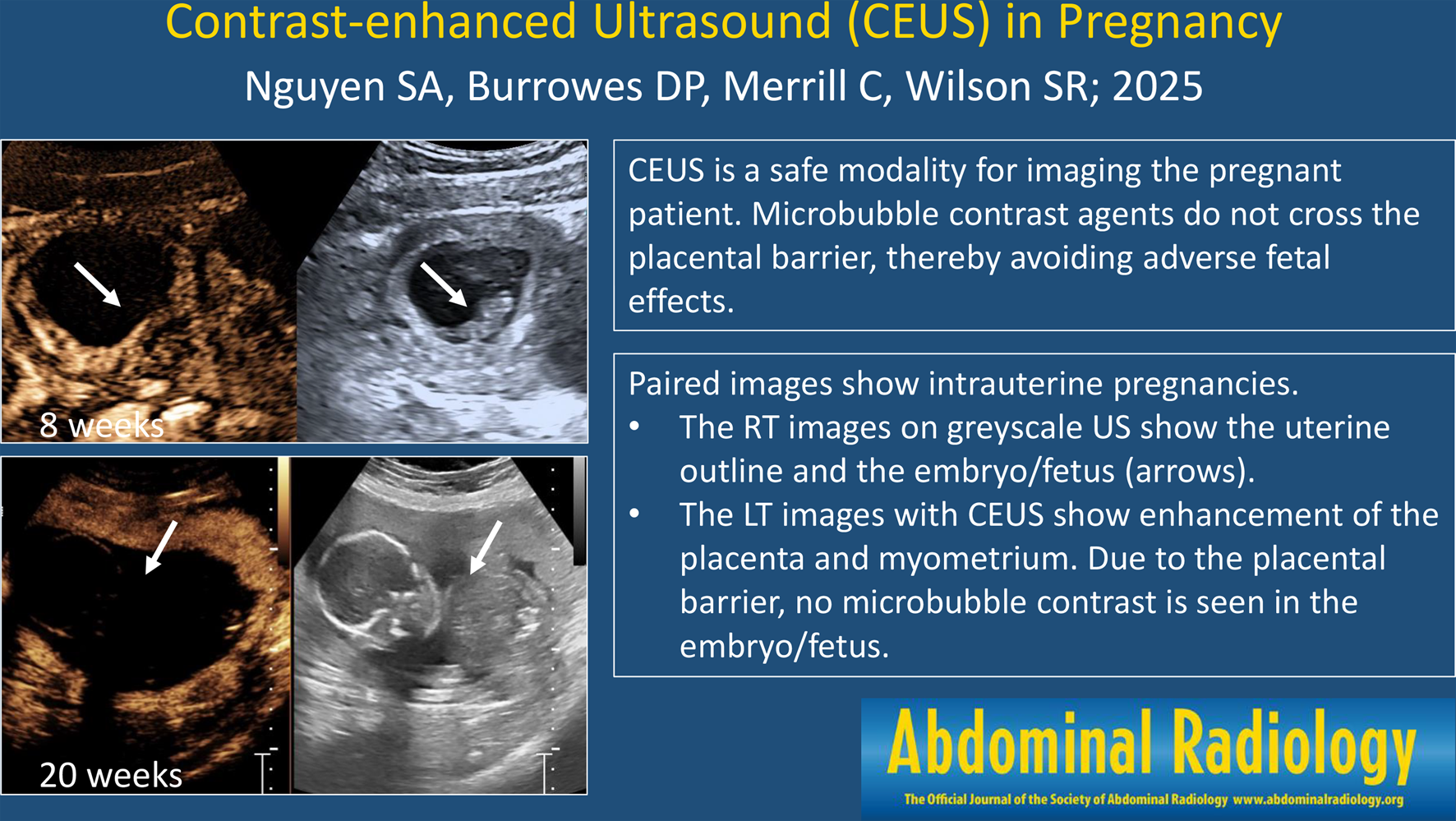
V. Schwarze, C. Marschner, G. Negrão de Figueiredo, J. Rübenthaler, and D.-A. Clevert, “Single-Center Study: Evaluating the Diagnostic Performance and Safety of Contrast-Enhanced Ultrasound (CEUS) in Pregnant Women to Assess Hepatic Lesions,” Ultraschall in der Medizin - European Journal of Ultrasound, vol. 41, no. 01, pp. 29–35, Feb. 2020, doi: https://doi.org/10.1055/a-0973-8517.
V. Schwarze, M. F. Froelich, C. Marschner, T. Knösel, J. Rübenthaler, and D.-A. Clevert, “Safe and pivotal approaches using contrast-enhanced ultrasound for the diagnostic workup of non-obstetric conditions during pregnancy, a single-center experience,” Arch Gynecol Obstet, vol. 303, no. 1, pp. 103–112, Jan. 2021, doi: https://doi.org/10.1007/s00404-020-05735-8.
P. S. Sidhu, D. Y. Huang, and C. Fang, “Contrast enhanced ultrasound (CEUS) in Pregnancy: Is this the last frontier for microbubbles?,” Ultraschall in der Medizin - European Journal of Ultrasound, vol. 41, no. 01, pp. 8–11, Feb. 2020, doi: https://doi.org/10.1055/a-0964-9827.
D. P. Burrowes, A. Medellin, A. C. Harris, L. Milot, B. C. Lethebe, and S. R. Wilson, “Characterization of Focal Liver Masses: A Multicenter Comparison of Contrast-Enhanced Ultrasound, Computed Tomography, and Magnetic Resonance Imaging,” Journal of Ultrasound in Medicine, vol. 40, no. 12, pp. 2581–2593, Dec. 2021, doi: https://doi.org/10.1002/jum.15644.
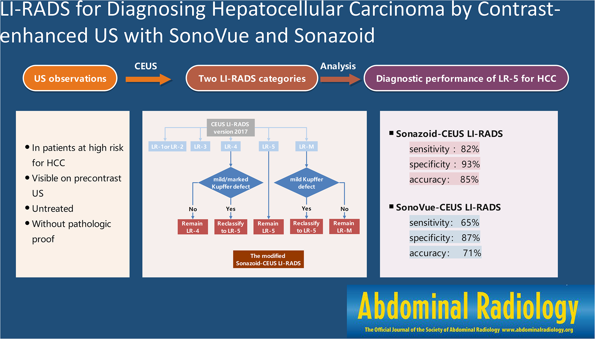
A. Makoyeva, T. K. Kim, H.-J. Jang, A. Medellin, and S. R. Wilson, “Use of CEUS LI-RADS for the Accurate Diagnosis of Nodules in Patients at Risk for Hepatocellular Carcinoma: A Validation Study,” Radiol Imaging Cancer, vol. 2, no. 2, p. e190014, Mar. 2020, doi: https://doi.org/10.1148/rycan.2020190014.
R. G. Barr, “Use of lumason/sonovue in contrast-enhanced ultrasound of the kidney for characterization of renal masses—a meta-analysis,” Abdominal Radiology, vol. 47, no. 1, pp. 272–287, Jan. 2022, doi: https://doi.org/10.1007/s00261-021-03295-2.
Article
PubMed
Google Scholar
A. Medellin, C. Merrill, and S. R. Wilson, “Role of contrast-enhanced ultrasound in evaluation of the bowel,” Abdominal Radiology, vol. 43, no. 4, pp. 918–933, Apr. 2018, doi: https://doi.org/10.1007/s00261-017-1399-6.
Article
PubMed
Google Scholar
K. Olinger et al., “Added Value of Contrast-enhanced US for Evaluation of Female Pelvic Disease,” RadioGraphics, vol. 44, no. 2, Feb. 2024, doi: https://doi.org/10.1148/rg.230092.
V. Miele, C. L. Piccolo, M. Galluzzo, S. Ianniello, B. Sessa, and M. Trinci, “Contrast-enhanced ultrasound (CEUS) in blunt abdominal trauma,” Br J Radiol, vol. 89, no. 1061, p. 20150823, May 2016, doi: https://doi.org/10.1259/bjr.20150823.
Article
PubMed
PubMed Central
Google Scholar
D. Cozzi et al., “Contrast-Enhanced Ultrasound (CEUS) in Non-Traumatic Abdominal Emergencies,” Ultrasound Int Open, vol. 06, no. 03, pp. E76–E86, Dec. 2020, doi: https://doi.org/10.1055/a-1347-5875.
Article
Google Scholar
“ACR-AIUM-SPR-SRU Practice Parameter for the Performance of Contrast Enhanced Ultrasound,” 2023.


Comments (0)