
Remember me

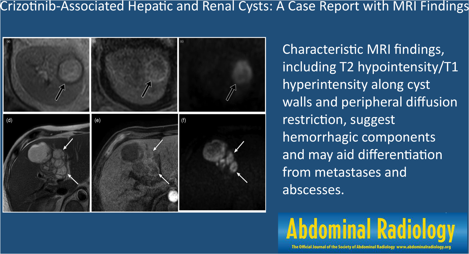

We report a case of a man in his 60s with ROS1 fusion-positive lung adenocarcinoma who developed multiple cystic lesions in both the kidneys and liver during crizotinib treatment. The patient presented with fever, abdominal pain, and oliguria. Contrast-enhanced CT revealed new cystic lesions in both the kidneys and liver, with some showing thick walls, septa, and hyperdense contents. MRI demonstrated heterogeneous signal intensities on T2-weighted images and mixed low to high signal intensities on T1-weighted images. Some cysts displayed T2 hypointense and T1 hyperintense areas along the cyst walls, with diffusion restriction predominantly at the periphery of the masses. Percutaneous drainage of the cysts was performed, and cytology revealed an increase in the number of cells, including neutrophils. However, no malignant cells or microorganisms were detected. Based on these imaging findings and laboratory results, the patient was diagnosed with crizotinib-associated renal and hepatic cysts. Symptoms improved after crizotinib discontinuation, and follow-up imaging showed cyst regression. This is the first report describing MRI findings for crizotinib-associated hepatic cysts. Characteristic MRI findings, such as T2 hypointensity/T1 hyperintensity along the cyst walls and diffusion restriction predominantly at the periphery of the masses, suggesting hemorrhagic components, may help differentiate crizotinib-associated cysts from other lesions, including metastases and abscesses.
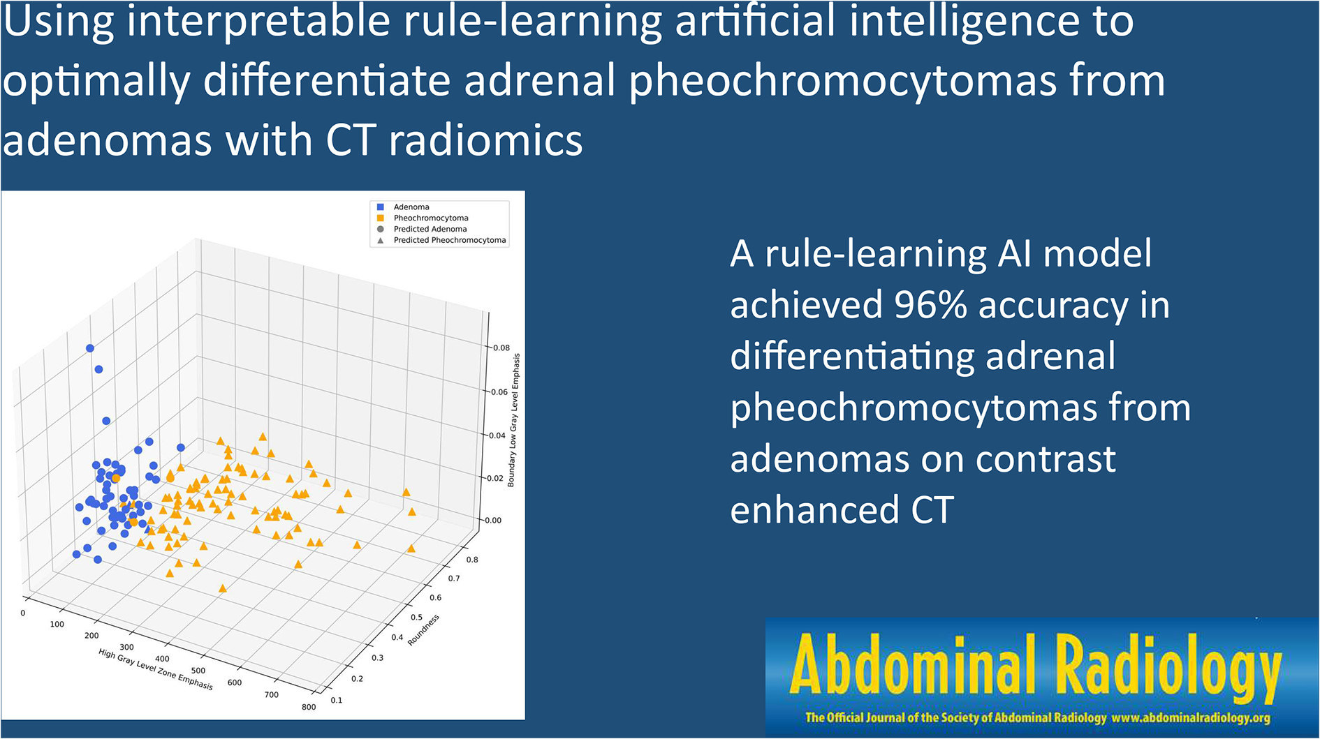
Graphical abstract
Comments (0)