
Remember me

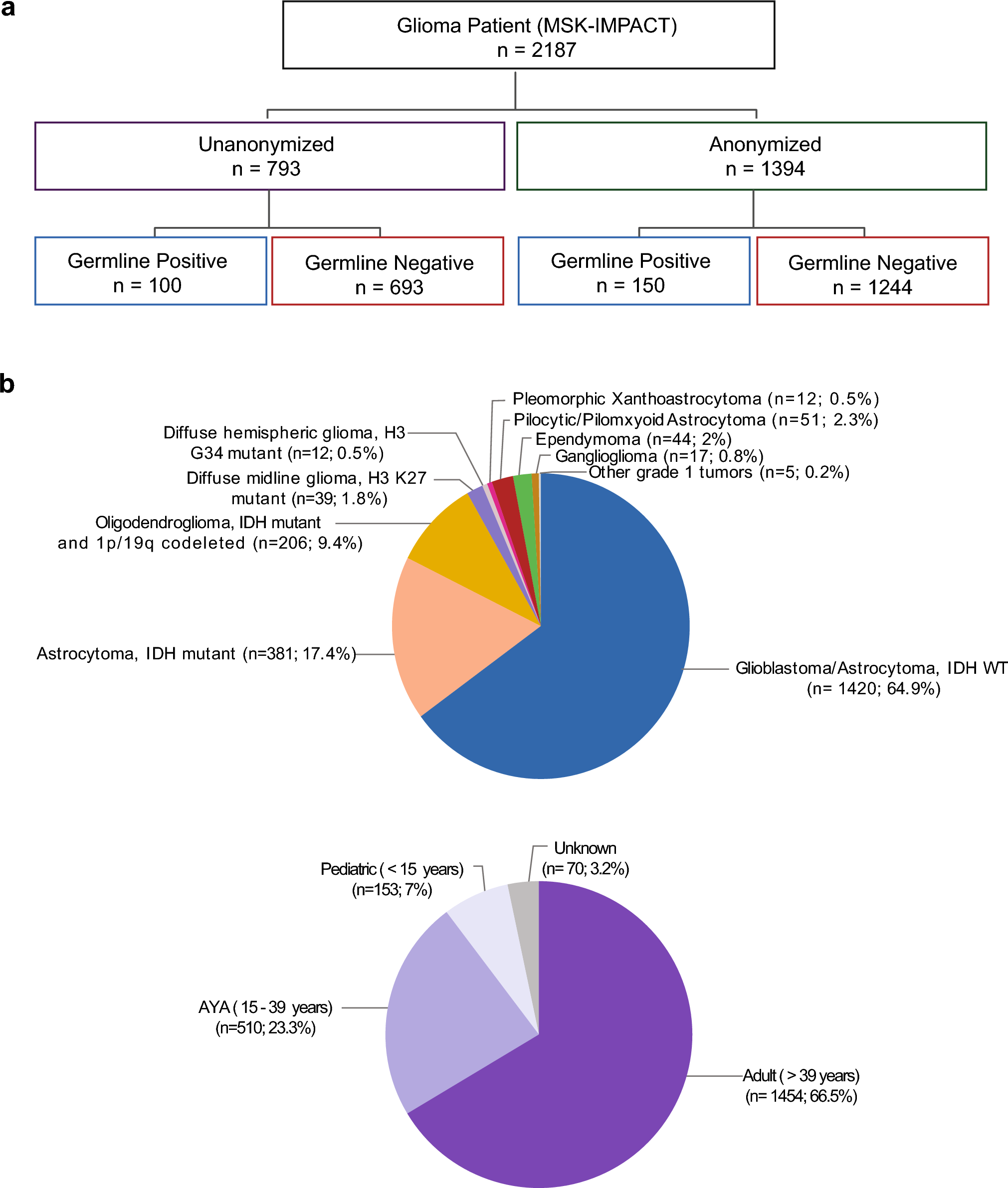


During routine diagnostics of brain tumours, we encountered an index case featuring “small blue round-cell” morphology, expression of TTF-1 and diffuse expression of synaptophysin. Histologically, the case resembled a small cell carcinoma metastasis. However, high-molecular-weight cytokeratin staining was negative (Fig. 1). After molecular analyses and further immunohistochemical staining, an IDH1 R132H mutation and a DNA methylation class prediction of “(high-grade) astrocytoma IDH-mutant” (in Heidelberg classifier v12.5) were obtained. Sampling of additional material identified additional areas of a diffusely infiltrating glioma consistent with an astrocytoma. We subsequently clustered the case with 176,857 DNA methylation profiles of tumour samples and control brain tissue samples (including local and external cases submitted through molecularneuropathology.org) and found a larger group of cases that were clustering in close relationship to the initial sample, most often with the highest score for IDH-mutant astrocytoma. For multiple cases, the referred diagnoses suggested a primitive neuroectodermal tumour or suspicion for metastasis. In total, we identified 51 samples with such a distinct methylation profile (Fig. 2).
Fig. 1
a The index case presents itself with a small blue round cell morphology with mitotic figures and occasional cell wrapping. b TTF-1 immunohistochemistry is expressed in the tumour cells. c High-molecular-weight cytokeratins were not expressed. d Synaptophysin is expressed. Scale bar denotes 200 µm
Fig. 2
Methylation data: in a tSNE analysis with selected reference entities from our database (n = 310), astrocytoma, IDH-mutant, PNC separates clearly from the other entities. PMMRDIA, primary mismatch-repair-deficient IDH-mutant astrocytoma; MB SHH IDH, medulloblastoma, SHH-activated, IDH-mutant; GBM PNC, glioblastoma, IDH-wildtype, with primitive neuronal component. The methylation class astrocytoma, IDH-mutant, high grade consists of 28 grade 4 tumours and 3 grade 3 tumours. The methylation class astrocytoma, IDH-mutant, low grade consists of 2 grade 4 tumours, 13 grade 3 tumours and 17 grade 2 tumours
After re-analysing the identified samples using the latest (v12.8) version of the Heidelberg brain tumour classifier, most samples (n = 39/51, 76.5%, Online Resource in supplementary Table 1) were successfully allocated to the methylation class of astrocytoma, IDH-mutant; high grade (calibrated score > 0.9). Because of the presence of a primitive neuronal component in all of these tumours, we named the distinct methylation group “astrocytoma, IDH-mutant, with primitive neuronal component” (ASTRO PNC). This methylation group was clearly distinct from the earlier described methylation classes of glioblastomas with primitive neuronal component (GBM PNC) and high-grade IDH-mutant astrocytomas, that include grade 4 IDH-mutant astrocytomas [(n = 28/31), Fig. 2].
ASTRO PNCs express TTF-1To assess the histology of the new methylation group, we collected tumour samples from 21 patients. All tumour samples (partially) presented with areas of astrocytic differentiation with a fibrillary matrix formed by astrocytic cell processes, a pattern that is usually observed in IDH-mutant astrocytomas. The round to oval nuclei showed nuclear atypia regarding chromatin density and size (Fig. 3). Using immunohistochemical staining, expression of mutant IDH1 (R132H) was detected in the majority of cases (n = 18/20, 90%) and a loss of ATRX expression (n = 15/16, 94%) was equally common. The p53 protein was accumulated in most observed cases (n = 14/15, 93%) (Fig. 4). For the two cases lacking IDH1 R132H expression, different IDH1 R132 substitutions were detected in the sequencing analysis (IDH1 R132S in patient 10 and IDH1 R132S in patient 3; Online Resource in supplementary Table 1).
Fig. 3
a All cases investigated by histology harbour a primitive neuronal component with small blue round tumour cells. In addition, in this case, cell wrapping (arrow) can be observed. b In most cases, a sharp demarcation of the primitive neuronal component and the astrocytic component can be found. c Presence of tumour growth inside the lumina of two vessels (asterisks). Despite reported extra-axial spread in this tumour entity, this case does not have reported metastases: d some cases present themselves with rosettes (arrows). Scale bar denotes 60 µm in a, b and d. Scale bar denotes 600 µm in c
Fig. 4
a TTF-1: the primitive neuronal component expresses TTF-1 (using TTF-1 clone EP229). In a subset of cases, the expression is limited to a small number of cells. b IDH1 R132H: most tumour cases strongly express IDH1 R132H. c ATRX: nuclear ATRX expression is lost in most of the cases with blood vessels suited as positive internal controls. d GFAP: GFAP expression is lost in the primitive neuronal component. e Olig2: a subset of the primitive tumour cells expresses Olig2. f AE1/3: the tumours do not express high-molecular-weight cytokeratins stained with AE1 and/or AE3. g Ki-67: the proliferation index is high in the primitive neuronal component, whilst in most cases, the glial component does not show an elevated proliferation index. h p53: p53 is strongly expressed in the primitive tumour cells. i Synaptophysin: synaptophysin is expressed in the primitive tumour cells. Scale bar denotes 300 µm
In contrast to the vast majority of IDH-mutant astrocytomas, all cases also showed a primitive neuronal component with strong nuclear expression of TTF-1, diffuse synaptophysin positivity and in a subset of cases positivity for NSE and Neu-N (Online Resource in supplementary Table 1). The morphology of the primitive neuronal component was heterogeneous with rosettes, cell wrapping and a small-blue-round-cells (Fig. 3). In addition, a high proliferation index above 85% was generally observed in the primitive area of the tumour. The PNC showed clear differences regarding the Ki-67, TTF-1 and GFAP expressions levels compared to the astrocytic component (Fig. 4). Similar to the astrocytic component, IDH1 R132H was expressed and p53 was accumulated, whilst ATRX expression was lost in the primitive component. In line with the expected glial origin, Olig2 was expressed in a subset of primitive cells as well. To check for TTF-1 cross-reactivity, we stained several non-neoplastic tissue and other samples to investigate TTF-1 expression in non-neoplastic CNS tissue and IDH-mutant astrocytomas. In line with our expectations, TTF-1 was not expressed in non-neoplastic CNS tissue and only 3.5% (1/28) of high-grade astrocytoma CNS WHO grade 4, with one case showing single tumour cells being positive
Necrosis was present in n = 17/20 (85%) cases. Microvascular proliferation was present in 18/20 (90%) cases. Multiple mitoses could be observed in 19/20 (95%) cases. Thus, based on the WHO CNS5 grading criteria, all tumours presented with histological criteria warranting an IDH-mutant astrocytoma CNS WHO grade 4 designation due to either presence of necrosis or vascular proliferation (Online Resource in supplementary Table 1).
ASTROs PNC show recurrent mutations in RB1To evaluate the mutational profile of our cohort, we performed next-generation sequencing with a custom Illumina Panel on 21 tumour samples. Besides the characteristic IDH1 mutation (n = 21/21, 100%, thereof n = 19/21, 90% IDH1 R132H mutant), alterations were detected in TP53 (n = 20/21, 95%) and in the telomere maintenance mechanism (TMM) including ATRX (n = 18/21, 86%) and the TERT promoter (pTERT) (n = 1/21, 5%). For IDH-mutant astrocytomas without a primitive neuronal component, the usual TMM alteration is a loss of function event on the ATRX gene. In addition, we observed mutations in RB1 in 12/21 cases (57%, Fig. 5a).
Fig. 5
a Oncoprint of 21 samples for which DNA targeted panel sequencing was performed, including genetic mutations, immunohistochemistry (IHC) and copy number variations calculated from the DNA methylation array. b The copy number summary plot (n = 42) reveals a focal deletion at the RB1 locus in addition to several chromosomal gains and losses
ASTROs PNC show recurrent copy number alterations in RB1 and MYCNCopy number profiles based on DNA methylation array analysis were performed on all 51 samples and frequently showed deletion of chromosome arm 13q with deletions of RB1 (homozygous deletions in n = 5/51, 10%; hemizygous deletions in n = 38/51, 75%). A gain of chromosome 1 and a loss of chromosome arms 3p, 4q, 16, 19 and 22q were commonly observed as well (Fig. 5b). MYCN was frequently altered in ASTRO PNCs, with a subset of cases showing MYCN amplifications in the copy number variation profile. One case harboured a pathogenic MYCN mutation (MYCN alterations n = 14/51, 27%). RNA sequencing performed on six samples did not show any relevant fusion events.
In contrast to GBM PNC, the PTEN locus was mostly balanced in ASTRO PNC, with only two cases showing a homozygous PTEN deletion (n = 2/51, 4%). PTEN deletions are rarely observed in IDH-mutant astrocytomas. High-grade IDH-mutant astrocytomas often show cell cycle deregulation due to homozygous CDKN2A/B deletions or amplifications of CDK4 or CDK6. However, these were only rare events in ASTRO PNCs with only a single case showing homozygous CDKN2A/B deletion (n = 1/51, 2%) and three cases with CDK4 amplification (n = 3/51, 6%). CDK6 amplifications were not detected in our cohort.
Tumour location shifts from the frontal to the temporal lobe during progressionWe also investigated whether, in addition to a distinct DNA methylation profile, ASTRO PNCs are associated with different clinical features compared to IDH-mutant astrocytomas without a primitive neuronal component. Therefore, we collected clinical data for 31 patients with ASTRO PNCs and compared them to clinical data from 38 patients with IDH-mutant astrocytomas CNS WHO grade 4 without a primitive neuronal component. Whilst IDH-mutant gliomas are predominantly located in the frontal lobe, ASTRO PNCs were more often located in the temporal lobe as well as the frontal lobe (Fig. 6a). For primary lesions (n = 15, 50%), the frontal lobe was the preferred location (Fig. 6a, left side), whilst recurrent tumours (n = 10, 90%) were enriched in the temporal lobe (Fig. 6a, right side), similar to our observations in IDH-wildtype glioblastomas with primitive neuronal components (52% located in the temporal lobe [18]). As we did not have any matched pairs of primary and recurrent tumours in our cohort, we do not know if primary lesions of recurrent tumours were also described to have a primitive neuronal component. We did not observe any frontal-to-temporal lobe transition during recurrence in a reference cohort of IDH-mutant gliomas (including 56 non-PNC astrocytomas and 48 oligodendrogliomas).
Fig. 6
a Left side: the most common site of origin for primary diagnoses of ASTRO PNC is the frontal lobe followed by the temporal lobe. Right side: for recurrent ASTRO PNC cases, the most common site is the temporal lobe. b The tumours show a predisposition for the male sex. c Age distribution in ASTRO PNC (n = 30, median age = 38). d Kaplan–Meier estimate indicates no significant difference to IDH-mutant astrocytomas WHO grade 4 (p = 0.24). In contrast, there is a significant difference to IDH-mutant astrocytomas WHO grade 2 and 3 (p(A2) = < 0.0001; p(A3) = < 0.0001)
Sex, age and MGMT promoter methylation similar to CNS WHO grade 4 IDH-mutant astrocytomasFor further characterisation, we compared median age and sex distribution of patients with ASTRO PNCs to those of patients with IDH-mutant astrocytomas CNS WHO grade 4 without a primitive neuronal component. Similar to IDH-mutant astrocytomas, we observed a male predominance (61%, Fig. 6b). Median age at diagnosis of an ASTRO PNC was 38 years compared to 42 years in an internal reference cohort of 38 patients with IDH-mutant astrocytomas CNS WHO grade 4 without a primitive neuronal component (p = 0.08, [11], Fig. 6c). The MGMT promoter was methylated in 90% of ASTRO PNCs (n = 46/51).
Of note, looking at methylation of all CpGs included on the methylation array, we observed a high rate of unmethylated CpG probes in ASTRO PNCs, which may indicate a more aggressive clinical course in IDH-mutant astrocytomas (G-CIMP low), comparable to IDH-wildtype glioblastomas and high-grade IDH-mutant astrocytomas (Supp. Figure 1).
Frequent leptomeningeal dissemination and poor survival similar to patients with CNS WHO grade 4 IDH-mutant astrocytomasTo examine the prognostic relevance of these tumours, we evaluated survival data for 23 patients. We performed a Kaplan–Meier estimate and compared our cohort to an internal cohort of 209 IDH-mutant astrocytomas of CNS WHO grades 2, 3, or 4 (Fig. 6d). The overall survival of patients with ASTRO PNC and IDH-mutant astrocytoma CNS WHO grade 4 patients showed no significant difference (p = 0.2446) but were significantly different to patients with IDH-mutant astrocytomas of CNS WHO grades 2 or 3 (p(A2) = < 0.0001; p(A3) = < 0.0001). Furthermore, IDH-mutant astrocytomas with primitive neuronal component showed a higher risk of leptomeningeal spread with seven patients developing leptomeningeal and/or subarachnoidal dissemination intracranially and in the spine during progression. Two patients had metastases outside of the central nervous system (Table 1).
Table 1 Clinical data of the patients of the core cohortTable 1 Progression-free survival (PFS), overall survival (OS) both rounded, survival status, localisation, age, extra-axial manifestations and leptomeningeal spread.
Comments (0)