SFRPs gene family expression in the GEPIA, TCGA, and GEO
The GEPIA (Gene Expression Profiling Interactive Analysis, http://gepia.cancer-pku.cn.) database was used to investigate the expression of SFRPs in CRC, drawing on data from TCGA and the Genotype-Tissue Expression (GTEx, https://www.genome.gov/Funded-Programs-Projects/Genotype-Tissue-Expression-Project.). Expression levels of each SFRP family member were analyzed in cancer and normal tissues within GEPIA. Additionally, mRNA expression data from normal colorectal tissues and CRC tissues were retrieved from the GEO database (https://www.ncbi.nlm.nih.gov/geo), using datasets GSE23878, GSE113513, GSE79793, and GSE156355. The limma package in R language was used to analyze the mRNA expression levels of SFRP1, SFRP2, FRZB, SFRP4, and SFRP5, producing a differential expression heatmap.
Survival analysis
RNAseq data from the TCGA-COAD and TCGA-READ STAR pipelines were downloaded and collated from the TCGA database (https://portal.gdc.cancer.gov), with data extracted in TPM format, along with associated clinical information. Using R, the survival package was applied to perform proportional hazards assumption testing and fit a survival regression model. Results were visualized with the Survminer and ggplot2 packages. For optimal grouping, the cutpoint function in the Survminer package was used to identify the best cutoff point for group stratification.
Analyze the molecular interactions through STRING and KEGG/gene ontology (GO) analysis
The molecular interactions and correlations between SFRPs and related proteins were analyzed using the STRING (https://string-db.org/) online interactive tool. It should be noted that the input genes used to identify the internal communications were as follows: ARMC8; RANBP9; GID8; SFRP2; RMND5A; MAEA; RMND5B; DVL2; FZD1; FZD7; FZD6; WNT1; LRP5; FZD4; FZD2; ROR2; WNT3A; WNT5A; FZD5; LRP6; FRZB; WNT2; WNT7A; SFRP1; RYK; NDP; FZD8; FZD9; MET; ROR1; TNFSF11; SFRP4; SFRP5; TNFRSF11A; and TNFRSF11B. The network contained 35 nodes and 470 edges, with a local clustering coefficient of 0.883 and a PPI (protein–protein interaction) enrichment p-value of less than 1.0e-16.
For output data analysis, Cytoscape software was used for further experimental examination. The candidate genes were analyzed using the DAVID online tool (https://david.ncifcrf.gov/) for GO and KEGG pathway analyses. GO analysis categorized the candidate genes by molecular function, cellular location, and biological pathways. The “ggplot2” package was employed to create a bubble chart.
Analyze the immune infiltration through the TIMER and TISIDB analysis
The relationship between the expression of various molecules in tumors and immune-related markers was evaluated using TIMER (Tumor IMmune Estimation Resource, https://cistrome.shinyapps.io/timer), based on data from The Cancer Genome Atlas (TCGA). TIMER was used to analyze the infiltration of SFRP family members in relation to CD4 + T cells, CD8 + T cells, macrophages, neurons, and B cells. Additionally, we used the TISIDB platform (http://cis.hku.hk/TISIDB/index.php) to analyze gene expression and examine the relationship between methylation levels and tumor-infiltrating lymphocytes, specifically focusing on the methylation status of SFRP1 and SFRP2 and their association with immune infiltration.
Methylation data and processing
DNA methylation data were derived from RNAseq and Illumina human methylation data in the TCGA database. Using NCBI, we identified the 2000-bp upstream promoter region and the 200-bp downstream region of the SFRP1 and SFRP2 genes. MethPrimer (http://www.urogene.org/cgi-bin/methprimer/methprimer.cgi) was then used to predict CpG islands within the promoter regions of these genes. Additionally, we analyzed the TCGA database to explore the relationship between SFRP1/2 expression and the expression of DNA methyltransferases (DNMTs), including DNMT1, DNMT3A, and DNMT3B.
Cell culture
CRC cell lines (SW620, HCT116, LoVo, and COLO205), as well as the normal colon cell line NCM460, were obtained from Procell in Wuhan, China. These cell lines were cultured in DMEM medium supplemented with 1% penicillin–streptomycin and 10% fetal bovine serum (both from Gibco, Thermo Fisher Scientific, Grand Island, NY). Standard cell culture protocols were followed for cryopreservation, resuscitation, passage, culture, and identification. Plasmid transfection was conducted using Lipofectamine 3000 as the transfection reagent.
RNA extraction and qRT-PCR
The processed cells were lysed in 1 ml of Trizol for 5 minutes, followed by the addition of 200 ml of chloroform. The cells were inverted approximately 30 times, and then centrifuged at 12,000 rpm for 5 min at 4°C. The upper aqueous layer was carefully transferred to an enzyme-free EP tube, and an equal volume of isopropanol was added. After centrifuging for 14 min at 12,000 rpm, the upper liquid was removed, leaving the white RNA pellet at the bottom.
RQ1 RNase-Free DNase (Promega, Madison, WI) was used to treat the RNA. The reaction mixture included 10 μl of solution containing 1 μl of 10 × buffer, 2 μl of DNase, 2 μg of RNA, and diethylpyrocarbonate (DEPC) water. The solution was incubated at 37°C for 30 min, and then immediately placed on ice. Next, 1 μl of RQ1 DNase Stop Solution was added to each tube, followed by incubation at 65°C for 10 min. The RNA was then reverse-transcribed into cDNA, which was incubated at 37°C for 2 min, 42°C for 60 min, and 70°C for 5 min, before being stored at 4°C.
The obtained cDNA was subjected to quantitative reverse transcription PCR (qRT-PCR). The PCR primer sequences are depicted in Table 1. The qRT-PCR was performed under the following conditions: initial denaturation at 95°C for 10 min, followed by 40 cycles of denaturation at 95°C for 15 s and annealing/extension at 60°C for 30 s. The details of the reaction system are provided in Table 2. The Ct values were then calculated to determine the relative mRNA expression levels.
Demethylation of CRC cells and colon cells by 5-azaD (5-aza-2-deoxycytidine)
5-azaD (Sigma-Aldrich, St. Louis, MO) was dissolved in water and then diluted with the cell culture medium. The cell lines were treated with 5-azaD for 4 d, with the medium being replaced daily. After treatment, the demethylated cells were directly collected for total RNA isolation, CCK-8 assays, and apoptosis analysis. Cells cultured under normal conditions and treated with 0.2% DMSO served as the control group.
MSP (methylation-specific PCR) assay
Genomic DNA was extracted from cells and colon tissues using the Genome DNA Purification Kit (Tiangen, Beijing, China). The DNA was then modified according to the instructions provided with the EZ DNA Methylation Kit (ZYMO RESEARCH, Orange, CA). Primers for MSP were designed as follows:
The PCR conditions were as follows: initial denaturation at 95°C for 5 min, followed by 30 cycles of denaturation at 95°C for 20 s, annealing at 60°C for 30 s, extension at 72°C for 20 s, and a final extension at 75°C for 5 min. The PCR products were then resolved on a 2% agarose gel, stained with ethidium bromide, and visualized under ultraviolet light.
BSP (bisulfite sequencing PCR) Assay
DNA was extracted from normal colon cells and CRC cells using the DNA Extraction Kit (Axigen, Los Angeles, CA). The extracted DNA was then treated with hydrogen sulfate. PCR products were excised from the gel and ligated into the pMD® 18-T Vector (D101B; Takara, Kusatsu, Japan). The ligated plasmid was transformed into DH5α bacteria, which were cultured overnight. Plasmid DNA was then isolated, and at least ten individual clones were selected for sequence analysis.
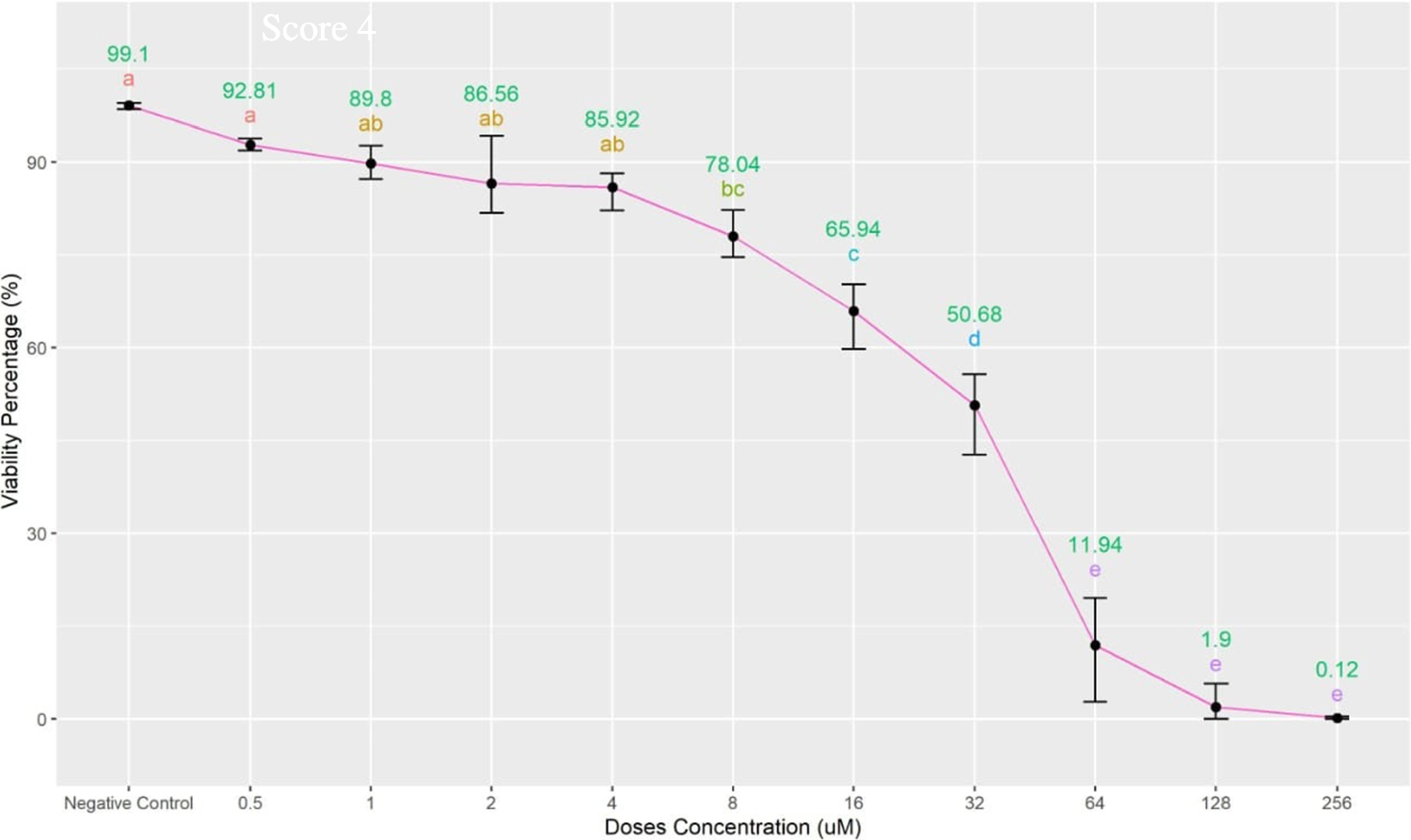
Detection of proliferation in CRC cells by CCK8 assay
SW620 and HCT116 cells were seeded into a 24-well plate at a density of 5 × 10⁵ cells/ml, with a blank control group included. After 12 h of incubation, the control group was removed, and the remaining cells were exposed to plasmids for 48 h (SW620) and 72 h (HCT116). Following treatment, 25 µl of CCK-8 reagent was added to each well. The wells were protected from light and incubated at 37°C for 1.5 h. Absorbance at 450 nm was measured using a microplate reader, and the cell growth rate was calculated using the following formula:
$$\text\left(\right)=\frac}_}-}_})}}_}-}_})}\times 100\%$$
(1)
where A represents the absorbance values obtained from the measurements.
Detection of apoptosis in CRC cells by flow cytometry
SW620 and HCT116 cells were seeded at a density of 5 × 10⁵ cells/ml in a 6-well plate. After incubation, the cells were washed with pre-cooled PBS, and trypsin was added for digestion. The cells were collected, centrifuged, and resuspended in PBS. To assess cell viability, PI and Annexin V-FITC antibodies were added to the samples. Flow cytometry analysis was then performed in a dark environment.
Statistical analysis
Statistical analysis was performed using SPSS 21.0, and the data are presented as mean ± standard deviation. Differences among the groups were compared using one-way ANOVA, and pairwise comparisons between groups were evaluated using the t-test. A p-value of less than 0.05 was considered statistically significant. All experiments were conducted independently at least three times.


Comments (0)