An exploratory in vivo trial in five female pigs (59–69 kg) of sterile bare iron oxide particles [25], 2 mL 25 g/L, suspended in deionized water and a magnetic probe was conducted to evaluate safety endpoints (acute systemic toxicity, side effects and adverse events). 4 mL 0.9% sterile isotonic saline solution per mL of particles was used to reconstitute or “activate” the particles, which begins the process of agglomeration. A magnetic probe with 9 Fr NdFeB magnets are used for the extraction.
Study Endpoints: The primary endpoints of this study were to assess the safety and tolerance of the magnetic particle suspension and magnetic probe during Retrograde Intrarenal Surgery (RIRS) in an animal model. Safety evaluation focused on the potential occurrence of adverse events, including complications such as haemorrhage, hypotension, or any toxic/anaphylactic shock syndrome associated with the application of magnetic particles during surgery. The absence of any adverse events or complications would be indicative of positive safety and tolerance results. Toxicity assessment was based on histological and biological (urinalysis and hemogram) findings in the two groups, and the absence of significant differences would indicate that the application of magnetic particles and the use of magnetic probe were non-toxic.
Animal Model: Pigs were chosen due to the anatomical similarity of their kidneys to human kidneys. The volume of the porcine kidney pelvis is much smaller than in humans and the blood of pigs is, compared to humans, hypercoagulable [26]. Despite the differences, the clinical risks, including mechanical damage to the urothelium as well as signs of sensitivity, are transferable from pigs to humans. The considerations and setup of this study were in accordance with Good Laboratory Practices (GLPs), closely adhering to guidelines in ISO 10993-2:2006 for animal welfare requirements and ISO 10993-11:2017 for acute systemic toxicity. All procedures were carried out under general anaesthesia.
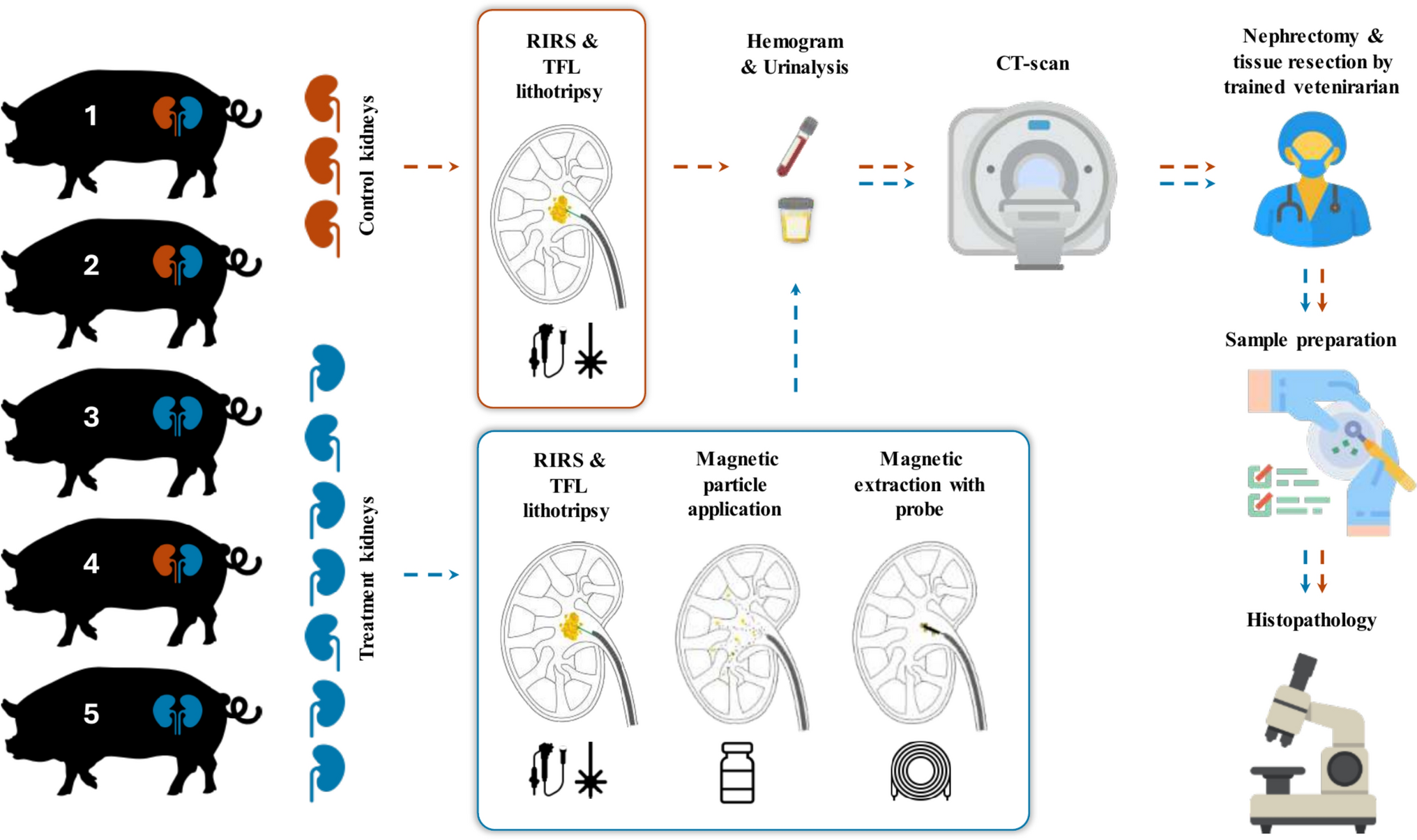
The study involved ten kidneys, with three pigs having intracorporeal control on one side and receiving treatment with the magnetic extraction method on the other side. The sample size of ten kidneys is defined by the endpoints which are acute complications during the acute treatment. Two pigs were treated bilaterally with the magnetic extraction method to attain sufficient samples for histopathology. All kidneys but two received a lithotripsy, where heterogenous small residual fragments (SRF)s were directly implanted. The size of these residual fragments varied between 3 to 20 mm. The fragments were prepared to fit through a 10/12 Fr ureteral access sheath (UAS) (ReTrace®, Coloplast, Denmark). Urine and blood samples were taken from each model before and after the surgery. Following the procedures, the animals were euthanized, and a high-dose CT scan with 0.7 mm section thickness was performed to assess the remaining fragments within the kidney’s collecting system. Subsequently, a bilateral nephrectomy was performed to evaluate the impact of the magnetic particle suspension and the magnetic probe on the kidney tissue by histopathological analysis. The nephrectomy procedure was conducted by a certified veterinarian. The blinded clinical pathology, i.e. hemogram and urinalysis, by IMD Labor Oberland (Am Kleistpark 1, 15,230 Frankfurt (Oder), Germany) and SYNLAB vet. Labor Berlin (Am Borsigturm 42, 13,507 Berlin, Germany) respectively.
Control RIRS: Standard RIRS procedures abiding international guidelines for kidney stone management were performed with the pigs in the dorsal lithotomy position [4, 27]. A cystoscopy was carried out to identify the ureteral orifices. Two hydrophilic guidewires (HiWire®, Cook Medical, USA) were placed under fluoroscopy into the renal pelvis. A 12/14 Fr UAS (Coloplast) was inserted to facilitate stone placement in mid-lower pole calyces, lithotripsy and a 10/12 Fr UAS (Coloplast) for extraction of the stone fragments (0.15 g, respectively ~ 4 mm, rehydrated overnight in 0.9% NaCl). Following a 60-min break to facilitate ureteral dilation, a 7.5 Fr flexible uretero-renoscope (PU3033A, Pusen Medical, China) was used to inspect the calyces. The stones (5–10 stones from human samples in the size range of 1–3 mm) were then positioned using a nitinol grasper (Cook Medical) or water propulsion through the UAS with a syringe. After the stones were introduced, we performed lithotripsy to create heterogenous dust, most of which was ≤ 1.5–2 mm in size. A Thulium fiber laser (Quanta Systems and Coloplast) with a 200 µm fiber was used in a low-energy dusting setting (0.4 Joules, 20 Hz, short pulse). Once lithotripsy was concluded, the urinary tract was occluded with a catheter (IMP Occlusion catheter 6 Ch, Germany) or the UAS to prevent expulsion of fragments. The procedure was concluded when the urinary tract was closed.
Treatment RIRS (With Magnetic Particle Application): The treatment RIRS procedures followed the same steps as the control RIRS till the fragmentation stage. After lithotripsy, we applied 10 mL magnetic particle suspension. After a 1-min incubation, they were extracted using a magnetic probe. The probe was backloaded onto the uretero-renoscope to inspect the kidney and passively retrieve the SRFs. If fragments were still present at the end of the magnetic removal, a second application of magnetic particles (5 mL) and extraction were performed. After completing the treatment, the urinary tract was occluded.
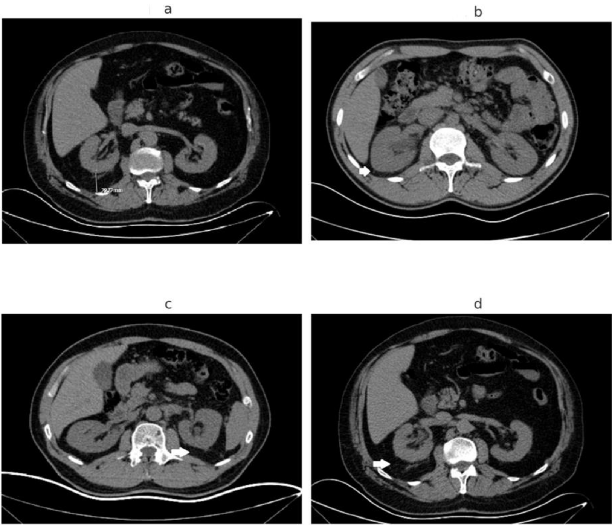
Computed Tomography Workup: The CT-examination was conducted by Radiologisch Nuklearmedizinische Praxis Beck & Kollegen (Am Stadtpark 5, 15,517 Fürstenwalde/Spree, Germany). Post-euthanization, the kidneys of the animal models were imaged using a Siemens Healthineers SOMATOM-CT scanner with the models in a supine position. We performed a preliminary fast scan to verify model positioning before performing the final images. The resulting data and metadata were exported in DICOM format for analysis, which was carried out using Slicer 5.2.2 (www.slicer.org).
Histopathological Workup: All kidneys were evaluated by an independent, accredited veterinarian pathologist (blinded), Dr. med. Vet. Wolfram Haider, Veterinary Physician for Animal Pathology at the Institute of Animal Pathology (Schönhauser Straße 62, 13,127 Berlin, Germany). Following nephrectomy, cranial, medial and caudal tissue sections were resected from the renal medulla, pelvis, vein and ureters from each kidney and embedded in paraffin for histopathological examination. Subsequently, a 2 µm-thick section was prepared from each paraffin block, and these sections were stained with hematoxylin and eosin for microscopic examination. Microscopic examination was conducted at 50x, 100x, 200x, and 400 × magnifications to photograph different levels of detail. The samples were investigated for potential toxicity and the occurrence of pathological events was compared between the control and treatment groups as a low sample t test. Due to the low sample size in both groups, the difference in occurrences is reported as confidence intervals at 95% confidence with the assumption that the mean difference is zero, since no conclusions about statistical significance can be drawn with high power in this setup. These confidence levels only serve to indicate safety and justify benefit-risk for a clinical investigation.

Comments (0)