Determining the aggressiveness of a tumor or predicting the course of localized low-grade prostate cancer is a challenging task. This is because there is a range of treatment options available, from immediate definitive therapy to active surveillance. To facilitate treatment decisions, molecular biomarkers in addition to established serological, radiological, and histological parameters are desired. At the ISUP consensus conference on molecular biomarkers in genitourinary tumors in 2019, Ki-67 and PTEN were identified as the most promising markers [6].
However, the application of Ki-67 as a biomarker is complicated by interobserver variability due to the reflective nature of the proliferative activity, which requires a cut-off value to determine prognostic subgroups [36,37,38]. On the other hand, PTEN is one of the most frequently altered genes in prostate cancer, and its prognostic and potential therapeutic implications have been demonstrated in multiple retrospective studies [20,21,22,23,24,25, 39]. PTEN appears to be a more reliable biomarker than Ki-67 because its readout is dichotomous, meaning it is either retained or lost. Immunohistochemistry is the preferred modality for assessing PTEN status due to its easy interpretation, inexpensiveness, and wide implementation in pathology laboratories.
PTEN loss in prostate cancer is mostly due to PTEN biallelic genomic deletion whereas PTEN mutations occur less frequently and in association with hemizygous deletions [7,8,9,10,11,12,13]. Gene rearrangement comprising the PTEN locus is reported as an additional mechanism of PTEN inactivation [14]. PTEN loss or downregulation via promotor hypermethylation, miRNAs or other mechanisms is less well studied [15, 16]. Due to an association with Gleason score and tumor stage, the reported prevalence of PTEN loss in prostate cancer is variable across different cohorts and is reported in about 20% of primary and in about 40% of metastatic tumors [7, 8, 10, 11, 17,18,19].
Apart from its role as a prognostic biomarker, it is expectable that PTEN status might have a role as a predictive biomarker in PTEN-deficient advanced prostate cancer. Promising treatment options focus on the blockage of the PI3K-AKT-mTOR pathway in combination with established therapeutics [26]. An ongoing phase III randomized double-blind study (IPATential150) testing the AKT-inhibitor ipatasertib with abiraterone and prednisone compared with placebo plus abiraterone and prednisone in patients with metastatic castrate-resistant prostate cancer with PTEN loss showed an improved radiographic progression-free survival in ipatasertib-treated patients [27].
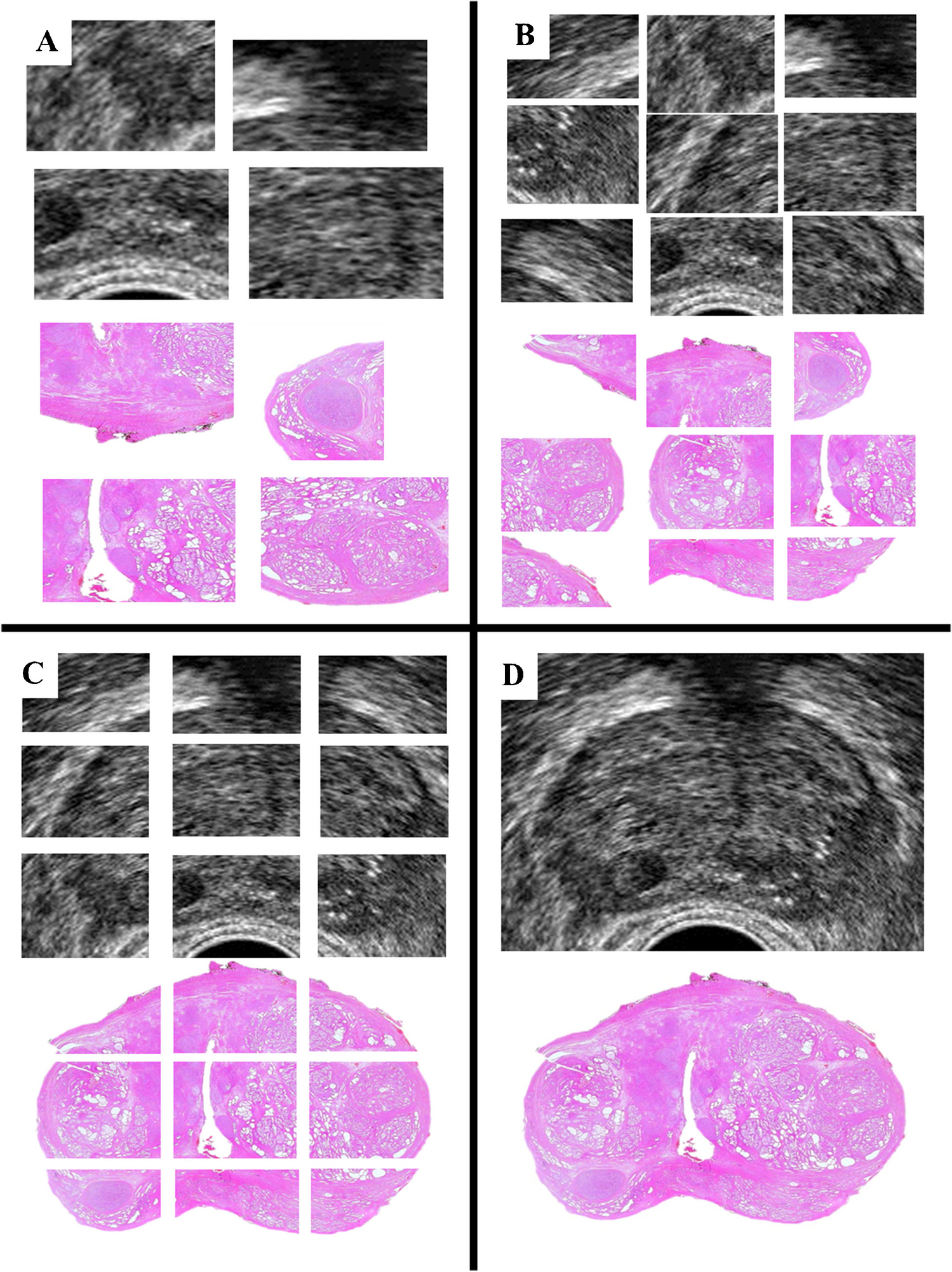
In preparation of a prospective multicentric collection of molecular prostate cancer data including PTEN immunohistochemistry, a ring trial was conducted to determine the status quo of the diagnostic accuracy of PTEN immunohistochemistry in localized low- to intermediate-risk prostate cancer among ten eminent university pathology institutes in Germany. Unexpectedly, the interpretation of PTEN immunohistochemistry in this study showed marked variation (12.5–51.2% PTEN loss rates) in an identical cohort of prostate cancer. This may not be surprising, as no harmonization of the PTEN assay was conducted prior to this ring trial. While there was a trend towards harmonization in the interobserver agreement in a subsequent smaller ring trial following feedback to the participants, even these results require further improvement; hence, we think this data argues against the uncritical use of PTEN immunohistochemistry to define PTEN status for clinical routine use. So far, the interobserver variability of PTEN immunohistochemistry has been studied in only a few other entities. Because of the frequent PTEN loss in endometrial cancer, PTEN immunohistochemistry is well implemented as a routine diagnostic biomarker in this entity. Comparing the interobserver variability of PTEN loss in endometrial cancer between two institutes, it was found to be highly reproducible with a kappa value of 0.8 [40].
FISH analysis of our cohort revealed PTEN hemizygous deletions in 5.5% (5/90) of all cases, which is in contrast to the reported prevalence of PTEN loss in about 20% of primary prostate cancers [7, 8, 10]. However, this finding is likely to be explained by the selection of only grade group 1 and 2 tumors [20, 23, 41].
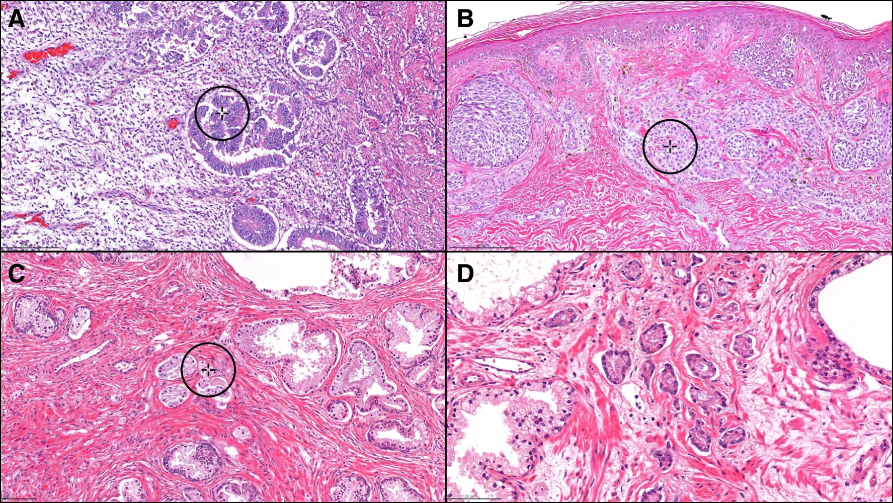
In direct comparison of immunohistochemical results with FISH analysis, the positive predictive value of the genomic alteration ranged from 11.9 to 45.5%. All cases with a hemizygous PTEN deletion were totally negative on immunohistochemistry, and conversely, any positive PTEN immunoreactivity ruled out a genomic PTEN loss (sensitivity 100%). However, negative (loss) immunohistochemistry was rather unspecific for an underlying genomic deletion. Then, 6.7% (6/90) of the tumors with a loss immunophenotype showed no genomic loss in the FISH analysis, and therefore, alternative mechanisms of PTEN inactivation such as gene rearrangement, gene mutation, epigenetic silencing, and posttranscriptional or posttranslational regulation are likely present in these cases. It remains elusive whether FISH analysis should, as proposed, be defined as the gold standard in equivocal immunohistochemistry cases, because PTEN loss by other mechanisms than genomic loss could be missed.
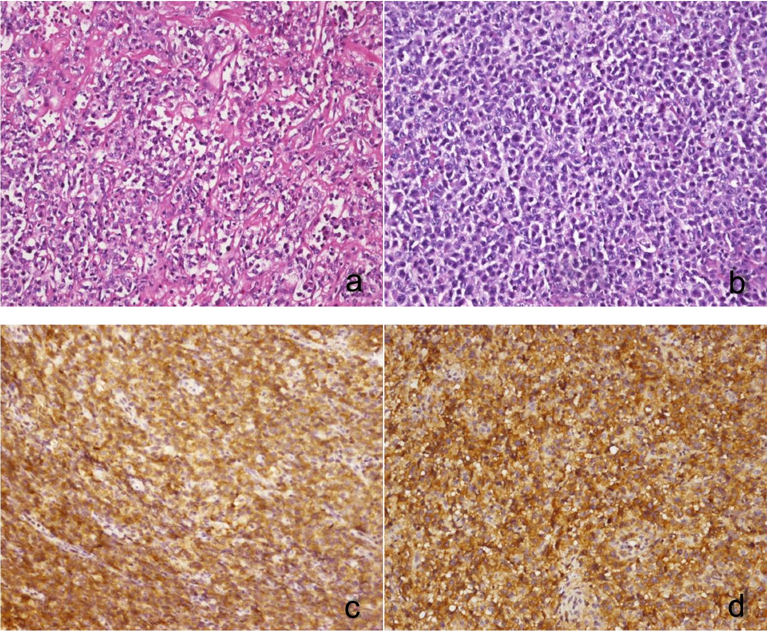
To enhance the diagnostic accuracy of determining PTEN status by immunohistochemistry, we explored potential surrogate markers for PTEN loss in prostate cancer. We focused on the activation of the PI3K-AKT pathway using S473-pAKT immunohistochemistry, which is indicative of PTEN loss [31, 32]. In a cohort of 20 prostate cancer cases, we found that S473-pAKT demonstrated a sensitivity of 77.8% and specificity of 90.9% for detecting PTEN loss. Consequently, S473-pAKT may serve as a surrogate marker for PTEN loss in cases where PTEN immunohistochemistry results are equivocal. Importantly, the sensitivity was markedly decreased when tested on samples from radical prostatectomy specimen, because being a phosphoprotein, S473-pAKT is an unstable protein modification that is sensitive to prolonged cold ischemia time. Therefore, short times to fixation is of paramount importance in the correct evaluation of S473-pAKT. In addition, we tested CD24 and GP2 expressions as additional immunohistochemical surrogate markers for PTEN loss. However, CD24 expression was heterogenous and showed a rather low specificity compared to S473-pAKT immunohistochemistry. GP2 expression was only found focally in one case of PTEN loss. Thus, S473-pAKT but not CD24 or GP2 may serve as an additional marker in cases where PTEN immunohistochemistry results are unclear.
Strengths of this study are its participants that reflect a representative fraction of academic pathology institutes in Germany and the deliberate selection of low-grade prostate cancer cases—exactly the group in which prognostic biomarkers are most urgently needed. Weaknesses of this study are its small to medium cohort size (n = 90), the use of a tissue microarray which limits the amount of tumor that can be studied, and the lack of follow-up data.
In summary, this ring trial investigates the variability of PTEN immunohistochemistry in low- to intermediate-risk prostate cancer. The data recommends that every institute should establish a PTEN immunohistochemistry protocol in close correlation with PTEN genomics and genetics. It also indicates that only a complete loss of immunoreactivity is diagnostic of a PTEN loss, as even the faintest positivity, that may be perceived as background, is indicative of a retained PTEN status. In addition, in equivocal cases, S473-pAKT immunohistochemistry may serve as an ancillary surrogate marker for PTEN loss. Regular interlaboratory ring trials are necessary to further improve the reliability and reproducibility of the PTEN assay.


Comments (0)