In this study, men had a greater OV than women (Table 1). A study using computed tomography [19] reports that the OV was greater in men than in women. In the present study, there was no sex-related difference in the GV, but the OV was greater in men, which resulted in a greater GOR in women. This indicates that women are more prone to globe and orbit imbalances. AL did not differ between sexes, but the EAR was closer to a long sphere-like shape with a lower EAR in women because of their lower ED. This is supported by Pope et al. [20] reporting that emmetropic and myopic eyes are spherical in men and assume the shape of a long sphere with the ocular axis as the long axis in women.
Furthermore, AL in highly myopic adult patients, particularly those with staphyloma, tends to increase with age [21]. In comparing age, older patients had a longer AL, but there was no age-related difference in ED, which resulted in a smaller EAR in the older group (Table 2). This indicates that older patients tend to have longer spheres in the long axis direction in strabismus cases with high myopia.
If the AL (major axis) of a long sphere is the same as the AL of a sphere, the equatorial length (minor axis) of the long sphere is smaller than of the corresponding sphere. If an extension of a globe in the direction of the AL causes the posterior part of this globe to exit the muscular cone toward the orbital wall, it can be inferred that dislocation from within the muscular cone is easier when the equatorial length is smaller for eyes with the same AL. A long sphere-like shape, which is anatomically more likely to cause dislocation from the muscle cone and which is characteristic of older patients and women, may contribute to the high prevalence of highly myopic strabismus in older adults and women [4].
Comparisons among the three groups of strabismus types show significant differences in the amount of horizontal strabismus, ED, EAR, and DA, but not age, AL, GV, OV, and GOR (Table 3). In particular, the ET group had a smaller ED and EAR than the NES group, i.e., the globe was more elongated in the direction of the major axis. Thus, ED and EAR might be factors determining the strabismus type.
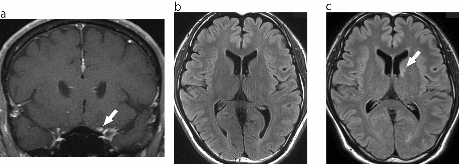
Detailed analyses of the EAR in the RS group show that women, who comprised 80.0% (12 eyes) of the RS group, had a mean EAR of 0.87 ± 0.08, and older women, who comprised 66.7% (10 eyes) of the RS group, had a mean EAR of 0.85 ± 0.08, both of which were smaller than the mean EAR (0.89) for the whole RS group. A small EAR increases the risk of developing strabismus with ocular motility disorders during aging even in the absence of ocular motility limitations at a younger age; female sex is an additional risk factor. In contrast, the two eyes of the elderly RS group had EARs of approximately 1.0. It is interesting to note that both ellipsoids and spheres may lead to the development of restrictive strabismus. For easier calculation of EAR (i.e., the ratio ED/AL) in clinical practice, it is recommended to use the most reliable variables of the following: the ED of the slice with the largest cross-section of the eye in coronal sections, the maximum ED and AL (Fig. 1, ED, AL) in axial or sagittal sections, and the AL determined with an AL-measuring device because the cross-section of the eye in coronal images is often oval in high myopia.
An imbalance between eye size and OV (i.e., a larger GOR) can cause compression of the external ocular muscles by the eye [22] and abnormal positioning of the external ocular muscles [23], resulting in oculomotor disturbances and strabismus. In HES, characterized by progressive restrictive esotropia and hypotropia among strabismus types with high myopia, the imbalance between eye size and OV is often evident on MRI [22]. Although the mean esodeviation was larger in the order of RS, ET, and NES, the variables GV, OV, and GOR were not significantly different among these groups. Thus, not only the progressive imbalance between eye size and OV but also other etiologies and facilitating factors, such as the weakening of the orbital connective tissue including the orbital pulley [11, 24] due to age-related changes and mechanical compression and changes in eye shape due to AL elongation, may lead to an increased strabismus angle and eye movement disorders. The mean esodeviation of the RS group was greater than the ET group, but this difference was not significant. The alternate prism cover test was not performed in patients with large angle esotropia in the RS group, so these data were missing, which may be the reason for the nonsignificant difference. Considering these factors, esodeviation would have increased in the order of NES, ET, and RS group.
The ET group had a greater DA than the NES group, but no difference was found between the ET and RS or NES and RS groups. Therefore, the RS group was divided into two subgroups: an RS-mild subgroup consisting of 12 eyes with mild abduction restriction (grade −1) and an RS-severe subgroup consisting of one eye with severe abduction restriction (grade −3) and two eyes with previous restrictive strabismus surgery. The ranges for the 12 eyes in the RS-mild subgroup were 0.83–1.02 for EAR and 0.31–0.46 for GOR. The ranges for the three eyes in the RS-severe subgroup were 0.74–0.82 for EAR and 0.39–0.40 for GOR; the EAR values were smaller than the mean values of the NES and ET groups, whereas the GOR values were larger than the mean. In the RS-severe subgroup, DA (175.9°) was greater than that of the ET group, although only one eye had an abduction restriction of grade −3. Although DA is used in clinical practice to quantify posterior ocular prolapse and is essential for the diagnosis and treatment of high myopic strabismus, our study showed no DA difference between the RS group and the other two groups. We consider three reasons: 1) the overall number of patients was small; 2) in the RS group, 80% of the eyes had mild abduction restriction (grade −1), and there were few patients with severe abduction restriction; and 3) the inclusion of 11 (37.9% of the total) patients with SES [17, 18]—1 (14.3%) and 10 (76.9%) in the NES and ET groups, respectively. In SES, an LR inferior shift is observed, therefore, DA is calculated larger even without SR nasal shift [3, 17], which is a characteristic change of HES. Therefore, the DA of the ET group, which had a higher SES frequency, might have been greater than the NES group, which had a lower SES frequency, and not significantly different from that of the RS group.
The characteristics of these disease types can be summarized as follows. The NES group had a more spherical ocular shape, whereas the ET group had a more long-spherical shape and a higher frequency of SES; the EAR was smaller and the DA larger in the ET group than in the NES group. EAR, DA, and GOR did not differ among the RS with mild restrictive strabismus, ET, and NES groups, but the EAR was smaller and GOR and DA were larger in the RS with severe restrictive strabismus group than in the ET and NES groups. When clinically evaluating ocular motility limitations associated with posterior prolapse of the globe in strabismus with high myopia, attention should be paid not only to the DA value but also to the presence or absence of disproportion between the globe and orbit, shape of the globe, degree of inferior deviation of the LR muscle, nasal deviation of the SR muscle, and pressure findings of the globe on the external ocular muscle, among others.
In all eyes with strabismus and myopia, as well as the three strabismus type groups, we examined the relationships between EAR, GOR, and DA (Figs. 3, 4 and 5, Table 4) and found that EAR was correlated with DA and GOR in the entire study population and even stronger correlated in the RS group. In the ET group, EAR was correlated only with GOR, whereas EAR and DA were not correlated. Only in the NES group, EAR and GOR were not correlated. With increasing AL, GOR increases and EAR decreases, leading to a negative correlation between EAR and GOR. Thus, AL may be related to the ET and RS, but not to the NES, groups. However, DA and GOR were not correlated in the entire study population and in the groups by disease type.
In conclusion, in the evaluation of strabismus with myopia, especially restricted strabismus, EAR (evaluation of eye shape), which correlates with DA, may be more useful than GOR (evaluation of imbalance between GV and OV), which does not correlate with DA.
This study had some limitations. First, the study was retrospective, and the number of cases was small. Second, two patients with a history of strabismus surgery were not excluded from the analysis, which may have biased the evaluation of eye movements and the analysis results. Third, AL was measured using different methods and apparatus depending on the participants. Since the EAR was calculated using ED and AL, measurement errors in the value of AL could have induced errors in the EAR. Finally, because esotropia and cyclovertical strabismus with myopia, especially in middle-aged and older patients, would include patients with HES or SES, comparing the rectus muscle pulley positions and LR-SR band among groups is important [3]; however, this was not investigated.
Among patients with strabismus and high myopia, older and female patients with ET have a smaller EAR and longer sphere shape with AL as the axis of rotation. The EAR, which can be measured relatively more easily than GV, might be a useful index for evaluating the pathogenesis of strabismus associated with high myopia using MRI. In patients with late-onset strabismus and high myopia, a small EAR increases the risk of developing strabismus with ocular motility disorders during aging, even in the absence of ocular motility limitation at present; female sex is a further risk factor.

Comments (0)