The appearance or the severity of the clinical symptoms of children with ASD is primarily affected by the size of defect. In infancy, the thickness of left and right ventricular wall is similar, with a minimal difference in pressure, meaning that the left-to-right shunt is not very pronounced and generally does not produce any noticeable clinical signs. With age, the compliance of the right ventricle wall increases, the pressure of the systemic circulation increases, and the horizontal shunt increases, resulting in pulmonary congestion and reduced amount of blood in the systemic circulation. Pneumonia is a common affliction of children with ASD, causing retardation in their development. As family living conditions improve, parents may not take into account the children’s slow weight gain and excessive sweating. Finding these signals is challenging, most people with ASD can still lead a normal life when they reach adulthood.
In recent years, there has been an upsurge in the amount of children in outpatients clinics experiencing chest tightness, sighing, chest pain and other primary indicators, and chest DR has become a typical initial diagnostic tool in children, which is straightforward and economically sensible. This examination provides significant insight into pediatric chest pathology. Nonetheless, analyzing children’s chest X-ray images can be challenging. In addition to lung imaging, physicians are more attuned to the shape of the heart shadow with more recognizing experience [11]. Echocardiography has often been used to screen for ASD from abnormal performers. For many children with ASD, there are no visible changes in the hemodynamic system, and chest radiographs usually appear normal. However, other physicians who are not cardiovascular experts or those with lack experience may have difficulty identifying any abnormal cardiac changes on chest radiographs. In addition, echocardiography is expensive, as routine screening for chest abnormalities is difficult to accept by ordinary families in developing countries, resulting in delay in diagnosis for children with ASD. Machine learning, especially deep learning, presents a great opportunity to improve medical imaging, expediting diagnostics and normalizing interpretation. Based on what we know now, this is the first time deep learning has been demonstrated that could effectively assist in the early detection of ASD in children through chest radiographs in our study.
Utilizing CNN is a common approach of constructing a deep learning system. CNN can accumulate data from each component of the picture and have potent feature learning aptitude. ResNet has become a well-known deep learning model for image classification because of its capacity to overcome the vanishing gradient problem, which is often encountered during training of traditional convolutional neural networks (CNN) using residual mapping. ResNets is utilized to draw out image features sans interference from shape, rib and other related noise. The ResNet mechanism has also been incorporated into more machine learning models, greatly improving the model ability to identify different types of lung diseases with chest X-rays.
In the past 5 years, there has been a rapid expansion in the application of artificial intelligence technology in examining chest X-ray images, the majority of which have been concentrated on adults, and its application in pediatric is still scant [12]. It is common knowledge that the imaging manifestations of children’s chest will alter throughout their normal growth and development. Physiological modifications in chest wall, lung, ossification center, and thymus width in children. Pathological issues of distinctive deformities such as congenital pulmonary cystadenoma. The above issues present additional challenges in the development of artificial intelligence models based on chest X-ray images for identifying lung diseases in children [13].
Luo et al. [14] showed that a new model based on YOLOv5 and ResNet-50 to identify abnormal chest radiographs, mean average precision (mAP) was 0.010, 0.020 and 0.023 higher than the mAP values of YOLOv5, Fast RCNN and EfficientDet models, respectively. And, the precision of the new model were 0.512, 0.018, 0.027 and 0.033, higher than YOLOv5, Fast RCNN and EfficientDet. The classification accuracy of the training model ResNet-50 based on CNN for lung cancer, pneumothorax, tuberculosis, pneumonia and other chest diseases was 96.15%, and that of Vgg-19 and Inception V3 were 95.61% and 95.16%, respectively [15].
Because the heart is positioned and shaped in a relatively stable manner within the chest cavity, it is less affected by the development of the lungs and bones. Thus, future research on the use of artificial intelligence technology for the diagnosis of chest X-ray images will concentrate primarily on the cardiovascular diseases [16]. Through the use of deep learning, research into cardiovascular diseases has been done by evaluating chest X-rays, such as automatic detection of cardiomegaly, valvular disease and cardiac function, classification of pulmonary hypertension and atrial fibrillation and prediction of pulmonary to systemic flow ratio in patients with congenital heart disease [6, 17,18,19,20]. The findings of this research point to the potential of deep learning-based methods to objectively and quantitatively evaluate cardiac morphology and pulmonary circulation blood volume as seen on chest X-rays, allowing for the distinction or diagnosis of heart conditions. Ueda et al. [21] first explored research on create and validate ResNet-50 for mitral regurgitation through chest radiographs recognition. The area under the curve, sensitivity, specificity, accuracy, positive predictive value and negative predictive value of the artificial intelligence model were 0.80 (95% CI: 0.77, 0.82), 71% (95% CI 67, 75), 74% (95% CI 70, 77), 73% (95% CI 70, 75), 68% (95% CI 64, 72) and 77% (95% CI: 73, 80), and the model has the potential to distinguish between patients with and without mitral regurgitation by means of the areas of the left atrium, left ventricle, and superior vena cava in chest radiographs [21]. Lee et al. [8] used ResNet-18 model to identify chest radiographs to diagnose acute thoracic aortic dissection. The accuracy was 90.20%, the precision was 75.00%, the recall was 94.44% and the F1-score was 83.61%, the model offers excellent performance and is able to accurately detect acute thoracic aortic dissection based on plain chest radiography.
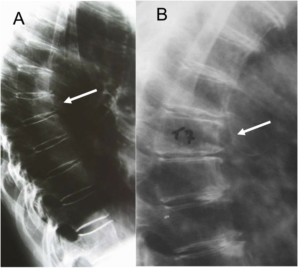
In children with ASD, due to increased left-to-right shunt, the right cardiac preload increases, leading to enlargement of the right atrium and right ventricle, increased pulmonary circulation blood volume and pulmonary artery dilation. Chest radiographs demonstrate a more prominent cardiac shadow, which can be observed as a higher cardiothoracic ratio. It is generally observed that the right side of the heart is more saturated in children with ASD. In the eventuality of pulmonary congestion, the pulmonary artery section is conspicuously projecting, the hilar shadow is dilated, the lung texture is bloated and the vascularity is particularly pronounced in the lower right lung [2]. Consequently, with the modifications in the characteristics of ASD chest radiographs, our study is bringing in artificial intelligence technology to upgrade the recognition accuracy of chest radiographs and serve as a premise for echocardiography, thus enabling an accurate diagnosis of ASD as quickly as possible without raising the financial cost for the family. With the comparison of a variety of models, this study revealed that the recognition accuracy of the ResNet-10t was exceptionally high, reaching 92% after training, notably better than other models. In this study, the Grad-CAM visual perception hotspot analysis of the ResNet-10t is carried out, and it was found that the local features capturing the model focused on the cardiac margin right and the pulmonary arterial segment of the heart shadow, which further confirms the high credibility of the model performance.
Limitations
All participants were over 1 year of age, mainly because of the substantial heart shadow in the first year of life and the cardiothoracic ratio of 0.55 in the chest radiograph diagnostic criteria. Children diagnosed with ASD in this age group tend to have large defects, typical clinical manifestations and imaging manifestations, potentially leading to an erroneous boost in the accuracy of the learning model. Thus, this study shall encompass this age group in the foreseeable future, and it is necessary to adjust the learning model algorithm or develop a new learning model directed to differentiate. The thymus is a significant lymphatic organ located in the front middle part of the chest cavity that grows throughout childhood and peaks in adolescence. Consequently, the thymus shadow in the chest radiographs is a soft tissue density with a smooth border, making it hard to separate from the heart shadow. This investigation is uncertain of the composition of thymus interference factors in image recognition, improving the accuracy of recognition is a main focus of the following research.
In conclusion, the ResNets model is feasible for identifying ASD through children’s chest radiographs. The ResNet-10t in this study was more effective than the main detection models when used on chest DR images of ASD. In subsequent studies, transformer models will be designed based on this model to expedite the identification and examination of chest DR images of ASD across all ages, which has great clinical and societal value.


Comments (0)