Long-Term Radiologic Healing After Enucleation of a Large Dentigerous Cyst Associated with the Mandibular Second Molar in a Child
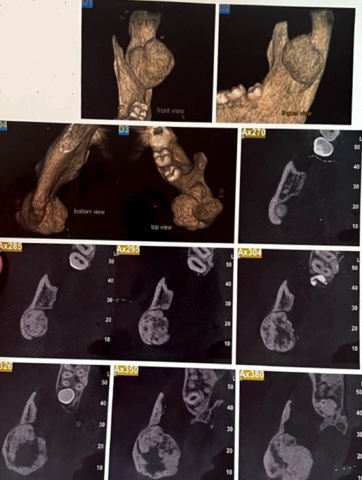
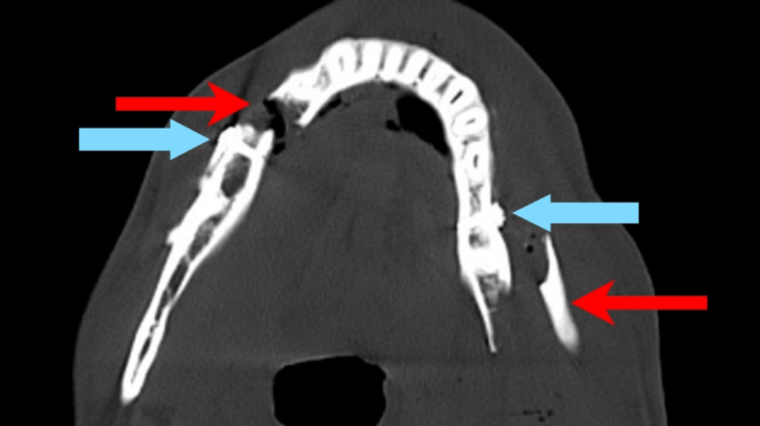
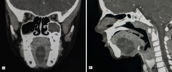
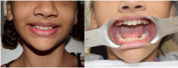
Dentigerous cysts most commonly involve mandibular third molars, whereas association with mandibular second molars in children is uncommon but documented in literature. Extended radiologic documentation of postoperative bone regeneration and inferior alveolar nerve (IAN) canal repositioning in such cases is infrequently illustrated. We report an 11-year-old girl who presented with a large unilocular dentigerous cyst in the left posterior mandible measuring 34 × 32 × 26 mm, associated with an unerupted mandibular second molar and accompanied by cortical thinning and inferior displacement of the IAN canal. Panoramic radiography and cone-beam computed tomography (CBCT) established the diagnosis and, importantly, guided surgical planning by defining the three-dimensional extent of the lesion, the degree of cortical thinning, the relationship to the IAN canal, and the eruptive prognosis of the involved teetth. Based on lesion size, eruption potential, marked bony expansion, and the need for a definitive single-stage approach, complete enucleation was performed under general anaesthesia. Histopathological examination confirmed a dentigerous cyst. Serial radiographic follow-up at 3, 6, 12, 24, and 36 months demonstrated progressive trabecular bone fill, cortical consolidation, complete osseous regeneration, absence of recurrence, and gradual radiologic repositioning of the IAN canal toward normal symmetry, without postoperative neurosensory deficit. This case highlights the regenerative capacity of the paediatric mandible and supports carefully planned enucleation as a predictable treatment option in selected large mandibular dentigerous cysts when eruptive prognosis is poor.

Comments (0)