
Remember me
Background:
Methyl acetate (MA) is a common industrial solvent that causes rapid blindness in large exposures. Its toxicologic mechanism is not fully elucidated currently. The currently used clinical marker for MA poisoning, formic acid, is unable to differentiate between MA exposure and methanol exposure, which hinders accurate diagnosis and exposure source tracing, and impairs the development and implementation of front-end preventive and control measures.
Objective:
This study utilized a cross-species, untargeted metabolomics approach, combining data from animal models and human cohorts, aiming to identify potential biomarkers for MA poisoning and provide new insights into its toxicological mechanisms.
Methods:
Subacute poisoning rat models of MA and methanol were established via gavage administration (n = 6 per group) and urine samples were collected. Meanwhile, 8 occupationally exposed MA-intoxicated patients and 10 healthy controls were enrolled, with their urine samples also being collected. All samples underwent untargeted metabolomic analysis using UPLC-QTOF/MS for comparative profiling among MA-exposed rats versus control rats, MA-exposed rats versus methanol-exposed rats, and MA-exposed patients versus healthy controls.
Results:
A total of 41 and 16 significantly altered metabolites were identified in MA-exposed rat models and occupationally exposed human subjects, respectively. Pathway enrichment analysis further revealed key pathways including the tricarboxylic acid (TCA) cycle, purine metabolism, glutathione metabolism, cysteine and methionine metabolism, and one-carbon metabolism, suggesting conservation of MA-induced toxic responses across species. These results indicate that MA toxicity involves not only classical TCA cycle inhibition but also close association with systemic oxidative stress. 20-carboxy-leukotrieneB4 (20-COOH-LTB4) and S-adenosylhomocysteine (SAH) were significantly elevated in the MA exposure group in both rat and human samples, but were not detected in the methanol exposure group, showing high specificity and cross-species conservation.
Conclusion:
This study reveals MA toxicity mechanism via oxidative stress, aids in developing therapies and enhancing MA exposure risk management. And the study identifies 20 - COOH - LTB4 and SAH as potential and sensitive biomarkers for MA intoxication, offering a tool for differentiating MA from methanol exposure clinically.
1 IntroductionMA is a green organic ester solvent with both lipophilic and hydrophilic properties. Owing to its low volatility, excellent solubility, and compliance with volatile organic compound (VOC) emission limits, MA is widely used in industrial and consumer fields such as coatings, resin processing, pharmaceuticals, and cosmetics (Huss et al., 2003; Heldreth et al., 2012). China is a major global producer and consumer of MA, with an annual production capacity exceeding one million tons (Jin, 2019). MA is also naturally released during wood processing, further expanding the exposure pathways for occupational populations (Bleich et al., 1998; Qin et al., 2021). With the expansion of its applications, the risk of MA exposure has become increasingly prominent, and poisoning incidents occur frequently. Occupational exposure is the primary route. Workers exposed to high concentrations of MA-based adhesives have developed severe symptoms including metabolic acidosis and visual impairment, with exposure levels at implicated sites far exceeding safety limits (Li et al., 2024). Multiple occupational poisoning incidents have been reported in workshops across various regions (Tang et al., 2021; Lyu et al., 2025). Long-term exposure has also been associated with malignant nasal tumors (Bleich et al., 1998). Additionally, acute exposure can cause blindness (Ogawa et al., 1988), and even accidental ingestion of small amounts in daily life can lead to adverse effects (Minns et al., 2013). Given its diverse exposure pathways, both occupational and general populations face health risks, making MA toxicity a public health issue requiring urgent attention.
Toxicokinetic studies have confirmed that MA is rapidly hydrolyzed to methanol (ME) upon entering the body. ME is further oxidized to formaldehyde and finally converted to formic acid (Kazunori et al., 1992; Heldreth et al., 2012; Nekoukar et al., 2021; Liberski et al., 2022). Both clinical case reports (Ogawa et al., 1988; Minns et al., 2013; Li et al., 2024) and animal experiments (Liesivuori et al., 1987) have verified that the accumulation of formic acid is the key mediator of toxicity, leading to metabolic acidosis and optic nerve damage. Consequently, blood and urinary levels of ME and formic acid are widely used as biomarkers for MA exposure, as with methanol exposure. Of critical importance, the misclassification between MA poisoning and methanol (ME) poisoning represents a substantial and underrecognized challenge in both clinical management and public health risk control. Clinically, MA and ME poisoning present with highly overlapping manifestations, and their core treatment regimens are largely analogous. Importantly, while such misclassification does not markedly compromise immediate therapeutic efficacy, it severely impairs accurate diagnosis and exposure source tracing. This confusion significantly obstructs the development and implementation of targeted front-end preventive and control measures specific to MA or ME exposure. Failure to accurately distinguish the true exposure source not only obscures the genuine epidemiological characteristics of MA poisoning but also limits the capacity to carry out targeted interventions for specific risk factor elimination, thereby posing a sustained threat to public health security. Compounding this dilemma, current diagnostic practice relies exclusively on blood and urinary levels of ME and formic acid as biomarkers for both forms of poisoning. These shared metabolic indicators lack the specificity to differentiate whether the toxic exposure originates from MA or ME, thereby exacerbating diagnostic ambiguity and perpetuating challenges in precise exposure attribution. Furthermore, these indicators only reflect exposure status and cannot reveal the molecular mechanisms of MA toxicity or support early warning (Kazunori et al., 1992; Nekoukar et al., 2021). Most existing research is limited to the linear metabolic pathway (MA → ME → formic acid), with insufficient investigation into how MA exposure reprograms broader metabolic networks involving carbohydrates, lipids, and amino acids. These gaps have constrained a comprehensive understanding of MA toxicity mechanisms and hindered the development of highly specific biomarkers.
This study established a MA intoxication model in Sprague-Dawley (SD) rats and performed untargeted metabolomics analysis on urine samples from the model rats. From the perspective of metabolic pathway disturbance, we explored the toxic mechanism of MA exposure, and screened potential biomarkers that could distinguish MA intoxication from methanol (ME) intoxication. We then conducted biochemical experiments to validate the key metabolites involved in dysregulated metabolic pathways. Furthermore, we performed urine metabolomics verification using an independent cohort of humans with occupational MA exposure. These findings address the limitations of conventional indicators (methanol and formic acid) that fail to discriminate MA and ME exposure, and offer novel molecular support for the early diagnosis of MA intoxication, exposure source tracing, and the refinement of health risk assessment systems.
2 Materials and methods2.1 Chemicals and reagentsMethyl acetate (purity ≥99%) was purchased from MACKLIN Reagent Co., Ltd. (Shanghai, China). Olive oil (pharmaceutical grade) was obtained from Innochem Technology Co., Ltd. (Beijing, China). Methotrexate was purchased from AbMole Biotechnology Co., Ltd. (Shanghai, China). Methanol (LC-MS grade) was acquired from Anpel Experimental Technology Co., Ltd. (Shanghai, China). Distilled Water was supplied by Guangzhou Watson’s Food & Beverage Co., Ltd. (Guangzhou, China).
2.2 Animals and treatmentThirty 10-week-old healthy male Sprague Dawley rats were obtained from the Guangdong Institute of Biotechnology (China). After 1 week of adaptive feeding, 30 rats were randomly divided into 5 groups (n = 6 per group): a control group, low-dose MA exposure group, medium-dose MA exposure group, high-dose MA exposure group, and the methanol exposure group. To model human formic acid metabolism, all rats received daily intragastric administration of methotrexate (0.3 mg/kg) for 7 days to induce a low-folate state. Following model establishment, from day 8–10, the control group received olive oil daily, while the low-, medium-, and high-dose MA exposure groups were administered MA in olive oil at 2, 3, and 4 g/kg/day, respectively. The methanol group received an aqueous methanol solution at 1.5 g/kg/day. On day 10, following the final administration of MA, methanol, or olive oil (control), rats were returned to their regular cages for routine feeding and housing. At 24 h post the final dose (i.e., on day 11), urine samples were collected manually as single spot samples from each rat immediately after voiding. The collected urine was promptly aliquoted into pre-labeled cryovials and stored at −80 °C. Following urine collection, rats were deeply anesthetized with isoflurane. Blood samples (both anticoagulated and coagulated) were collected via abdominal aortic puncture. Rats were then dissected on ice, and eyeballs were promptly harvested, rinsed with ice-cold PBS (pH 7.4), flash-frozen in liquid nitrogen, and stored at −80 °C. All procedures complied with national regulations on animal administration and were approved by the Ethics Committee of the Guangdong Institute of Biotechnology (Approval No. IACUC2025121).
2.3 Sample collectionUrine samples were collected from 8 patients with acute methyl acetate (MA) poisoning during the acute phase of hospitalization and 10 healthy occupationally - matched controls (Approval No. 2022104). All participants were recruited from the same workplace. Urine samples were collected from patients immediately upon hospital admission and prior to any therapeutic intervention. For both patients and controls, samples were collected during working hours (non-first-morning voids) to control for circadian variations. The samples were immediately aliquoted and stored at −80 °C until subsequent analysis.
2.4 Experimental methods2.4.1 Determination of methanol and formic acid concentrationsEach sample (plasma or eyeball homogenate) was derivatized by mixing with the derivatization reagent (10% sulfuric acid, 20% isopropanol, 70% pure water) at a 1:1 volume ratio. Gas chromatography-mass spectrometry (GC-MS) analysis was conducted using an Agilent 7890B GC system interfaced with an Agilent 5977B quadrupole MS detector. The system was equipped with an automatic headspace sampler and a RESTEK Rxi-624Sil capillary column (60 m × 250 μm × 1.4 μm). The injector was operated at 180 °C in split mode with a 10:1 split ratio. The column temperature was programmed as follows: initial temperature 45 °C held for 6 min, increased to 210 °C at 30 °C/min, and held for 0.5 min. The mass spectrometer was operated in electron impact (EI) mode with the ion source temperature set to 230 °C. Data were acquired in both full-scan (m/z 10–350 amu) and selected ion monitoring (SIM) modes. Characteristic ions and specific time windows were optimized for methanol, ethanol, formic acid, and acetic acid. The analytical method for determining methanol and formic acid concentrations via HSGC-MS was detailed in our earlier work (Wu S. et al., 2024).
2.4.2 Pathological H&E stainingThe right eyeball of each rat was rinsed with PBS and immediately fixed in a dedicated FAS eye fixative (protected from light) for 48 h. The fixed tissues were embedded in paraffin and sectioned consecutively at a thickness of 4 μm using a microtome. The sections were baked at 60 °C for 2 h to enhance adhesion. Conventional hematoxylin and eosin (H&E) staining was performed according to standard procedures. Briefly, after dewaxing and rehydration, the sections were stained with hematoxylin, differentiated, blued, and counterstained with eosin. Finally, the sections were dehydrated, cleared in xylene, and mounted with neutral gum. Staining results were examined under an optical microscope, with nuclei appearing blue and the cytoplasm red.
2.4.3 MetabolomicsThe frozen samples were thawed at 4 °C. An aliquot of 200 μL was then combined with 800 μL of pre-cooled methanol (−20 °C) and mixed gently. The mixture was centrifuged at 12,000 rpm for 10 min at 4 °C to precipitate proteins. A volume of 150 μL of the clear supernatant was transferred to a vial for subsequent detection. A quality control (QC) sample was generated by pooling equal aliquots (50 μL) of the supernatant from every sample. Chromatographic analysis was conducted using an Agilent 1,290 Infinity II liquid chromatography system. Separation was carried out on a Poroshell 120 EC-C18 column (3.0 × 150 mm, 2.7 μm) maintained at 40 °C. The mobile phase was composed of (A) water containing 0.1% formic acid and (B) acetonitrile, with a flow rate of 0.400 mL/min. The injection volume was 3.0 μL. The reversed-phase chromatographic separation employed the following gradient program: 0–1.5 min, 5% B; 1.5–15 min, 5% → 55% B; 15–20 min, 55% → 95% B; 20–30 min, 95% B; 30–30.1 min, 95% → 5% B; 30.1–35 min, 95% B. Mass spectrometric analysis was performed on an Agilent 6545B Q-TOF spectrometer equipped with a Dual AJS electrospray ionization (ESI) source operating in positive ion mode. The capillary voltage and fragmentation voltage were set to 3500 V and 140 V, respectively. Ion source parameters were as follows: drying gas temperature 350 °C (flow rate 8 L/min); sheath gas temperature 350 °C (flow rate 2 L/min); nebulizer pressure 40 psig. The all-ions MS/MS acquisition spanned m/z 50–1,100 at collision energies of 0, 10, and 40 eV. A quality control (QC) sample was analyzed after every 6-8 experimental samples to monitor instrument stability and reproducibility.
2.5 Determination of plasma TNF-α, IL-1β and MDA levelsInflammatory cytokine levels in plasma, including TNF-α and IL-1β, were measured using ELISA kits (Rat IL-1β ELISA Kit, Cat. No. AD3032Ra, AndyGene Co., Ltd.; Rat TNF-α ELISA Kit, Cat. No. AD3238Ra, AndyGene Co., Ltd.) following the manufacturer’s protocols. Meanwhile, malondialdehyde (MDA) content in plasma was detected using ELISA kits (Rat SOD ELISA Kit, Cat. No. AD2872Ra, AndyGene Co., Ltd.) in accordance with the manufacturer’s guidelines.
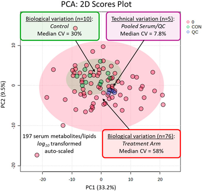
2.6 Statistical analysisThe normality of data distribution was assessed via the Shapiro-Wilk test, and variance homogeneity via the Brown-Forsythe test. Statistical analyses were tailored to data types: continuous variables were analyzed with t-tests/ANOVA (one-way ANOVA and Bonferroni for normal/homogeneous data, Welch ANOVA and Games-Howell for normal/heterogeneous data, Kruskal–Wallis H and Dunn for non-normal data); categorical rates used Fisher’s exact test; correlations used Spearman’s rank correlation test. For the LC-QTOF-MS data processing, raw data were first processed using Agilent MassHunter Profinder software to perform initial peak detection, feature extraction, and reverse peak alignment. The resulting data matrix was processed in Mass Profiler Professional (MPP) for retention time alignment, and 75th percentile shift normalization was subsequently applied to account for potential variations in urine concentration/dilution across samples prior to statistical analysis. Strict quality control was applied to the processed data. Features were retained only if they were detected in more than 70% of the samples within at least one experimental group and demonstrated a coefficient of variation (CV) below 30% across the quality control (QC) samples.
Multivariate statistical analyses were employed to interrogate the metabolomic data. Unsupervised principal component analysis (PCA) and sample correlation analysis were first performed using MPP software to assess intrinsic data clustering and overall trends. Subsequently, supervised orthogonal partial least squares-discriminant analysis (OPLS-DA) was conducted in SIMCA to maximize group separation and identify candidate biomarkers. Differential metabolites were selected based on a fold change (FC) ≥ 2.0 and a p-value <0.05 from Student’s t-test. For metabolite identification, features were first queried against databases via IDBrowser using a mass accuracy threshold of 50 ppm. Putative identifications were then confirmed by comparing experimental MS/MS spectra against public databases (KEGG, HMDB, MassBank) with a mass tolerance of 50 ppm, using MassHunter Qualitative Analysis. Pathway enrichment analysis of the confidently identified metabolites was performed using MetaboAnalyst (version 6.0), with a p-value <0.05 deemed significant. Finally, The diagnostic efficacy of the potential biomarkers was assessed using receiver operating characteristic (ROC) curve analysis, with the area under the curve (AUC) quantifying their discriminatory power.
3 Results3.1 Rat body weight, methanol/formic acid levels in blood and eyeballs, and retinal histological changesAfter 3 days of MA exposure, there was no statistically significant difference in body weight gain between MA-exposed groups (low-dose LMA, medium-dose MMA, high-dose HMA) and the control group, but a decreasing trend was observed in the MA-exposed groups (Figure 1A). Plasma analysis showed that methanol concentrations in all MA-exposed groups (LMA, MMA, HMA) were significantly higher than that in the control group. Among them, the methanol concentration in the MMA group was significantly higher than that in the LMA group, while no statistically significant difference was found between the HMA and MMA groups, despite a numerical increasing trend (Figure 1B). For formic acid levels, only the MMA and HMA groups were significantly higher than the control group, with no significant difference between the LMA group and the control group (Figure 1C). Descriptive statistics only were performed for methanol and formic acid contents in eyeball tissues, with no intergroup significance testing conducted. Observational results showed that methanol content in the eyeball tissues of the MMA and HMA groups was higher than that in the control and LMA groups (Figure 1D), while formic acid content in the eyeball tissues of all MA-exposed groups was higher than that in the control group (Figure 1E). Both metabolites exhibited a clear increasing trend with the elevation of exposure dose. Additionally, histological analysis of retinal tissues via H&E staining revealed obvious abnormalities (Figure 1F). Under 200× magnification, fields of view were randomly selected for cross-group comparison to evaluate gross morphological changes, while 800× magnification revealed marked structural alterations in the retinas of MA-exposed animals compared with the intact, well-organized structure in controls. These changes were characterized by cytotoxic edema, manifested as swollen ganglion cell bodies with vacuolated cytoplasm; densely packed nuclei in the inner and outer nuclear layers with loss of intercellular gaps; and compaction of the photoreceptor inner and outer segments, which lost their normal loose organization. Notably, the methanol group (ME) exhibited similar changes to the MMA group in all above indicators.

General toxicological indicators in rats after methyl acetate exposure. (A) Body weight change from Day 1 pre-exposure to Day 4 post-exposure. (B) Log-transformed plasma methanol concentration (log(methanol concentration +1)). (C) Log-transformed plasma formic acid concentration (log(formic acid concentration)). (D) Methanol concentration in eyeball tissues. (E) Formic acid concentration in eyeball tissues. (F) Histopathological analysis of retinal tissues. The first row displays a 200× view of the overall retinal structure. The second row presents an 800× magnified view of the photoreceptor layer. Black arrows: normal ganglion cells; red arrows: swollen cells with cytoplasmic vacuolation. Data are shown as mean ± SD. Sample sizes: n = 6 for Con, LMA and ME, n = 4 for MMA and HMA (A–C); n = 3 for Con, LMA and ME, n = 2 for MMA and HMA (D–E). *p < 0.05, **p < 0.01, ***p < 0.001, vs. Con; #p < 0.05, ##p < 0.01, ###p < 0.001, between exposure groups. Abbreviations: Con, control; LMA, low-dose methyl acetate; MMA, medium-dose methyl acetate; HMA, high-dose methyl acetate; ME, methonal.
3.2 Metabolomics analysis3.2.1 Total ion chromatogram (TIC) of QC samplesThe total ion chromatograms (TICs) of the quality control (QC) samples demonstrated highly overlapping peaks (Figure 2), indicating excellent instrumental stability and high reproducibility throughout the UPLC-QTOF analysis. To further confirm the reproducibility of the analytical method, the coefficient of variation (CV) was calculated for each detected signal across all QC samples. A large proportion of signals exhibited CV values within an acceptable range (below 30%), further supporting the good stability and reliability of the metabolomics platform. The detailed intensities and CV values of signals in QC samples are presented in Supplementary Table S1.

Representative TIC of QC samples.
3.2.2 Principal component analysis (PCA) and metabolic profile correlation analysisAlthough individual variations resulted in some dispersion within groups, unsupervised PCA clearly distinguished the MA group from the control group in both rat (Figure 3A) and human (Figure 3B) samples. Furthermore, Pearson correlation coefficients were calculated to assess the similarity of metabolic profiles across all samples in both species and visualized using heatmaps (Figures 3C,D). High correlations were observed among samples within each group (MA or control) in both species. In contrast, low inter-group correlations highlighted a clear distinction in metabolic profiles between MA-exposed and control conditions. These correlation-based findings are consistent with the PCA results across species, confirming that MA exposure induces significant and consistent alterations in metabolic profiles in both rats and humans. This indicates that the metabolic remodeling effect of MA is conserved across species.

Multivariate analysis of metabolic profiles in rat and human urine samples. (A,B) PCA score plots derived from the urine metabolome of (A) rats (n (MA) = 14, n (Con) = 6) and (B) humans (n (MA) = 8, n (Con) = 10). (C,D) Pearson correlation heatmaps for (C) rat and (D) human samples. The color gradient from blue (0, no correlation) to red (1, strong positive correlation) represents the magnitude of the correlation coefficient.
3.2.3 Orthogonal partial least squares discriminant analysis (OPLS-DA) and univariate analysisTo maximize the discrimination between the MA group and control group, based on metabolic profiles, orthogonal projections to latent structures-discriminant analysis (OPLS-DA) models were established. The resulting models clearly separated the MA-exposed group from the control group in both rat (Figure 4A) and human (Figure 4B) samples. To validate model reliability, 200 permutation tests were performed (Figures 4C,D). The validation results showed a negative y-intercept of the Q2 regression line, and all permuted Q2 values (left) were lower than the original point (right), confirming model robustness without overfitting. These validated models were subsequently used for differential metabolite screening.

Multivariate analysis and differential metabolite screening in rat and human urine metabolomes following MA exposure. (A,B) OPLS-DA score plots demonstrating distinct separation between the control and MA-exposed groups in (A) rats (n (MA) = 14, n (Con) = 6) and (B) humans (n (MA) = 8, n (Con) = 10). (C,D) Permutation test results (200 permutations) validating the OPLS-DA models for (C) rat and (D) human data. The original model’s actual values (right points) exceed all permuted R2 and Q2 values (left), confirming model robustness without overfitting. (E,F) Volcano plots visualizing significantly altered metabolites in (E) rat and (F) human samples. Metabolites are color-coded based on fold change (FC) and statistical significance (p < 0.05): red (significantly upregulated, FC ≥ 2.0), blue (significantly downregulated, FC ≤ 0.5), orange (marginally upregulated, 1.0 < FC < 2.0), light blue (marginally downregulated, 0.5 < FC < 1.0), and gray (non-significant, p ≥ 0.05).
Based on p-values from univariate t-tests and fold changes, a total of 1,602 (rats) and 815 (humans) significantly differential metabolites were screened using the criteria “p < 0.05, FC ≥ 2.0”. The distribution of these differential metabolites was visualized via volcano plots (Figures 4E,F). Further identification via database matching (HMDB, KEGG, MassBank) and MS/MS fragmentation analysis yielded 41 (rats) and 16 (humans) differential metabolites, with 10 metabolites listed for each group in Table 1 and the remaining ones provided in Supplementary Table S2.
Metabolite of ratMetaboliteKEGG IDLog2 FC3-SulfinylpyruvateC055274.005-OxoprolineC018794.00SuccinateC000424.00MethylglyoxalC005464.00DihydroxyfumarateC009754.005-AminolevulinateC00430−2.24L-homoserineC00263−2.11L-GlutamineC00064−1.86D-SerineC00740−1.81L-CysteineC00097−1.70Metabolite of humanMetaboliteKEGG IDLog2 FCCys-glyC014194.49HomocitrateC012514.00SarcosineC002134.0015-Keto-prostaglandin I2C048354.00DehydroalanineC022184.00S-adenosyl-L-homocysteineC000212.5120-COOH-leukotriene B4C059502.262-OxobutanoateC001091.492,8-DihydroxyadenineC225001.33CreatineC00300−1.98L-cystathionineC02291−4.52Differential metabolites identified in rats and humans following methyl acetate exposure.
3.2.4 Key differential metabolic pathways perturbed by MA exposureTo systematically characterize the metabolic alterations induced by MA exposure, KEGG pathway enrichment analysis was performed on differential metabolites in rat and human urine. The results revealed that MA-induced toxicity led to highly consistent disruptions in metabolic pathways between the two species (Table 2).
Metabolite of ratPathway nameHitRaw pFDRImpactPurine metabolism7/714.3258E-050.00255280.05548CystEine and methionine metabolism5/338.2461E-050.00255280.1396Glycine, serine and threonine metabolism5/349.5731E-050.00255280.28464One carbon pool by folate4/260.000446690.00893370.07357Pyruvate metabolism3/230.00418310.066930.10512Glutathione metabolism3/280.00737080.0842380.09925Alanine, aspartate and glutamate metabolism3/280.00737080.0842380.33734Glyoxylate and dicarboxylate metabolism3/320.0107350.107350.11333Arginine biosynthesis2/140.0172460.15330Pantothenate and CoA biosynthesis2/200.0341150.248110Citrate cycle (TCA cycle)2/200.0341150.248110.08276Metabolite of humanPathway nameHitRaw pFDRImpactCysteine and methionine metabolism4/335.0962E-050.00230180.26496Glycine, serine and threonine metabolism4/345.7546E-050.00230180.08874One carbon pool by folate3/260.000635570.0169480.1504Purine metabolism3/710.0117040.234090.06814Glutathione metabolism2/280.0156940.25110.06389Metabolic pathways disturbed by methyl acetate exposure in rats and humans.
In the rat model, 11 significantly enriched pathways were screened (p < 0.05), among which purine metabolism, cysteine and methionine metabolism, glycine, serine and threonine metabolism, and one-carbon pool by folate exhibited the most pronounced enrichment significance (Figure 5A). In human samples, the same four core pathways were also significantly enriched, along with pathways strongly associated with oxidative stress, such as glutathione metabolism (Figure 5C). Notably, although the false discovery rate (FDR)-corrected p-values for some pathways did not meet the stringent threshold of <0.05, these pathways—including pyruvate metabolism and the citrate cycle (TCA cycle) in rats—are biologically closely related to the core perturbed pathways, collectively forming an integrated metabolic response network. Their uncorrected p-values (p < 0.05) still provide strong evidence of biological relevance. To visualize functional interactions among these pathways, metabolite pathway interaction networks were constructed for rats (Figure 5B) and humans (Figure 5D). Both networks clearly demonstrated that the consistently enriched core pathways were closely interconnected through key differential metabolites (e.g., glycine, serine), forming a conserved functional module. This high degree of cross-species consistency strongly supports the conclusion that MA exerts its toxic effects by disrupting this core metabolic network.

(A,C) Significantly disturbed metabolic pathways in (A) rats (n (MA) = 14, n (Con) = 6) and (C) humans (n (MA) = 8, n (Con) = 10) after methyl acetate exposure. Bubble size represents the impact value derived from topological analysis, while color indicates the enrichment significance (-log10 (p-value)). (B,D) Pathway interaction networks for (B) rats and (D) humans. Node size and color scheme are consistent with panels (A) and (C). Small square nodes denote shared metabolites, and their size is proportional to the number of connected metabolic pathways (larger squares represent more pathway connections). Connecting lines represent metabolite-mediated interactions between pathways, illustrating the integrity and coordination of the metabolic network response.
3.2.5 Screening of potential biomarkers for methyl acetate intoxicationA multi-step screening strategy was used to identify MA-potential biomarkers. Differential metabolite lists were generated from pairwise comparisons among the rat medium-dose MA, normal control, and methanol control groups (a methanol dose equivalent to the medium-dose MA on a molar basis), designated as list A, list B, and list C (Figure 6A). The intersection of list A and list C, excluding metabolites overlapping with list B, produced list D, representing candidates related to MA exposure rather than methanol exposure (Figure 6A). Finally, the intersection of list D with list E, which contained differential metabolites from the human cohort, selected biomarkers with cross-species conservation (Figure 6B). This process identified 20-COOH-LTB4 and SAH as the core biomarkers, corresponding to the intersecting area in Figure 6B.
Comments (0)