
Remember me
Acquired spatial representations are not static. Each re-exposure to the spatial environment stimulates retrieval of the stored experience followed by information re-encoding, including updating if the environment has changed. It remains unclear if the same neurons are involved in these three events. Here, we used a multiplexed fluorescence in situ hybridization (FISH) approach that detected “time-locked” nuclear immediate early gene (IEG) expression to identify hippocampal neuronal ensembles that were engaged in the acquisition of a spatial representation, as well as its subsequent stabilization and/or updating. Responses were assessed in distal CA1 (dCA1) and proximal CA1 (pCA1) of the dorsal hippocampus of male rats. Homer1a was used to detect neuronal recruitment triggered by novel learning of a holeboard environment (HB). cFos and Arc expression were used to detect ensemble stability and/or expansion, or ensemble remodeling, respectively, that was triggered by animal exposure to the now familiar HB that included novel objects (HBO) 25 min after the initial HB exposure. Novel HB exposure resulted in nuclear Homer1a expression in both dCA1 and pCA1. Subsequent HBO triggered significant cFos and Arc expression only in dCA1. IEG co-labeling (Homer1a/cFos, Homer1a/Arc and Homer1a/cFos/Arc) was also only evident in dCA1, reflecting both re-iteration and remodeling of dCA1, but not pCA1 ensembles.
In sum, we show that the contiguous acquisition and updating of spatial representations recruits distinct populations of CA1-neurons reflecting ensemble selection and stabilization, as well as ensemble remodeling. Moreover, whereas dCA1 and pCA1 are involved in the acquisition of the original spatial representation, only dCA1 is engaged in representation updating related to changes in spatial content information.
1 IntroductionThe acquisition of the initial scaffold of a spatial and/or associative experience can occur in matter of seconds (Piette et al., 2020). However, the acquisition of more detailed spatial representation and/or its updating, is a process that requires time spent in, and movement through, the environment, re-exposure to the same or the updated environment, as well as perception of the saliency and valence of spatial details (Caragea and Manahan-Vaughan, 2021; Griesbauer et al., 2022; Nyberg et al., 2022; Tse et al., 2023). In rats, this process of context- and experience-dependent spatial information acquisition and adaptation is reflected by the stabilization, rate remapping and global remapping of hippocampal place fields (Colgin, 2020) and in the enablement of hippocampal long-term potentiation (LTP) and long-term depression (LTD) by the learning and/or updating of specific components of a spatial representation (Hagena and Manahan-Vaughan, 2024).
Although neuronal ensembles have been reported following one-trial aversive learning in rodents (Kupke and Oliveira, 2025), the reactivation of which leads to reinstatement behavioral indicators of memory retrieval (Vasudevan et al., 2024), and neuronal populations that express Arc or cFos have been identified during non-aversive learning conducted in mice (Kwapis et al., 2019; Autore et al., 2023; Chen et al., 2022), little is known about how neuronal ensembles contribute to non-aversive forms of spatial learning in rats. Moreover, it has been proposed that rather than have designated neuronal populations that retain specific elements of a spatial representation, the hippocampus utilizes manifold representations of similar (Stacho and Manahan-Vaughan, 2022) or different spatial experiences (Fenton, 2024) that allows the disambiguation, storage and retrieval of context-dependent space. Here, fluorescence in situ hybridization (FISH), used in a multiplexed approach, to detect experience dependent nuclear expression of different immediate early genes triggered by novel learning and information updating, may help resolve some of these controversies.
Subfields of the hippocampus show specialization for the storage, disambiguation and retrieval of different aspects of spatial experience. It has been controversially discussed that different subfields of the cornus ammonis (CA) region of the hippocampus proper, as well as the dentate gyrus, support pattern completion and pattern separation (Kesner, 2013; Quian Quiroga, 2020; Suthana et al., 2021; Yassa and Stark, 2011). However, evidence also exists that the dentate gyrus and proximal CA1 region (pCA1) support pattern separation, whereas the distal CA1 region (dCA1) supports pattern completion (Lee et al., 2020). It has also been proposed that information about non-spatial (e.g., item) identity (“what”) and allocentric space (“where”) is delivered to the hippocampus by an “offshoot” of the dorsal and ventral visual streams (de Haan and Cowey, 2011), whereby spatial information is delivered by temporoammonic afferents to pCA1 from the medial entorhinal cortex (EC) and non-spatial information is transferred to dCA1 by lateral EC afferents, resulting in the corresponding functional compartmentalization of the response of dCA1 and pCA1 to spatial and non-spatial experience (Allison et al., 2023; Vandrey et al., 2021). This interplay was confirmed in studies where nuclear expression of immediate early genes (IEG) was used to study how neurons of dCA1 and pCA1 are engaged in registering novel spatial information: whereas an overt spatial change triggered nuclear IEG expression in both dCA1 and pCA1, the inclusion of novel items into a known spatial environment triggered IEG expression in dCA1 (Hoang et al., 2018).
In the present study, our goal was to examine whether differentiated nuclear expression of IEGs resulting from the initial exposure of rats to an overt change in the spatial environment (introduction of a novel holeboard), followed by insertion of novel physical objects into the holeboard holes, can reveal information encoding and updating dynamics in the dorsal CA1 region of the hippocampus. In particular, we examined the extent to which the same neurons are engaged in information acquisition and updating of a representation of the same spatial environment. Our strategy was based on the latency required for specific IEGs to reach peak nuclear expression after a behavioral learning event (Guzowski et al., 1999; Minatohara et al., 2015). We used FISH to detect neuronal Homer1a expression as a biomarker of novel exposure of adult rats to a holeboard. This IEG reaches peak nuclear expression 30–40 min after a specific experience (Bottai et al., 2002; Hoang et al., 2018, 2021; Hoang and Manahan-Vaughan, 2024; Vazdarjanova et al., 2002). To identify neurons that were engaged in subsequent information encoding and/or updating we used FISH to detect nuclear expression of cFos and the activity-regulated cytoskeleton-associated protein (Arc), both of which show peak nuclear expression 5–6 min after a specific experience (Guzowski et al., 1999; Hoang et al., 2018; Saidov et al., 2018; Vazdarjanova et al., 2002). Whereas Homer1a and cFos are transcription factors, Arc is a cytosolic-associated protein (Lyford et al., 1995; Yelhekar et al., 2024). Homer1a plays a role in rendering excitatory synapses amenable for synaptic plasticity (Clifton et al., 2019) and nuclear expression of Homer1a in the hippocampus is increased by induction of long-term potentiation (LTP) and long-term depression (LTD) (Hoang et al., 2021), cellular mechanisms that support spatial learning (Hagena and Manahan-Vaughan, 2024). Associative learning experiences trigger expression of cFos, whereby most studies have examined the role of cFos-expressing neuronal ensembles in the acquisition and retrieval of fear memory (Minatohara et al., 2015). A role for Arc in restructuring dendrites and dendritic spines has also been described (Chowdhury et al., 2006; Peebles et al., 2010; Mikuni et al., 2013; Okuno et al., 2012, 2018). We thus, used Homer1a as a biomarker of information encoding resulting in the recruitment of neurons into an ensemble, whereas cFos and Arc indicated whether these neurons were re-engaged during information updating following object insertion into the holeboard, or whether new neurons were recruited into the ensemble. Our findings reveal the dynamic nature of ongoing spatial information encoding and indicate that this process is likely to comprise both the strengthening and weakening of hippocampal neuronal networks.
2 Materials and methods2.1 AnimalsThe study was conducted in accordance with the European Communities Council Directive of September 22nd, 2010 (2010/63/EU) for care of laboratory animals. The experiments were approved in advance by the local state authority [Landesamt für Verbraucherschutz und Ernährung (LAVE), North Rhein-Westfalia]. All efforts were made to minimize the number of animals used for this study, specifically by conducting power calculations to establish the minimal cohort size for meaningful statistical analyses. The animals also served as their own controls. They were housed in sibling groups in a temperature and humidity-controlled vivarium (Scantainer Ventilated Cabinets, Scanbur A/S, Denmark) with a constant 12-h light–dark cycle (lights on from 7 a.m. to 7 p.m.), controlled temperature (22°C ± 2°C) and humidity (55% ± 5%). Food and water were available ad libitum throughout all experiments. In total, 24 male Wistar rats (7–8-weeks old) were used for this study. Female rats were not used because adult female rats become stressed by the presence of male rats (DeBold and Miczek, 1981) and this could alter the outcome of IEG expression and data interpretation. This was also part of our strategy to keep animal numbers to a minimum (reduction of variability of responses) and minimize stressed-related elevations of IEG in the hippocampus (Figure 1A; see, also, comments on FISH strategy, below).

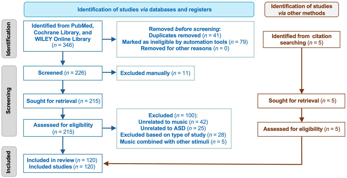
Novel spatial experience significantly increases nuclear expression of Homer1a, Arc and cFos in the CA1 region. (A) Definition of regions of interest in the CA1 region (left). Image (right) shows a DAPI-stained section at the level of CA1 region of the dorsal hippocampus [3.6 mm posterior from Bregma as determined by a rat brain atlas (Paxinos and Watson, 2014)]. Z-stacks were obtained in the distal and proximal subcompartments of the CA1 region (black and white squares). Scale bar: 200 μm. (B) Experimental design: After having resided in the same experiment room overnight, animals underwent 1 h-habituation in the test chamber on the day of the experiment. During the first event, an empty holeboard was introduced to the animal in the chamber. After 10 min exploration time, the holeboard was removed. Animals rested in the chamber for 25 min. Then, the same holeboard was re-inserted to the recording box, but this time, it contained 3 small objects that were placed inside 3 of the 4 holeboard holes. After 5 min of exploration, brain extraction occurred. (C) Bar chart shows the percentage (mean ± SEM) of total exploration time for the exposure to empty holeboard (EHB) during the first holeboard exposure event, and holeboard with objects (HBO) during the 2nd exposure event. During each exposure event, animals exhibited a high exploration percentage. (D) Bar charts show the relative percentage (mean ± SEM) of nuclear Homer1a (red bars), cFos (yellow bars) and Arc (green bars) mRNA expression in neuronal nuclei of the distal (left) and proximal CA1 (right). Novel holeboard (HB) exposure led to a significant increase in Homer1a expression in both distal and proximal CA1 compared to controls (no exploration event). Re-exposure to the holeboard containing novel objects (HBO) significantly triggered cFos expression in the distal and proximal CA1 compared to controls. Significant increase in nuclear Arc expression was only evident in the distal but not the proximal CA1 after HBO (***p < 0.001, *p < 0.05). (E) Representative images of nuclear Homer1a, cFos and Arc mRNA expression in the distal CA1 of a rat that underwent exploration tasks (HB, bottom row) and in a control rat (control, no exploration event, upper row). Nuclear Homer1a, cFos and Arc mRNA signals are indicated by red, yellow and green dots, respectively. Nuclei were counterstained with DAPI. White arrows indicate IEG positive nuclei. Images were acquired using a wide-field fluorescence microscope at the final magnification of 63×. Scale bars: 20 μm.
2.2 Behavioral experimentsThe behavioral habituation and learning protocols were conducted as previously described (Hoang et al., 2018). Animals were handled by the experimenter for 15 min per day for a minimum of 5 days. Then they were habituated to the experiment room and the test chambers for 1 h per day, for 3 consecutive days prior to the commencement of the experiments. The test chambers were 40 × 40 × 40 cm in size, open at the top, and were made of gray washable acrylic panels (Perspex®, polymethylmethacrylate, PMMA). The chamber interior was accessible via a translucent PMMA front wall that was held in place by means of tracks installed at the ends of the abutting chamber walls. The front wall could be moved upward by sliding it along these tracks. After the final habituation day, animals remained in the same experiment room overnight. On the day of the first experiment, the animals were placed in the chamber for 1 h before the acquisition phase commenced. The front wall of the chamber was moved upward so that a holeboard (HB) could be inserted gently into the chamber. The HB was 39.8 × 39.8 × 39.5 cm in proportions (gray PMMA) and included four holes (with a closed base) that were 5.5 cm in diameter and 5 cm deep and were placed equidistantly 2 cm from the edges of HB (Figure 1B).
2.3 Acquisition-reexposure experimentBehavioral learning events comprised two separate events: The first event comprised exposure to the novel empty HB. Here, the front panel of the chamber was moved upward so that the HB could be inserted. The animals hopped onto the HB as it was moved into the chamber until one side touched the back wall. The HB was left in place for 10 min and then removed (Figure 1B). After HB removal, the animals typically moved to a corner of their chamber and closed their eyes or rested with eyes open.
The second event comprised the re-introduction of the, now familiar, HB into the chamber, whereby this time it included three novel objects (4 × 2 × 2 cm) placed (one each) into three of the four HB holes (HBO) (Figure 1B). The total exposure time to HBO was 5 min. Events were timed so that HB exposure was aligned with the subsequent detection of peak nuclear Homer1a expression (Hoang et al., 2018) and HBO exposure was aligned with peak expression of Arc (Hoang et al., 2018) and cFos (see below). For this, HB and HBO exposure were separated by 25 min (i.e., 10 min HB exposure plus 25 min pause), meaning that HBO began 35 min after commencing HB. In the interval between HB and HBO, animals resided undisturbed in the recording chamber. The objects used for HBO were distinct from one another and did not extend above the surface of the holeboard. The animals approached the holes, inserted their noses inside the holes to examine the objects and sometimes lifted them out of the holes with both paws. Their typical behavior was to return the objects to the same holes if they had lifted them outside the holes. The configuration of the objects was randomly assigned for each rat.
Immediately after the conclusion of HBO (40 min after starting HB exposure) brains were rapidly removed in a smooth movement that involved taking the animals out of the chamber and placing them in a guillotine. This movement took no longer than 3 s. Brains were immediately shock frozen in iso-pentane (placed in a small metal container and maintained at a temperature of −60°C), surrounded by dry ice, and then stored at −80°C until further processing. An aged-matched control group of male rats (n = 6) was included in the study. These animals underwent the same handling and habituation procedures as the test animals. On the day of the experiment, they resided undisturbed in the chamber for at least 1 h. No learning events were implemented. Their brains were quickly removed and flash frozen using the same timeline as described above.
2.4 Acquisition-only experimentIn this set of experiments, animals explored a novel holeboard containing novel small items (HBOa) for 10 min, as described above (Supplementary Figure 1A). Then the holeboard was removed from the recording chamber. Animals resided in the chamber for further 30 min. Brain removal occurred 40 min after the start of the exploration. In this experiment, HBOa was the only behavioral condition experienced. Homer1a was used as a biomarker to detect neurons activated by HB, or HBOa, whereas Arc and cFos served as biomarkers to detect neurons that were activated by HBO 40 min after the start of novel HB exposure.
Holeboard environment, HBOa and HBO exploration were video-monitored, and the experiment was discontinued, or the data discarded, if an animal spent less than 5 min exploring the empty HB, HBOa or less than 3 min exploring during HBO.
The exploration time was recorded when the animals first started moving after HB insertion and used their noses to explore the environment. Later, the percentage of exploration time was calculated as exploration time divided by the total exposure time to the HB, HBO or HBOa and multiplied by 100 (Figure 1C, Supplementary Figure 1B).
2.5 Multiplexed fluorescence in situ hybridizationUnder RNAse free conditions, brains were sectioned using a cryostat (Leica CM 3050S, Leica Biosystem GmbH, Wetzlar, Germany). Coronal slices (20 μm) containing hippocampus (from ca. 3.0 to 4.0 mm posterior from Bregma) were collected, mounted directly on slides (superfrost plus® Gerhard Menzel GmbH, Braunschweig, Germany) and stored at −80°C until further processing.
Activity-regulated cytoskeleton-associated protein, Homer1a and cFos cDNA plasmids were prepared commercially (Genscript Biotech, Piscataway Township, New Jersey, USA) using transcripts described by Lyford et al. (1995), Brakeman et al. (1997) and Saidov et al. (2018). The cRNA probes were prepared using a transcription kit (Invitrogen Ambion Maxiscript Kit, ThermoFischer Scientific Waltham, USA) and a premixed RNA labeling mix containing Digoxigenin-11-UTP (Roche Diagnostics, Basel, Switzerland) or Fluorescein-12-UTP (Roche Diagnostics, Basel, Switzerland) or Biotin-16-UTP (Roche Diagnostics, Basel, Switzerland). For this study, Arc RNA was labeled with Biotin, Homer1a RNA with Fluorescein and cFos RNA with Digoxigenin. The generated RNA probes were purified using an RNA CleanUp Kit (Monarch®RNA Cleanup Kit, New England BioLabs, Ipswich, USA). Yield and integrity were verified using gel electrophoresis and the concentration was measured by using a QuantiFluor®RNA system (Promega, Madison, USA).
From each animal, we chose 3 consecutive dorsal hippocampal brain sections (ca. 3.6 mm posterior to Bregma) and left the slides at room temperature (RT) until they were defrosted. Later, slides were fixed for 10 min in ice-cold 4% paraformaldehyde in fresh filtered phosphate buffered saline, quickly washed in 2-fold concentrate saline-sodium citrate (2xSSC) buffer (RNAse free), incubated in acetic anhydride solution and briefly washed in 2xSSC (RNAse free) at RT. Then, the slides underwent a prehybridization process for 15 min in a mixture of 4xSSC and Formamide (1:1) (RNAse free) at 37°C. The mixture of labeled RNA probes (Arc-Biotin, Homer1a-Fluorescein and cFos-Digoxigenin) was diluted with a concentration of 1 g/1 ul in 1× hybridization buffer (Sigma-Aldrich, St. Louis, USA), treated at 90°C for 5 min and then quickly put on ice. After the prehybridization, the slides were incubated with the hybridization buffer containing RNA probes for the hybridization process in a humid chamber that contained filter paper soaked with 2xSSC/50 deionized Formamide (Sigma-Aldrich, St. Louis, USA) in a dilution of 1:1. The hybridization process in the humid chamber lasted ca. 17 h at 56 °C.
Additionally, a negative control, FISH test, was included to verify the specificity of the hybridized signal. For this, no RNA probes were added to this brain slide (data not shown). After the hybridization, slides underwent stringent washing steps: thrice in 2xSSC at 56°C, then in 2xSSC containing RNAse A at 37°C, again in 2xSSC at 37°C, twice in 0.5xSSC at 56°C, 0.5xSSC at RT, twice in 1xSSC at RT and finally thrice in tris-buffered saline (TBS) at RT. Slides were then treated for 15 min with 3% H2O2 solution at RT in order to block the endogenous peroxidase.
The Arc-Biotin protocol was previously established (Hoang et al., 2018). Arc-Biotin signal detection was conducted using the following steps:
70 min incubation in TBS-Tween (0.05%, Polysorbate 20) containing 1% Bovine Serum Albumin (BSA) and 20% Streptavidin (Vectorlabs, SP2002, Burlingame, USA).
90 min incubation in TBS-Tween (0.05%) containing 1% BSA and 20% Biotin (Vectorlabs, SP2002, Burlingame, USA) and streptavidin-peroxidase (1:2000, Jackson Immuno Research, Scottsdale, Arizona, USA).
Washed 4 times in TBS (5 min each).
Incubation for 20 min in TBS containing 1% biotinylated tyramine and 0.01% H2O2.
Washed 4 times in TBS (5 min each) and 60 min incubation in TBS-Tween (0.05%) containing streptavidin CF488 (Biotium, Biotrend GmbH, Cologne, Germany).
Sections were left in TBS at 4°C and protected from light overnight until the next detection.
On the next day, the Homer-Fluorescein signal was detected. The protocol to detect the Homer1a-Fluorescein signal was previously established (Hoang et al., 2018) and comprised the following:
Incubation for 70 min in TBS-Tween (0.05%, Polysorbate 20) containing 10% n-Goat serum (Histoprime, Biozol, Hamburg, Germany) and 20% Streptavidin (Vectorlabs, SP2002).
90 min incubation in TBS-Tween (0.05%) containing 1% n-Goat serum (Histoprime, Biozol, Hamburg, Germany) and 20% Biotin (Vectorlabs, SP2002, Burlingame, USA) and anti-fluorescein-peroxidase (1:2000, Jackson Immuno Research, Scottsdale, Arizona, USA).
Washed 4 times (5 min each) in TBS.
Incubation for 20 min in TBS containing 1% biotinylated tyramine and 0.01% H2O2.
Washed 4 times (5 min each) in TBS and 60 min incubation in TBS-Tween (0.05%) containing 1% n-Goat serum and streptavidin Cy5 (Jackson Immuno Research, Scottsdale, Arizona, USA).
Sections were left in TBS at 4°C and kept away from light overnight until the next detection.
On the next day, the cFos-Digoxigenin signal was detected as follows:
Incubation for 70 min in TBS-Tween (0.05%, Polysorbate 20) containing 10% n-Goat serum (Histoprime, Biozol, Hamburg, Germany) and 20% Streptavidin (Vectorlabs, SP2002, Burlingame, USA).
Incubation for 90 min in TBS-Tween (0.05%) containing 1% n-Goat serum (Histoprime, Biozol, Hamburg, Germany) and 20% Biotin (Vectorlabs, SP2002, Burlingame, USA) and anti-digoxigenin-peroxidase (1:2000, Jackson Immuno Research, Scottsdale, Arizona, USA).
Washed 4 times (5 min each) in TBS.
Incubation for 20 min in TBS containing 1% biotinylated tyramine and 0.01% H2O2.
Washed 4 times (5 min each) in TBS followed by 60 min incubation in TBS-Tween (0.05%) containing 1% n-Goat serum and streptavidinCy7 (Invitrogen, Thermo Fischer Scientific, Waltham, Massachusetts, USA).
Later, slides were washed 4 times (5 min each) in TBS, rinsed in double-distilled water, quickly dipped in 70% ethanol and finally stained using 1% Sudan Black B (Merck KGaA, Sigma-Aldrich, St. Louis, USA) in 70% ethanol (Oliveira et al., 2010). Finally, slides were rinsed in distilled water, air dried and mounted in antifading mounting medium (immunoSelect®, Dianova, Hamburg, Germany) containing 4′-6-diamidino-2-phenylindole (DAPI).
Peak nuclear Homer 1a expression occurs 30–40 min after a specific induction event (Bottai et al., 2002). Peak nuclear Arc and cFos expression occur 5–6 min and 6–8 min after a specific induction event, respectively (Saidov et al., 2018; Vazdarjanova et al., 2002). Afterwards, the IEG diffuses into the cytoplasm (Bottai et al., 2002; Saidov et al., 2018; Vazdarjanova et al., 2002). For this reason, we assume that if we detect cytoplasmic expression of a given IEG at the time-point of nuclear detection, this is an indicator that the animals underwent another salient experience in the time before the experiment was started. Given that the animals resided in their homecages before and after HB/HBO exposure, this salient experience would have to be stress-related. This can be caused by unexpected noises (in the room or corridor outside the room), strangers entering the room, the presence of adult male rodents (for females), or females in oestrus (for males), or stress-related (22 Hz) vocalizations by animals in the proximity of the room (Cullinan et al., 1995; Inagaki and Ushida, 2017; Manahan-Vaughan, 2017, 2018; Schreiber et al., 1991). For this reason, we ensured that the animals were well-habituated to handling by the experimenter, that only male rats were used and that experiments were conducted under quiet and calm conditions.
2.6 Data analysisWe focused our analysis on the dorsal CA1 region, given that this subregion of the hippocampus expresses long-term potentiation (LTP) when novel HB exposure is coupled with afferent stimulation, whereas long-term depression (LTD) is facilitated by novel HBO exposure coupled with afferent stimulation (Kemp and Manahan-Vaughan, 2004). Moreover, novel HB and novel HBO exposure results in a distinct pattern of nuclear IEG expression in the distal and proximal parts of the dorsal CA1 region (Hoang et al., 2018). In other words, the CA1 generates distinct functionally relevant “encoding” responses to novel HB and novel HBO exposure.
Z-stacks were obtained in the distal CA1 (dCA1) and proximal CA1 (pCA1) at a 63× magnification using a widefield fluoresence microscope (Zeiss Apotome, Oberkochen, Germany) (Figure 1A). Region of interest (ROI) that contained dCA1 and pCA1 were determined based on their anatomical locations (Naber et al., 2001) and comprised a standardized area of 150 μm × 200 μm (Figure 1A). Three consecutive slices of each animal were used for the analysis, whereby we analyzed both hemispheres of each slice and calculated the mean of these three slices. Complete DAPI stained nuclei that showed no evidence of cutting on the edges (of the slides) either in the x, y or z planes, were marked using Fiji software (Schindelin et al., 2012). Neurons were distinguished from glial cells and endothelial cells on the basis of cell morphology and size (Félétou, 2011).
Neurons were checked for nuclear mRNA expression of Homer1a, Arc and cFos that peaked in the nuclei of the CA1 neurons, in an experimenter-blind manner. Based on the fluorescent label, these were detected as red punctae for Homer1a, green punctae for Arc and yellow punctae for cFos (Figure 1E). Percentages of IEG-mRNA positive cells were calculated per total counted neurons for each subregion of each rat. The designation “positive nuclei” was given to cells that contained intense intranuclear foci of IEG-mRNA fluorescent signals (Figure 1D). Nuclei that did not contain any intranuclear foci representing a fluorescent signal of IEG-mRNA were counted as negative. The total number of cells analyzed for each ROI of each slide of each animal was on average 80. Then, the relative percentage of single labeled IEG-mRNA positive cells was calculated relative to the total DAPI counts for each ROI. Additionally, to examine the contribution of the co-labeling of IEGs within the activated neurons, the number of neurons that expressed only Homer1a (H1a-only) or cFos (cFos-only) or Arc (Arc-only), were compared with co-labeling of IEG-mRNA. Here, percentages were calculated by dividing labeled counts of IEGs and total IEG positive counts. Final results are presented as average means of percentages ± standard error of the mean (SEM) for each group. Graphs were generated using GraphPrism (GraphPad Software Version 8, San Diego, California USA). Venn diagrams were created in Python 3.11.8 using a custom script created with the matplotlib_venn package.
2.7 Statistical analysisControl and test animals comprised n = 6 each. All values were verified for normal distribution using the Kolmogorov-Smirnow test with Lilliefors correction. Statistical analysis was performed using Statistica software (version 14.0.1.25, TIBCO Software Inc., Santa Clara, CA, USA). Multifactorial analysis of variance (mANOVA) was performed, followed by a subsequent Tukey HSD post-hoc test for pairwise comparison between factor groups or regions or IEGs. For multiple comparisons between co-labeled factors, Fisher’s LSD post-hoc test was performed and summarized in Tables 1, 2. The significance level was set at p < 0.05.
SummaryP-valuedH1a-only:control vs. pH1a-only:controlns0.7741dH1a-only:control vs. dcFos-only:control****<0.0001dH1a-only:control vs. dArc-only:control****<0.0001dH1a-only:control vs. d(H1a+cFos)-only:control****<0.0001dH1a-only:control vs. d(H1a+Arc)-only:control****<0.0001dH1a-only:control vs. d(H1a+Arc+cFos):control****<0.0001dcFos-only:control vs. pcFos-only:controlns0.1388dcFos-only:control vs. dArc-only:controlns0.4092dcFos-only:control vs. d(H1a+cFos)-only:control**0.0017dcFos-only:control vs. d(H1a+Arc)-only:control***0.0003dcFos-only:control vs. d(Arc+cFos)-only:control****<0.0001dcFos-only:control vs. d(H1a+Arc+cFos):control****<0.0001dArc-only:control vs. pArc-only:controlns0.8639dArc-only:control vs. d(H1a+cFos)-only:control*0.0191dArc-only:control vs. d(H1a+Arc)-only:control**0.0052dArc-only:control vs. d(Arc+cFos)-only:control**0.0052dArc-only:control vs. d(H1a+Arc+cFos):control***<0.0001d(H1a+cFos)-only:control vs. d(H1a+Arc)-only:controlns0.6410d(H1a+cFos)-only:control vs. d(Arc+cFos)-only:control*0.0157d(H1a+cFos)-only:control vs. d(H1a+Arc+cFos):control**0.0083d(H1a+cFos)-only:control vs. p(H1a+cFos)-only:controlns0.3489d(H1a+Arc)-only:control vs. d(Arc+cFos)-only:control*0.0499d(H1a+Arc)-only:control vs. d(H1a+Arc+cFos):control*0.0289d(H1a+Arc)-only:control vs. p(H1a+Arc)-only:controlns0.3489d(Arc+cFos)-only:control vs. d(H1a+Arc+cFos):controlns0.8183d(Arc+cFos)-only:control vs. p(Arc+cFos)-only:controlns0.8469d(H1a+Arc+cFos):control vs. p(H1a+Arc+cFos):controlns0.8777pH1a-only:control vs. pcFos-only:control****<0.0001pH1a-only:control vs. pArc-only:controlns0.6252pH1a-only:control vs. p(H1a+cFos)-only:control****<0.0001pH1a-only:control vs. p(H1a+Arc)-only:control****<0.0001pH1a-only:control vs. p(H1a+Arc+cFos):control****<0.0001pcFos-only:control vs. p(H1a+cFos)-only:controlns0.4436pcFos-only:control vs. p(H1a+Arc)-only:controlns0.0689pcFos-only:control vs. p(Arc+cFos)-only:control****<0.0001pcFos-only:control vs. p(H1a+Arc+cFos):control****<0.0001pArc-only:control vs. p(H1a+cFos)-only:controlns0.2106pArc-only:control vs. p(H1a+Arc)-only:control*0.0217pArc-only:control vs. p(Arc+cFos)-only:control****<0.0001pArc-only:control vs. p(H1a+Arc+cFos):control****<0.0001p(H1a+cFos)-only:control vs. p(H1a+Arc)-only:controlns0.2888p(H1a+cFos)-only:control vs. p(Arc+cFos)-only:control***0.0004p(H1a+cFos)-only:control vs. p(H1a+Arc+cFos):control***
Comments (0)