
Remember me
The selection of excipients in the development of dermal gel formulations is critical, as each component contributes specific functional and therapeutic advantages. The formulation described in Table 1 incorporates a combination of polymers, humectants, preservatives, and functional additives, each selected to optimize the performance, stability, and safety of the final product.
CMC and xanthan gum functioned as the principal gelling agents, establishing the structural matrix necessary for attaining the requisite viscosity, spreadability, and stability of the formulation. Their amalgamation guaranteed an optimal gel consistency and preserved homogeneity during application. Glycerin was added to enhance moisture retention in the gel, hence improving flexibility and user comfort. Sodium benzoate served as the preservative agent to maintain microbiological stability during storage, while limonene and rose water enhanced the sensory profile of the formulation, hence increasing consumer acceptability [35,36,37,38,39,40,41,42,43,44,45].
The chosen excipients successfully facilitated the development of a stable gel with appropriate rheological properties and favorable application qualities. Their synergistic functionality facilitated the creation of a formulation exhibiting consistent performance and satisfactory aesthetic attributes, in accordance with the planned application for dermal distribution.
Characterization of the Prepared GelFT-IR spectroscopy is a very common analysis method applied to characterize pharmaceutical formulations and identify any interactions between the drugs and excipients. Moreover, the successful entrapment of drugs into the formulation can be confirmed via FT-IR as many studies, identify [46]. Therefore, in this study, in order to confirm the successful incorporation of CAB into the hydrogel formulation and to investigate potential chemical interactions between the CAB and the gel matrix components, Attenuated Total Reflection Fourier Transform Infrared (ATR-FTIR) spectroscopy was performed. The spectra of the CAB, the blank hydrogel formulation (F1), and the CAB-loaded formulation (F1-CAB) were recorded in the 4000–650 cm⁻¹ region. The resulting spectra are shown in Fig. 1. The most significant bands of CAB, F1 and F1-CAB, are demonstrated in Table 2.
Fig. 1
FTIR spectroscopy analysis of CAB, F1 and F1-CAB
Table 2 Important bands according to FTIR analysisThe FTIR spectrum for pure calcium butyrate powder exhibits several distinct absorption bands characteristic of a carboxylate salt (Fig. 1). The most prominent peaks are observed in the lower wavenumber region. The peaks in the region of 2965 cm⁻¹, 2973 cm⁻¹, and 2911 cm⁻¹ are attributed to the asymmetric and symmetric stretching vibrations of the C-H bonds in the methyl (CH₃) and methylene (CH₂) groups of the butyrate alkyl chain. The most definitive evidence for the salt structure is the presence of two strong, sharp absorption bands corresponding to the carboxylate anion (COO⁻). The intense band at approximately 1546 cm⁻¹ is assigned to the asymmetric stretching vibration (νasym (COO−)), while the very sharp and strong peak at 1417 cm⁻¹ corresponds to the symmetric stretching vibration (νsym (COO−)). The significant separation between these two peaks is typical for ionic carboxylates. Additional peaks are noted in the fingerprint region, including those around 930 cm⁻¹ and 650 cm⁻¹, which relate to various rocking, wagging, and skeletal vibrations of the molecule. The absence of an intense broad band in the 3200–3600 cm⁻¹ range indicates the lack of significant hydroxyl groups or moisture in the pure CAB sample.
The spectrum of the F1, composed of polymers such as carboxymethyl cellulose and xanthan gum, shows features characteristic of polysaccharides (Fig. 1). The FT-R spectrum of Carboxymethylcellulose, is well documented [47]. Hidayat et al. (2018) reported the an abdoprtion band around 1600 cm-1 due to the streching vibration of the (COO-). The broad band around 3400 cm-1 is due to the stretching frequency of hydroxyl molecules (-OH). Similar results have been identified also in this study, where the formulation is based also in CMC. Xanthan gum is well studied polymer for producing hydrogels. Its FTIR spectrum according to Amaral et al. Depicts as peak around 3439 cm − 1, due tothe deformation of the O-H group. At 2800–2950 cm-1, was seen the the axial deformation of the C-H group [48].
According to already reported studies, herein, a very broad and high-intensity absorption band is dominant in the 3600–3000 cm⁻¹ range, centered at approximately 3350 cm⁻¹. This band is characteristic of the stretching vibrations of hydroxyl (O-H) groups present in the polysaccharide backbones and from intermolecular hydrogen bonding, as well as absorbed water within the hydrogel structure. A smaller, less defined peak is observed at approximately 2931 cm⁻¹, which is attributed to the C-H stretching vibrations of the alkyl groups within the polymer chains. A strong absorption peak is visible at 1589 cm⁻¹, corresponding to the asymmetric stretching of carboxylate (COO⁻) groups inherent to the chemical structure of the polymers (e.g., carboxymethyl cellulose). The region between 1450 cm⁻¹ and 1000 cm⁻¹ displays a series of complex, overlapping bands, with a particularly strong and broad peak centered around 1050 cm⁻¹. This area, known as the fingerprint region for polysaccharides, is associated with C-O stretching and C-O-C glycosidic bond vibrations.
The FTIR spectrum of the F1-CAB effectively represents a superposition of the spectra of the F1 and the pure CAB, confirming the successful physical entrapment of calcium butyrate within the hydrogel matrix (Fig. 1). The overall spectral profile is dominated by the features of the blank formulation (F1) due to the high concentration of the polymers relative to the bioactive. The broad O-H band centered at ~ 3350 cm⁻¹ and the C-H stretching peak at ~ 2931 cm⁻¹ are preserved. Crucially, the successful incorporation of the CAB is confirmed by the presence of its characteristic peaks in the F1-CAB spectrum. The highly characteristic, sharp peak corresponding to the symmetric stretching of the carboxylate group (νsym (COO−)) of CAB at 1405–1410 cm⁻¹ is clearly identifiable in the final formulation. A comparative overlay of the three spectra confirms these observations. No significant new peaks or major shifts in the positions of the primary functional group peaks are observed [49]. This lack of new chemical bands or shifting strongly suggests that the CAB is physically dispersed within the polymer network and has not formed any new covalent bonds with the hydrogel components. Similar results have been also reported [50].
In conclusion, the FTIR analysis validates that CAB was successfully entrapped within the hydrogel matrix, maintaining its chemical integrity through a process of physical entrapment rather than chemical interaction. Similarly, Preet et al. studied hydrogels loaded with oxytetracycline revealing that the successful encapsulation of the bioactive within hydrogel matrix was identified due to the presence of absorption bands of the bioactive in the spectrum [51].
Comprehensive characterization of the prepared gel formulations is essential to ensure their suitability for dermal application, stability, and therapeutic efficacy. In this study, F1 and F1-CAB gels were evaluated in terms of pH, viscosity, CAB content in formulation, and other critical quality attributes.
The pH of topical formulations is a crucial parameter, as it must be compatible with the physiological pH of the skin (typically 4.5–6.5) to avoid irritation and maintain the skin barrier function [40]. The F1-CAB gel exhibited a pH of 6.75 ± 0.08, while the blank gel (F1) had a pH of 7.22 ± 0.02 (Table 3). Although slightly above the average skin pH, these values are within the acceptable range for dermal products and are unlikely to cause irritation or disrupt the skin’s natural flora. Similar pH values have been reported in the literature for bioadhesive gels intended for wound healing, supporting the safety of the developed formulation [45].
Table 3 Characterization results of prepared gelViscosity is a key determinant of a gel’s spreadability, retention at the application site, and drug release profile. The F1-CAB formulation demonstrated a significantly higher viscosity (21.17 ± 0.13 P) compared to the blank gel (12.65 ± 0.06 P). Increased viscosity is generally associated with enhanced bioadhesion and prolonged residence time on the skin, which can improve therapeutic outcomes by maintaining sustained contact with the affected area [35]. However, excessively high viscosity may hinder spreadability and patient comfort. The values observed in this study are consistent with those reported for effective dermal gels, indicating a favorable balance between retention and ease of application [52, 53].
Textural Profile AnalysisTexture Profile Analysis (TPA) is a widely used technique to quantitatively assess the mechanical properties of semi-solid formulations, providing valuable information on parameters such as hardness, adhesiveness, resilience, and cohesion. These properties are not only critical for the physical stability of the gel but also play a pivotal role in user experience, drug release, and therapeutic efficacy [35, 52, 53].
In the present study, the F1-CAB formulation demonstrated a significant increase in hardness (11.261 ± 0.124 g) compared to the blank gel (6.526 ± 0.191 g). Hardness reflects the force required to deform the gel and is directly related to the gel’s internal network structure, which is influenced by the concentration and interaction of gelling agents and the presence of active compounds [37,38,39]. The higher hardness observed in F1-CAB can be attributed to the incorporation of calcium butyrate, which may enhance cross-linking within the polymer matrix, resulting in a denser and more robust gel structure (Table 4). The divalent Ca²⁺ ions presumably participated in electrostatic interactions with the polyanionic polysaccharide chains, thereby generating additional crosslinking sites within the gel. This molecular reinforcement resulted in enhanced network density, which was observed macroscopically as a more robust elastic response and a more significant solid-like characteristic.
Table 4 Textural profile analysis of prepared formulationsAdhesiveness, defined as the work required to overcome the attractive forces between the gel and a probe (or skin), was also markedly higher in F1-CAB (−15.252 ± 1.756 g.sec) than in F1 (−8.270 ± 0.772 g.sec). Enhanced adhesiveness is desirable for topical formulations, as it ensures prolonged contact with the skin, facilitating sustained CAB release and improved therapeutic outcomes [35]. Literature suggests that increased adhesiveness correlates with better bioavailability and patient compliance, particularly in wound care and regenerative medicine [45]. The observed increase in adhesiveness in F1-CAB is likely due to synergistic interactions between the gel matrix and calcium ions, which can promote stronger intermolecular bonding and mucoadhesive properties [36].
Resilience and cohesion are indicators of the gel’s ability to recover its original structure after deformation and to withstand repeated stress, respectively. The F1-CAB formulation showed slightly higher resilience (24.891 ± 0.839%) and cohesion (0.813 ± 0.007) compared to F1 (17.695 ± 0.580% and 0.804 ± 0.005, respectively). These properties are essential for maintaining the integrity of the gel during application and ensuring consistent dosing. According to [54,55,56], gels with higher resilience and cohesion are less likely to break down or lose their structure during use, which is particularly important for formulations intended for dynamic environments such as the skin.
When compared to similar studies, the mechanical properties of F1-CAB are within the optimal range for dermal gels. For instance [45], reported that bioadhesive gels with hardness values between 10 and 15 g and adhesiveness above − 10 g.sec provided optimal performance in terms of application and retention. The F1-CAB formulation not only meets but exceeds these benchmarks, suggesting its suitability for clinical use (Fig. 2).
Fig. 2
Detailed results of textural profile analysis of F1 and F1-CAB. *p < 0.05 is significant
In summary, the TPA results indicate that the incorporation of calcium butyrate into the gel matrix significantly enhances the mechanical strength, adhesiveness, and structural integrity of the formulation. These improvements are consistent with findings in the literature and are expected to translate into superior handling, increased patient compliance, and more effective dermal delivery of the active ingredient. The robust textural profile of F1-CAB supports its potential as an advanced topical formulation for skin regeneration and wound healing.
Spreadability AnalysisSpreadability is a critical parameter in the evaluation of topical gel formulations, as it directly influences patient compliance, dosing accuracy, and the overall therapeutic efficacy of the product. An ideal dermal gel should possess sufficient firmness to maintain its structure yet be easily spreadable to ensure uniform application over the target area [37,38,39, 41, 42]. In this study, the spreadability of both the blank (F1) and calcium butyrate-loaded (F1-CAB) gel formulations was assessed using a texture analyzer, with key parameters including firmness, work of shear, stickiness, and work of adhesion (Table 5).
Table 5 Spreadability results of prepared formulationsThe F1-CAB formulation exhibited higher firmness (1033.71 ± 15.00 g) and work of shear (923.06 ± 37.93 g.sec) compared to the blank gel (653.83 ± 3.54 g and 500.89 ± 15.11 g.sec, respectively). This increase in firmness and work of shear can be attributed to the denser gel network formed by the addition of calcium butyrate, which enhances the mechanical strength and stability of the formulation. Despite the increased firmness, the F1-CAB gel maintained adequate spreadability, as indicated by its stickiness (−653.61 ± 12.44 g) and work of adhesion (−315.39 ± 2.76 g.sec) values. These parameters reflect the gel’s ability to adhere to and be distributed across the skin surface without excessive resistance. According to [45], optimal spreadability ensures that the formulation can be applied in a thin, uniform layer, maximizing contact with the skin and promoting efficient drug delivery. The balance between firmness and spreadability is crucial. Excessively firm gels may be difficult to apply and uncomfortable for the patient, while overly soft gels may lack sufficient residence time and be easily removed from the application site [35].
The F1-CAB formulation’s spreadability profile aligns with the requirements for advanced topical gels, as described in the literature, where a moderate increase in firmness is often associated with improved bioadhesion and sustained release, provided that spreadability remains within acceptable limits [36, 54,55,56]. Furthermore, the pseudoplastic (shear-thinning) behavior imparted by the combination of carboxymethyl cellulose and xanthan gum allows the gel to become less viscous under the shear force of application, facilitating easy spreading, and then regain viscosity at rest, enhancing retention [37,38,39]. This rheological property is highly desirable for dermal formulations, as it combines user comfort with functional performance (Fig. 3).
Fig. 3
Detailed representation of spreadability results of F1 and F1-CAB, *p < 0.05 is significant
In summary, the spreadability analysis demonstrates that the incorporation of calcium butyrate into the gel matrix increases the mechanical strength of the formulation without compromising its ability to be easily and uniformly applied. These findings are consistent with previous reports on bioadhesive gels and support the suitability of the F1-CAB formulation for dermal drug delivery applications.
Rheology StudiesThe rheological characterization of the investigated formulations has provided comprehensive insights into the behavior of both the carrier gel matrix and the Ca-butyrate-loaded system, particularly with respect to flow properties, viscoelastic responses, and structural stability under varying conditions. In the case of the blank formulation, which was primarily composed of carboxymethyl cellulose (CMC) and xanthan gum and further stabilized by hydrogen bonding interactions in the presence of glycerin, the results indicated the development of a viscous network with limited strength, displaying the features of a relatively weak gel system. Although such a network was sufficient to provide apparent viscosity and an initial three-dimensional structure, its elasticity was restricted, and the system was therefore not fully able to resist deformation under stress. The incorporation of Ca-butyrate, however, distinctly altered the rheological properties of the matrix. The divalent Ca²⁺ ions were likely engaged in electrostatic interactions with the polyanionic polysaccharide chains, thereby creating additional crosslinking points within the gel. This molecular reinforcement led to an increased network density, which manifested macroscopically as a stronger elastic response and a more pronounced solid-like behavior. Thus, in addition to its therapeutic functionality, Ca-butyrate also appeared to play a structural role by fortifying the rheological framework of the formulation.
When the frequency sweep results were examined, it was observed that in the blank formulation, at both 25 °C and 32 °C, the storage modulus (G′) was consistently greater than the loss modulus (G″), demonstrating that gel-like properties predominated across the measured range. Nevertheless, at lower frequencies the gap between G′ and G″ progressively narrowed, suggesting a weak gel character, where viscoelastic balance tended to shift with increasing deformation time scales. In contrast, the Ca-butyrate-loaded formulation exhibited a marked increase in G′ values across the entire frequency spectrum (Fig. 4). This outcome confirms that Ca²⁺ ion incorporation strengthens the polymeric network by forming ionic bridges between polysaccharide chains, resulting in the transition from a weak gel to a strong gel system. Notably, the superiority of this elastic network was preserved even at 32 °C, which highlights the enhanced thermal stability of the CAB-loaded gel. These findings are in agreement with previous studies reporting the ability of calcium ions to increase crosslink density in CMC- and xanthan-based networks, thereby improving their durability and mechanical performance (Table 6) [57, 58].
Fig. 4
Frequency-dependent changes of viscoelastic properties of the formulations (n = 3). A: F1 at 25 °C, B: F1 at 32 °C, C: F1-CAB at 25 °C, D: F1-CAB at 32 °C
Table 6 Effect of temperature on the loss tangent tan(δ) and on the dynamic viscosity (η’) of the formulations at three representative frequenciesThe shear stress and viscosity measurements further corroborated these findings. Both blank and loaded systems exhibited typical shear-thinning (pseudoplastic) behavior, as viscosity decreased sharply with increasing shear rate. This rheological profile is particularly desirable for topical applications: while at low shear rates the formulation maintains high viscosity and structural stability, at higher shear rates-such as during spreading on the skin-the viscosity decreases, enhancing spreadability and user compliance [25, 26]. The Ca-butyrate-loaded system demonstrated higher shear stress and viscosity values than the blank formulation, further indicating network reinforcement by the active component (Fig. 5).
Fig. 5
Flow curves of blank (A: F1 at 25 °C and at 32 °C) and CAB loaded (C: F1-CAB at 25 °C and at 32 °C) and viscosity curves of blank (B: F1 at 25 °C and at 32 °C) and CAB loaded (D: F1-CAB at 25 °C and at 32 °C)
Analysis using the Herschel–Bulkley model supported these interpretations. The blank formulation at 25 °C showed negative yield stress (τ₀), implying the absence of a true yield point and reinforcing the weak gel characteristics observed in oscillatory testing. At 32 °C, however, a positive τ₀ value emerged, signifying the development of a more classical gel-like response at elevated temperature. Consistency indices (K) and flow indices (n < 0.2) confirmed highly shear-thinning behavior. In the Ca-butyrate-loaded formulation, although the fitting quality to the Herschel–Bulkley model was reduced (lower correlation coefficients), the higher K values and emerging τ₀ confirmed the strengthening effect of Ca²⁺ addition, even if its strongly elastic behavior could not be fully captured by the model equations (Table 7).
Table 7 Herschel–Bulkley model parameters for blank (F1) and Ca-butyrate-loaded (F1-CAB) formulations at 25 °C and 32 °CFinally, the G′–G″ crossover point analysis provided additional support for these conclusions. For the blank formulation, crossover occurred at approximately 80 Pa and 0.3–0.4 Hz, consistent with weak gel behavior. By contrast, in the Ca-butyrate-loaded gel, crossover occurred at much lower frequencies (~ 0.02–0.03 Hz) and lower modulus values (~ 35–44 Pa), meaning that elastic dominance was established almost immediately and maintained throughout the entire measurement window (Table 8). This is highly characteristic of a strong gel system, which resists viscous flow across a broad frequency range [59, 60].
Table 8 Crossover parameters derived from oscillatory rheological measurementsTaken together, these results clearly demonstrate that the addition of Ca-butyrate not only enhances the functional role of the formulation as a therapeutic gel but also significantly modifies its rheological framework. The Ca²⁺ ions act as ionic crosslinkers within the CMC/xanthan network, producing a denser and more elastic three-dimensional structure. As a result, the Ca-butyrate-loaded system exhibits strong gel characteristics (G′>> G″ across frequencies), prominent shear-thinning behavior favorable for patient use, and increased thermal stability, all properties that are highly advantageous for the development of robust and reliable topical gel formulations.
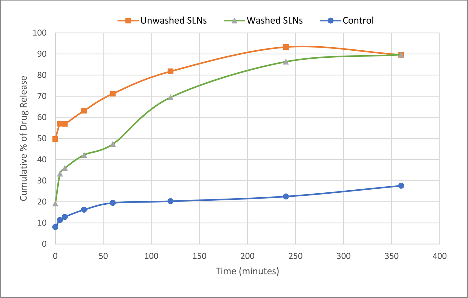
In Vitro Release Studies and Kinetic ModellingIn vitro release investigations of F1-CAB and the CAB solution were performed using dialysis membranes. Quantification of CAB in the release media was carried out by HPLC analysis of aliquots withdrawn at predetermined time intervals. The cumulative release profiles of both formulations are presented in Fig. 6. After 24 h, the overall release percentages were calculated as 100.002 ± 0.001% for F1-CAB and 99.179 ± 1.426% for the CAB solution. Examination of the release curves revealed that the CAB solution exhibited a rapid release pattern, with approximately 49.884 ± 1.845% of the bioactive released within the first hour and nearly complete release achieved by the fourth hour. In contrast, F1-CAB showed a markedly slower release, with 21.878 ± 6.246% released at the first hour and full release observed only at the eighth hour. These findings indicate that, unlike the immediate-release behavior of the solution, F1-CAB provided a sustained and controlled release profile (Fig. 6).
Fig. 6
In vitro release studies of F1-CAB and CAB Solution
The kinetic modelling of in vitro release studies presents a comparative kinetic evaluation of in vitro release data obtained from F1-CAB and CAB Solution. Release profiles were analyzed using Zero-order, First-order, Higuchi, Hixson–Crowell, and Korsmeyer–Peppas models. The main findings are summarized in Table 9. The gel formulation exhibited a controlled release profile with gradual bioactive liberation. Among the tested kinetic models, the First-order (R²=0.95) and Hixson–Crowell (R²=0.93) models provided the best fit. The Korsmeyer-Peppas model yielded an exponent n = 0.39, indicating Fickian diffusion as the primary mechanism. Higuchi modeling demonstrated moderate correlation (R²=0.80), while Zero-order kinetics showed poor fit (R²=0.61). Overall, the gel system combined concentration-dependent release with surface erosion effects, delivering a prolonged and controlled release profile. The solution form demonstrated very rapid and nearly complete release within the first hours. The First-order model provided the strongest correlation (R²>0.95), highlighting concentration-dependent kinetics. The Hixson–Crowell model also showed good agreement (R²>0.90), consistent with rapid dissolution phenomena. In contrast, Higuchi and Korsmeyer–Peppas models only moderately fit the data, confirming that diffusional control was not the governing mechanism. The n exponent in the Korsmeyer–Peppas model was < 0.45, which theoretically corresponds to Fickian diffusion, but in this case simply reflects the absence of any structural diffusion barrier in the solution matrix. When comparing both formulations, the gel system clearly behaved as a controlled release matrix, offering prolonged delivery governed by a combination of Fickian diffusion and surface erosion phenomena. The solution formulation resulted in immediate and complete bioactive availability, suitable for indications where a fast onset of action is required. In conclusion, the gel formulation demonstrated a prolonged release profile best desc
Comments (0)