Incidence
The incidence of likely NB-related cerebrovascular events within our NB cohort was 3.1% (13/413) overall and 4.5% for adults (12/267), which is markedly higher than the estimated 0.3-1% in older literature (Winter et al. 2024; Back et al. 2013; Artal 2016). This discrepancy may reflect regional variations, such as a higher prevalence of neuroinvasive Borrelia species in our area, but it more likely results from the increased use of high-quality brain and vascular imaging, which enhances diagnostic sensitivity and specificity of CNS vasculitis, the probable cause of these cerebrovascular events. In older NB studies, such as those by Bremell and Dotevall, and a recent systematic review by Knudtzen et al., NB encephalitis was reported as the most common CNS presentation (Knudtzen et al. 2022; Bremell and Dotevall 2014). Notably, the patients in these studies did not consistently undergo advanced imaging that might have revealed vasculitis. For comparison, in our entire NB cohort, only one patient was diagnosed with encephalitis (Rosendahl et al. 2023), whereas older studies reported an incidence of approximately 4–5% (Rauer et al. 2020; Oschmann et al. 1998). Notably, this rate closely matches our observed incidence of NB-associated ischemic stroke and TIA in adults, suggesting that underdiagnosis of vasculitis may have influenced historical data. Supporting this perspective, a recent German case series by Volk et al. identified vasculitis in 5 of 35 consecutive NB patients (14.3%) (Volk et al. 2024). This growing recognition of NB vasculitis aligns with the established pathophysiology of neuroborreliosis, where vasculitic and peri-vasculitic pathology are well-documented as key CNS features (Oksi et al. 1996; Miklossy et al. 1990). Thus, the predominance of vasculitis over encephalitis in modern cohorts may reflect improved diagnostic methodologies rather than a true shift in disease presentation.
In the prospective study by Arboix et al., 1,617 first-ever stroke/TIA patients were registered, of whom 70 (4.3%) were classified as having ischemic stroke of unusual cause (ISUC) (Arboix et al. 2001). Eleven of these 70 cases were attributed to infection, corresponding to 0.68% of all first-ever strokes/TIAs. This confirms that infection-related stroke is rare but recognized. In the present study we believe that NB-related stroke is likely underdiagnosed. Despite nearly half of all NB patients in our total cohort originating from North Zealand Hospital, only one of the 13 NB-associated stroke/TIA cases came from this centre. This disparity strongly suggests that recognition of NB-related vasculitic stroke requires a high index of suspicion, particularly in patients with subacute prodromal symptoms or unexplained infarction patterns.
Clinical presentation
More than half of our patients presented with gait difficulties, and some exhibited UMN signs, consistent with CNS manifestations of NB such as encephalomyelitis (Bajons et al. 2023; Artal 2016; Volk et al. 2024; Iversen et al. 2023; Dumic et al. 2019; Beuchat et al. 2018; Lindland et al. 2018). These findings are also commonly observed in non-infectious CNS vasculitis and NB-associated vasculitis (Bajons et al. 2023; Artal 2016; Volk et al. 2024; Iversen et al. 2023; Dumic et al. 2019; Lindland et al. 2018; Beuchat et al. 2018). In line with previous studies, approximately half of our patients suffered from a chronic prodromal phase of headache and / or cognitive symptoms prior to stroke (Artal 2016; Topakian et al. 2008; Back and Grünig 2016; Arboix et al. 2001). The symptoms of cases 3, 4, 6, 11, and 13 evolved insidiously, and the detection of acute diffusion-restricted lesions on MRI was unexpected. This spectrum mirrors primary angiitis of the central nervous system (PACNS), where initial stroke symptoms are reported in 11.5–51% across cohorts, lowest in biopsy-confirmed small-vessel disease (11–22%) and highest in large/medium-vessel PACNS (62%, LV-PACNS), and where walking disability, headache, and cognitive decline are frequent presenting or evolving features (Reddy et al. 2025; Sundaram et al. 2019; Paramasivan et al. 2024).
Notably, only patients 2 and 12 (Table 2) presented with acute stroke symptoms as the initial manifestation. Patient 7 (Table 2) initially exhibited acute 2-hour lasting focal neurological symptoms, and only after admission did her mother vaguely recall mild prodromal symptoms prior to admission.
These findings highlight the importance of actively inquiring about prodromal symptoms, particularly in cases of stroke without conventional vascular risk factors. The presence of UMN signs that cannot be explained by an acute stroke should prompt consideration of alternative stroke etiologies. Clinicians should also inquire about a history of rash or tick bites, as half of our patients reported one or both. However, it is important to note the variability in the literature regarding the frequency of prodromal symptoms and tick exposure preceding stroke in NB-associated vasculitis (Garkowski et al. 2017; Back et al. 2013).
Cerebrospinal fluid presentation
CSF analysis revealed lymphocytic pleocytosis in all but two of the 13 patients, with a median leukocyte count of 105/µL (range 1–832). This finding aligns with previous studies on NB vasculitis and did not differ significantly from the rest of the cohort (p = 0.83) (Garkowski et al. 2017; Winter et al. 2024). Pleocytosis is uncommon in ischemic stroke without inflammatory or infectious etiology (Rundblad et al. 2023).
CSF glucose levels were significantly lower in NB patients with cerebrovascular manifestations compared to the rest of the cohort. This may reflect more profound CNS inflammation, possibly driven by more pathogenic Bb species and/or longer symptom duration prior to diagnosis. However, we found no relationship between symptom duration and CSF WBC in these patients.
Consistent with Volk et al., Q-alb and IgG index, markers of blood-brain barrier disruption, were elevated and higher than the rest of the cohort (Volk et al. 2024).
Case 2 (Tables 1, 2, 3) highlights that CSF inflammation can persist for years after treatment, with strokes occurring months or even years later (Fig. 3). This may result from a combination of persistent vessel stenosis, scarring, and ongoing, albeit diminishing, inflammation. Case 5 (Tables 1, 2, 3) presented with acrodermatitis chronica atrophicans (ACA)-like rash 6 months before her first stroke and went on to experience two additional strokes over the subsequent 2 years. Despite biopsy-confirmed ACA, and a markedly elevated Bb IgG IT index, her CSF lacked pleocytosis. This may partly be explained by a 17-day course of penicillin prior to the CSF examination (Krogen et al. 2022). Furthermore, previous PCR and culture studies have demonstrated that Bbsl can be found in the CSF even in the absence of pleocytosis (Strle et al. 2006). This phenomenon is particularly notable in B. afzelii-associated NB, where CSF WBC appear to decrease as the disease progresses (Strle et al. 2006; Rožič et al. 2019; Busch et al. 1996; Ornstein et al. 2002). Non-infectious CNS vasculitis can present with normal CSF, and rare cases of NB vasculitis without pleocytosis have been described in the literature (Garkowski et al. 2017; Beuker et al. 2021; Riescher et al. 2023).
Imaging and stroke location
As in other reports of NB-associated ischemic strokes, the posterior circulation in our cohort was more commonly affected (9/13, 69%) compared with typical ischemic strokes, where posterior circulation strokes account for 20–25% of the total amount (Garkowski et al. 2017; Winter et al. 2024; Demir Unal 2023). This pattern is also observed in other unusual causes of ischemic stroke (Arboix et al. 2001).
While brain inflammation can produce a hyperintense signal on DWI, all patients in our case series who were classified as having MRI evidence of brain infarction, had hyperintense lesions on DWI with a characteristic appearance—appropriate location and ADC-confirmed diffusion restriction—consistent with acute or subacute ischemic stroke. Furthermore, 8/11 patients with DWI changes had follow-up MRIs and stroke was confirmed in all eight patients (Cases 2–6, 8, 12, and 13, Table 2).
The gold standard for diagnosing CNS vasculitis is DSA and meningocortical biopsy (MCB), yet their diagnostic yield is only in the 70–88% and 60–70% range, respectively (Sundaram et al. 2021). A promising alternative is HRVWI-MRI, a novel non-invasive technique for early detection of CNS vasculitis (Sundaram et al. 2021). In a study by Sundaram et al., a neuroradiologist correctly identified vessel wall abnormalities in 20 out of 21 patients with PACNS based on HRVWI-MRI (Sundaram et al. 2021). Another study demonstrated that HRVWI-MRI could detect medium-sized vessel CNS vasculitis with a specificity of 95% and sensitivity of 94% (Ferlini et al. 2022). Moreover, in direct comparisons with DSA, HRVWI-MRI has shown higher, albeit not statistically significant, sensitivity in diagnosing CNS vasculitis (Park et al. 2017).
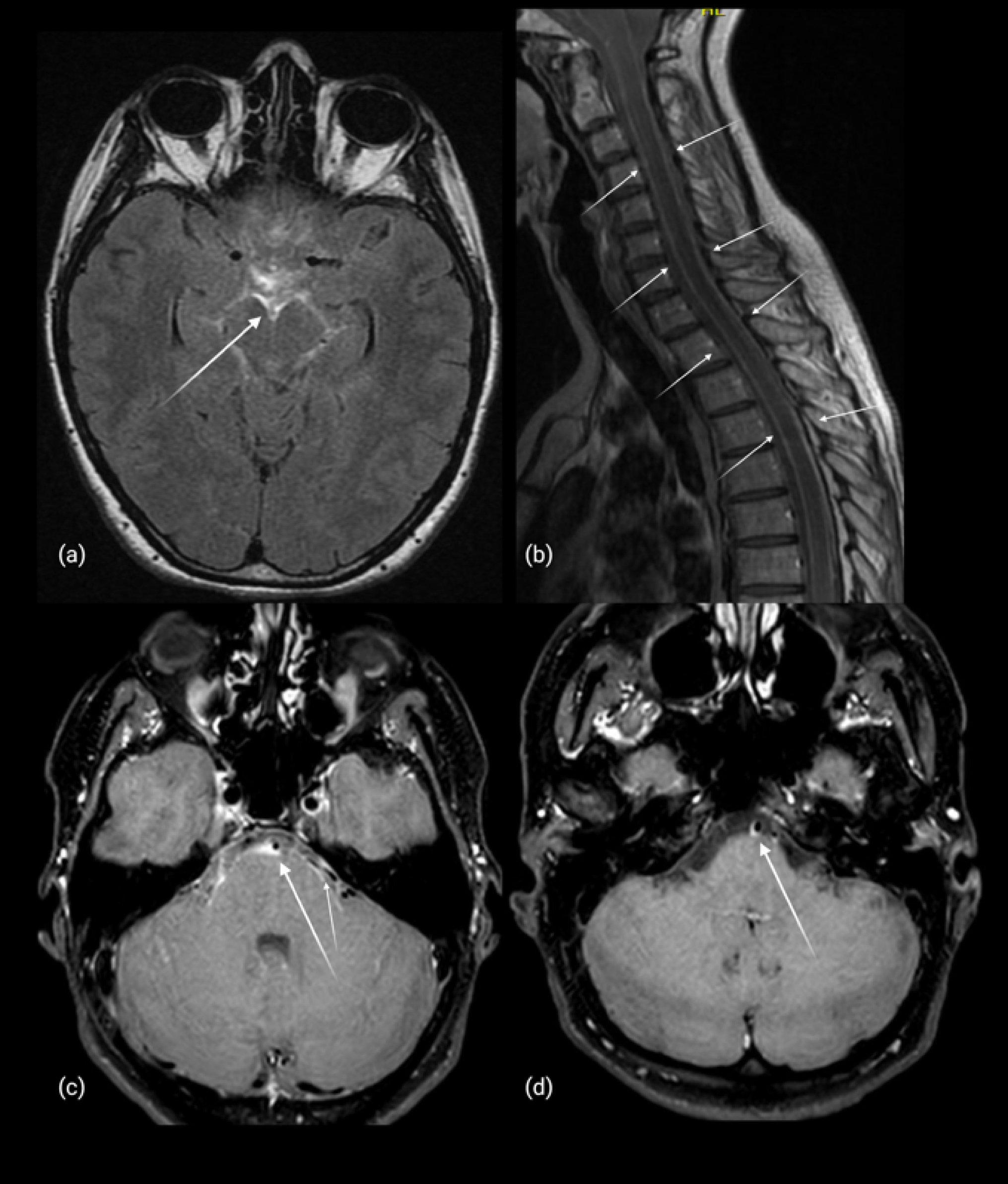
In this study, HRVWI-MRI demonstrated vascular abnormalities consistent with vasculitis in all three patients in whom it was performed. These findings align with results from Winter et al., where vascular concentric contrast enhancement was observed in all 40 patients examined (Winter et al. 2024).
Evidence on whether HRVWI-MRI can differentiate between perivascular inflammation in meningitis or inflammation of the vessel wall itself, is limited to small series and case reports. Importantly, our case 3 did show that vessel-wall enhancement with wall thickening on HR-VWI (Fig. 2c) corresponded at autopsy to a lymphocytic transmural vasculitis with significant structural vessel-wall changes (notably severe intimal hyperplasia). A case study by van Rooij et al. showed similar findings; however, their case 2 had positive HR-VWI but a negative biopsy despite compelling evidence for LV-PACNS (Van Rooij et al. 2018). Similarly, Zeiler et al. used HR-VWI to target brain/meningeal biopsies in suspected CNS vasculitis and found vascular inflammation in 8/9 biopsies; however, 3/8 showed only perivascular inflammation, suggesting that HR-VWI can overestimate true mural vasculitis (Zeiler et al. 2018). These findings are not surprising since large arteries with concentric enhancement were not biopsied; instead, more superficial linear areas of enhancement corresponding to leptomeningeal venules were sampled. Consistent with these sampling limitations, a meta-analysis by Beuker et al. found diagnostic biopsies in only ~ 8% of angiogram-confirmed LV-PACNS cases (Beuker et al. 2021).
Since HRVWI-MRI has only been available at our hospital since 2021, it was not performed on a larger number of patients. Nevertheless, given its non-invasive nature and seemingly high sensitivity, HRVWI-MRI may be considered as part of the diagnostic workup in patients with suspected NB vasculitis.
Despite multiple strokes, the 18F-FDG-PET of case 3 showed no sign of vasculitis. Although whole-body ¹⁸F-FDG-PET has high sensitivity in systemic vasculitis, the modality has not been systematically evaluated in NB-vasculitis or PACNS (Deb-Chatterji et al. 2025). In an unselected NB cohort (n = 23), most patients exhibited temporal hypometabolism and in some cases diffuse cortical hypometabolism, consistent with our case and with additional NB-vasculitis cases diagnosed at our institution after that study period (Newberg et al. 2002). In a 2025 series of 20 PACNS patients, all 18 examined were reported as PET-negative, although it was not specified how many underwent brain ¹⁸F-FDG-PET (Scoppettuolo et al. 2025). Conversely, and similar to our case 3, biopsy-proven granulomatous PACNS has been shown to exhibit hemispheric cortical hypometabolism on brain ¹⁸F-FDG-PET (Sagnelli et al. 2024). A likely explanation is that affected vessel calibers in CNS vasculitis are below the spatial resolution of ¹⁸F-FDG-PET and that detection is further challenged by high physiologic cortical background metabolism. Consequently, in CNS vasculitis, PET is mainly applied to exclude mimics (encephalitis, lymphoma, cancer, systemic vasculitis) rather than to confirm the vasculitis itself. Nevertheless, we believe that our PET findings provide mechanistic clues and may contribute to a better understanding of the complex pathophysiology in NB-vasculitis.
Other potential imaging findings of interest include cerebral white matter lesions (WML) and their possible link to NB-related cerebrovascular events. Although most studies have not found a link between WML and NB, a recent Norwegian longitudinal MRI study using quantitative white matter hyperintensities (WMHs) volumetry found a small reduction in WMH volume in treated NB patients over follow-up, whereas controls showed a slight increase (Andreassen et al. 2021; Aalto et al. 2007; Lindland et al. 2025). Yet, most published MRI series include all NB patients rather than only those with CNS involvement or late NB, which probably dilutes detection of NB-vasculitis–specific patterns. In several cerebral small vessel disease (CSVD) studies, diffusion tensor imaging (DTI) detected microstructural injury even in normal-appearing white matter on FLAIR and often preceded or exceeded visible WML progression (O’Sullivan et al. 2001; Maillard et al. 2011; De Groot et al. 2013; Tuladhar et al. 2015; Segura et al. 2010). On this basis, DTI may represent a more sensitive imaging method for detecting subtle NB-related microvascular or inflammatory injury, particularly in NB-associated vasculitis. To date, however, no systematic DTI studies have been performed in NB patients who have CNS manifestations and fulfill the EFNS criteria, representing a relevant future research avenue.
Treatment
The EFNS guidelines recommend either 14 days of IV ceftriaxone 2 g daily or oral doxycycline 200 mg daily therapy as treatment of NB vasculitis (Rauer et al. 2020; Mygland et al. 2010). This approach leads to complete recovery in 75% of cases, an incomplete but stable response with residual neurological sequelae in about 20%, and fatality in few cases despite appropriate therapy (4.7%) (Garkowski et al. 2017). Five out of thirteen of our patients were initially treated with high doses of ceftriaxone (4 g daily) while they awaited Intrathecal Bb tests and other causative agents were still being considered.
The progressive nature of NB vasculitis, even with treatment, was demonstrated by Winter et al., where modified Rankin scale (mRS) remained unchanged and NIH stroke scale scores (NIHSS) had worsened 1 year after admission (Winter et al. 2024). Both mRS and NIHSS were significantly worse at follow-up for NB vasculitis patients compared to matched stroke patients despite relevant antibiotic treatment (Winter et al. 2024). Some authors advocate adjunctive corticosteroids and prophylactic low-dose aspirin therapy although there is no high-level evidence supporting their benefit (Rauer et al. 2020). Importantly, in the study by Winter et al., no association was found between the type or duration of antibiotic or steroid therapy and clinical outcomes (Winter et al. 2024). We believe this progressive course (resembling PACNS) reflects prolonged inflammation and structural changes (stenosis) to vessel walls. In cases 1 and 4, follow-up HR-VWI demonstrated persistent, although gradually diminishing, vessel wall enhancement that took approximately 1 year to resolve, supporting a protracted inflammatory/structural course. Case 3 (Tables 1, 2, 3) ended in fatality despite relevant treatment with ceftriaxone, doxycycline, and MP/prednisolone. It could be argued that cyclophosphamide might have been considered as an adjunct therapy in this case. However, like corticosteroids, there is no randomized evidence supporting its use in this context. The available data is limited to rare case reports, which carry a significant risk of publication bias favoring successful outcomes (Bajons et al. 2023; Rauer et al. 2020; Komdeur 2001).
Neuropathology
NB vasculitis occurs due to inflammation in the meninges that extends into cerebral arteries, ultimately leading to vessel narrowing and thrombosis (Artal 2016). This mechanism closely resembles meningovascular neurosyphilis, an important differential diagnosis, where obliteration and thrombosis of the vessel lumen similarly results from an initial chronic meningitis (Miklossy et al.


Comments (0)