
Remember me

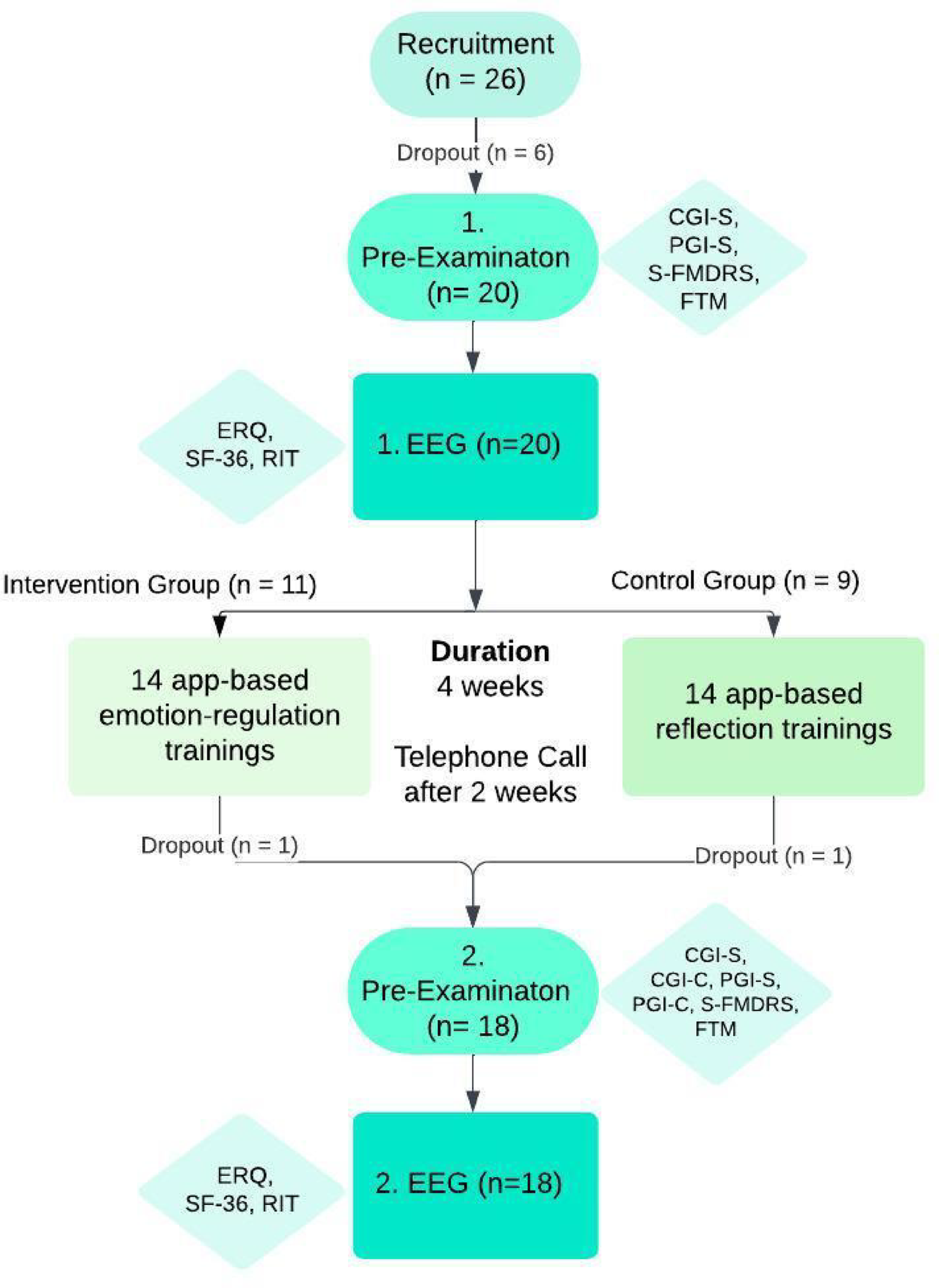
A total of 26 patients with clinically definite functional movement disorders were recruited between June 2023 and April 2024; 18 completed the study, while 8 dropped out for personal reasons. Inclusion criteria were age 18–80 and no contraindications for EEG (e.g., epilepsy, brain surgery) and a clinically definite FMD according to diagnostic criteria (Gupta and Lang, 2009) and best practice recommendations (Espay et al. 2018). Telephone follow-up took place 14 days after baseline, clinical follow-up after 4 weeks. As a pilot study, no formal sample size calculation was conducted; the sample size was based on feasibility and prior pilot work.
All patients underwent a detailed clinical examination at the Department of Neurology, Medical University of Graz, including the Fahn-Tolosa-Marin Clinical Rating Scale for Tremor (FTM), the Simplified Version of the Psychogenic Movement Disorders Rating Scale (S-FMDRS), and the clinical (CGI-S) and the patient (PGI-S) global impression of symptom severity/change scales (CGI-S, PGI-S, CGI-C, PGI-C) before and after the intervention (see Fig. 1). Clinician- and patient-rated versions were clearly distinguished throughout, with CGI scales completed by the investigator and PGI scales by the patients themselves.
Additionally, patients were asked during the telephone visit and the 2nd clinical visit if any adverse events had occurred. No harms were expected or systematically assessed due to the non-invasive nature of the intervention.
A placebo-controlled design was used to control for expectancy effects and nonspecific intervention factors. We also attempted double-blinding with our approach: Both groups received structurally similar app-based training tasks with identical duration and format, while participants as well as the physicians conducting the clinical ratings were unaware of group allocation. Participants were randomly assigned to the intervention or placebo group in a 1:1 ratio using simple randomization without blocking or stratification. Allocation was determined by drawing sealed, pre-labelled paper slips from a container, which were thoroughly mixed before each draw.
Although the participants were aware of the nature of their assigned training tasks (cognitive reappraisal vs. reflection), participants were not informed about the study hypotheses or which intervention was considered the “active” treatment, minimizing expectation bias. Full debriefing regarding group assignment occurred only after study completion. Thus, participants were blinded to the experimental condition throughout the intervention phase.
The lead experimenter (MS), who administered the EEG, RIT, SF-36, and ERQ assessments, was informed of group allocation for practical reasons. However, all clinical outcome ratings—including the FTM, S-FMDRS, PGI-C, PGI-S, CGI-C, and CGI-S—were performed by independent clinicians who were fully blinded to both group allocation and training content, thereby ensuring objectivity in the assessment of primary endpoints. Neither the rating clinicians nor the patients were aware of group assignments at any point during the study. The interventions were matched in format, duration, and delivery to ensure comparable user engagement across groups.
Finally, both training programs were matched for duration, mode of delivery, and overall user engagement, serving to equalize non-specific effects such as attention, time, and perceived support. Thus, while traditional double-blinding (in the pharmacological sense) was not feasible due to the nature of the interventions, the study design incorporated robust blinding procedures where possible and appropriate for a psychological intervention trial.
The following Flowchart in Fig. 1 gives an overview on the study design aswell as the questionnaires used (also see Supplemental material/methods). In the EEG laboratory, participants first completed questionnaires, followed by EEGmontage.
Fig. 1
Flowchart on the study design and utilized questionnaires. Clinical/Patient Global Impression – Severity/Change (CGI-S, CGI-C, PGI-S, PGI-C): A 5-point scale assessing the clinical impression of disease severity/change, completed by medical professionals/patients (Busner and Targum 2007; Guy 1976) – for the purpose of this study, three items have been created regarding mood, motor function and overall disease; Simplified Version of the Psychogenic Movement Disorders Rating Scale (S-FMDRS): A scale assessing various aspects of functional movement disorders, including movement quality, symptom distribution, severity, and daily life impairment (Nielsen et al. 2017); Fahn-Tolosa-Marin Tremor Rating Scale (FTM): A Scale assessing tremor severity and related motor impairment (Fahn et al. 1993); Reappraisal Inventiveness Test (RIT): A test evaluating the ability to reframe emotionally distressing situations (e.g., anger or fear) to reduce their emotional impact (Weber et al. 2014; Papousek et al. 2017; Perchtold et al. 2018, 2019); Emotion Regulation Questionnaire (ERQ): A questionnaire assessing emotion regulation strategies, including reappraisal and suppression (Abler and Kessler 2011); Level of Personality Functioning Scale - Brief Form (LPFS-BF): A short self-report questionnaire evaluating personality functioning across self- and interpersonal domains (Weekers et al. 2019); SF-36 (Short Form Health Survey − 36) A self-report questionnaire assessing health-related quality of life across 8 dimensions, including “General Health” and “Emotional Well-Being” (Brazier 1995).66
EEG procedure and cognitive reappraisal taskBrain activation at rest and during cognitive reappraisal generation was measured with EEG, and analyzed with regard to prefrontal EEG alpha asymmetry, indicating different activation patterns in the left and right prefrontal cortex, which emerged as a robust EEG correlate of cognitive reappraisal generation in previous studies (Papousek et al. 2017; Perchtold et al. 2018; Perchtold-Stefan et al. 2024).
A 2-minute resting EEG measurement was conducted while participants calmly focused on a green dot on the screen. They were then equipped with a chest-mounted microphone and introduced to the Reappraisal Inventiveness Test (RIT, Weber et al. 2014; Perchtold-Stefan et al., 2024) — a standardized task designed to assess individuals’ ability to flexibly generate cognitive reappraisals, i.e. alternative interpretations of emotionally distressing situations that help reduce their emotional impact. For the RIT, participants received written and verbal instructions, including a brief keyboard practice. In the RIT, they were presented with two anxiety-inducing and two anger-inducing scenarios (see SM for examples), each shown on the screen for 20 s and separated by a 5-second fixation cross. Participants were instructed to imagine themselves in each provided scenario and then, once a white question mark turned green upon pressing the space bar, to verbally generate as many different reappraisal strategies as possible — such as reframing the situation, altering its meaning, or focusing on positive aspects. They had up to three minutes per scenario and ended their response by pressing the space bar again. Participants answers were recorded via a microphone, transcribed, and subsequently analysed. After each scenario, participants rated how much anxiety or anger it had induced on a 6-point Likert scale. Two versions of the RIT (using different items) were administered during EEG recording, with version A given to half the subjects during the first appointment and version B during the second, and vice versa for the other half.
The administration of the RIT during EEG ensures that participants actively try to engage in cognitive reappraisal (and not any other emotion regulation strategy). As a quantitative metric of cognitive reappraisal success, we quantified RIT fluency, which is the total number of generated unique reappraisal ideas (see Papousek et al. 2017; Perchtold et al. 2018, 2020). This metric serves as an established indicator of successful cognitive reappraisal engagement, and measures individual differences in fluent and flexible cognitive reappraisal generation (see Papousek et al. 2017; Perchtold et al. 2018, 2019). RIT Fluency was determined by two independent raters (ICC two-way mixed, consistency, pre-test: 0.94; post-test: 0.87). However, as some cognitive reappraisal strategies may be more effective than others, RIT answers were also coded for the reappraisal strategies of problem-solving (ICC two-way mixed pre-test: 0.87; post-test: 0.80), positive re-interpretation (ICC two-way mixed pre-test: 0.79; post-test: 0.77), and relativizing (ICC two-way mixed pre-test: 0.71; post-test: 0.88) that reflect different qualities of reappraisal ideas (see Perchtold-Stefan et al.,2024).
Participant introduction to trainingFinally, the training introduction differed between the intervention and control group (see Supplementary Material for full protocol). The intervention group received a brief theoretical overview of cognitive reappraisal and was then guided through 14 daily app-based training sessions over a period of two weeks. Each session lasted approximately three minutes and involved emotionally evocative scenarios targeting fear or anger. Participants were instructed to generate as many alternative, less distressing thoughts as possible to reduce the emotional impact of the situation—mirroring the structure of the Reappraisal Inventiveness Test (RIT). For example, one scenario described walking home alone at night while being followed, prompting participants to reinterpret the situation in ways that would reduce anxiety (e.g., “Maybe the person lives nearby and is heading in the same direction”). Another anger-related scenario involved discovering that a friend had neglected to water one’s plants during a vacation. In contrast, the control group received an introduction to general reflection techniques and completed matched sessions in which they responded to neutral prompts without deliberate emotion regulation. These included tasks such as “What special thing happened today that you would like to record?” or “Think of a small, unexpected, positive event.” Both trainings were matched in frequency, duration, and format to control for expectancy effects and general engagement, allowing us to isolate the specific effects of cognitive reappraisal practice.
App data was analyzed to determine compliance with training. Over the 14-day intervention period, participants in the intervention group completed an average of 11.82 cognitive reappraisal responses (SD = 3.25, Min = 6, Max = 14), while those in the placebo group completed an average of 10.22 reflection responses (SD = 3.60, Min = 5, Max = 14).
EEG metricsEEG data were recorded with a Brainvision ActiCHamp Plus Research Amplifier (1000 Hz sampling rate, low cutoff DC, high cutoff 280 Hz) in a shielded room. Nineteen active electrodes (FP1, FP2, F3, F7, FZ, F4, F8, C3, CZ, C4, T7, T8, P3, PZ, P4, P7, P8, O1, O2) were positioned according to the international 10–20 system. The ground electrode was placed on the forehead and EEG was referenced to the nose and re-referenced offline using an average ear reference (see Perchtold-Stefan et al., 2024). Electrode impedances were kept below 30 kΩ for EEG electrodes and below 10 kΩ for ground and reference electrodes.
EEG pre-processing included removing low-frequency drifts (1 Hz filter), setting a notch filter (50 Hz), referencing to an averaged ear reference, and removing intervals that indicated ocular or muscle artifacts by visual inspection. Only intervals in which participants generated reappraisal ideas were analysed (i.e., idea generation), excluding reading, speaking, and rating intervals. Analyses focused on the EEG alpha band (8–12 Hz).
Laterality coefficients (LQ) were calculated as LQ = [(L + R)/(L - R)] × 100, where R is the right and L the left alpha power. Eight LQs were derived for frontopolar (Fp1/Fp2), dorsolateral frontal (F3/F4), ventrolateral frontal (F7/F8), central (C3/C4), temporal (T7/T8), parietal (P3/P4), ventrolateral parietal (P7/P8), and occipital (O1/O2) regions. Positive values indicate more left-hemisphere alpha power, reflecting higher right-hemisphere cortical activity due to the inverse relationship between alpha power and brain activity (see Papousek et al. 2017).
Comments (0)