Several retrospective studies have been reported on the prognostic impact of lymphadenectomy in advanced OCCC [12]. However, none have specifically addressed patients with complete macroscopic resection and no evidence of lymph node metastasis preoperatively or intraoperatively. Compared to HGSC, OCCC exhibits distinct biological characteristics, including a lower rate of lymphatic spread and greater resistance to chemotherapy [12]. These differences suggest that the rationale for systematic lymphadenectomy may not directly apply to OCCC. In the LION trial, which demonstrated no survival benefit from systematic lymphadenectomy in advanced ovarian cancer, only 2.2% of patients had clear cell histology [10]. Therefore, evidence specifically addressing the role of lymphadenectomy in OCCC may remain essential. Given the 7.8% lymph node involvement despite clinically negative nodes and the chemoresistance of OCCC, it remains unclear whether omitting lymphadenectomy compromises outcomes. Optimal surgical strategies for OCCC remain inadequately defined. This highlights the need to tailor treatment to the unique biology of OCCC. As described in the 2020 Japanese Society of Gynecologic Oncology guidelines for ovarian cancer, a prospective study would be required to establish the role of lymphadenectomy in advanced OCCC. [13]. However, no retrospective studies have focused specifically on patients with advanced OCCC and clinically negative lymph nodes to provide a foundation for such prospective research. To our knowledge, this is the first study to evaluate the prognostic impact of systematic lymphadenectomy exclusively in patients with advanced OCCC who underwent complete macroscopic resection and had no intraoperative evidence of lymph node metastasis—a cohort analogous to that studied in the LION trial.
Although the results of an earlier study showed that complete surgical staging involving systematic lymphadenectomy appeared to improve survival in patients with stage I OCCC [14], additional recent research has shown no such benefit [15]. Furthermore, in advanced OCCC, especially in cases with a macroscopically complete resection and with clinically negative lymph nodes, the prognostic impact of lymphadenectomy is unclear. Liu et al. reported, in a retrospective study using SEER and Chinese registry data, that the performance of lymphadenectomy in patients with OCCC did not demonstrate a significant impact on survival for either early or advanced stage patients [16]. The results of these studies are not limited to patients with macroscopically complete resection, as they also include patients with macroscopically residual tumors and those with clinically positive nodes. Gao et al. also reported that lymphadenectomy does not significantly impact survival in OCCC [17]. Furthermore, a sub-analysis of this study showed similar results in patients with clinically negative lymph nodes, but the analysis was performed for both early and advanced stage cases. In the surgical approach of OCCC, the significance to analyze lymphadenectomy in the early stage and advanced stage is different.
For OCCC patients with early stage, the frequency of lymph node metastasis is much lower than with other tumor subtypes, according to previous studies. Several previous studies have reported low rates of lymph node metastasis in patients with OCCC and at an early stage, from 0 to 11% [17,18,19,20,21]. Mahdi et al., in 2013 identified nearly 1900 OCCC patients restricted to the ovary through the SEER program. Among the 1359 cases that underwent lymphadenectomy, only 61 (4.5%) had positive lymph nodes [21]. Heitz et al. also showed that 3.6% of OCCC patients with clinically negative lymph nodes had lymph node metastasis, while the rate was 71.6% in patients with high-grade serous ovarian cancer [18]. On the other hand, Gao et al. reported high rates of lymph node metastasis of 58.8% in pT3 cases, but they included cases with clinically positive lymph nodes in these pT3 cases [17]. There are no reports on the rate of lymph node metastasis in advanced OCCC with clinically negative lymph nodes. In our study, the rate of lymph node metastasis in advanced OCCC with clinically negative lymph nodes was 7.8%, a very low rate compared to the 57% in the LION study, in which 85% of cases were HGSC [10]. Matsuo et al. reported that adequate dissection is achieved with at least 8–12 dissected lymph nodes in ovarian cancer [22]. The median number of lymph nodes removed in this study was 48.5 (range, 5–89), which we believe ensures the quality of skill of lymphadenectomy; the low rate of lymph node metastasis even in advanced OCCC with clinically negative lymph nodes may be the reason why lymphadenectomy does not have an impact on prognosis.
Interestingly, our study showed no difference in the frequency of single or multiple-site recurrences between the lymphadenectomy and no-lymphadenectomy groups. Peritoneal dissemination was the most common pattern of recurrence in both groups (38.7% vs. 40%), consistent with previous reports [23]. Notably, lymph node recurrence occurred only in the lymphadenectomy group (48%), and all were within the dissected regions. While the median number of dissected lymph nodes was high (48.5), suggesting that the extent and quality of lymphadenectomy were likely maintained, we acknowledge that there was no direct method to validate the adequacy or completeness of the procedure across institutions. This represents an important limitation of our study. Nevertheless, the occurrence of nodal recurrences within the dissected areas may imply that, in OCCC, even technically appropriate lymphadenectomy might be insufficient to fully eliminate microscopic lymphatic disease. This observation highlights the need for further investigation into the biological behavior of OCCC and the limitations of current surgical strategies.
Although this analysis was based on data from a randomized phase III trial, several limitations must be acknowledged. Additional limitations of this study include the lack of criteria for preoperative and intraoperative lymph node metastasis assessment criteria, the differences in the choice of adjuvant chemotherapy regimens at the time of this phase III trial, and the potential differences in the quality of care between the two treatment groups due to lack of randomization, e.g., quality of surgical skill, or surgical material and peri-operative care, and potential differences in patient’s background between two treatment groups, e.g., undocumented factors that can lead physicians to avoid lymphadenectomy. These limitations warrant caution in interpreting the results. To our knowledge, this study is one of the first to explore the prognostic impact of systematic lymphadenectomy in advanced OCCC patients with complete macroscopic resection and no clinical evidence of lymph node metastasis. To better understand surgical decision-making and clarify the role of lymphadenectomy in this population, further retrospective analyses with detailed clinical background data are warranted, which may ultimately inform future prospective trials. Moreover, the observed age imbalance between groups (median age 55 vs. 66) suggests possible selection bias, likely reflecting clinical decisions on surgical eligibility. Due to the small sample size and lack of detailed background data (e.g., performance status, comorbidities), we could not perform multivariable or propensity score-adjusted analyses. These limitations should be carefully considered. A separate retrospective study is underway to clarify surgical decision-making factors. Additionally, the extent of lymphadenectomy (e.g., pelvic and/or para-aortic) was not standardized within the JGOG3017 protocol and may have varied between institutions. This variability could influence both staging accuracy and survival outcomes, and represents another limitation of this retrospective analysis. Although the JGOG3017 trial was primarily conducted in Japan and the majority of patients were Japanese (93.5%), a small proportion (6.5%) were non-Japanese. Therefore, while our analysis population predominantly reflects Japanese clinical practice, the findings may have broader relevance, though generalizability should still be approached with caution. However, given that the biological characteristics of OCCC—such as low lymphatic dissemination and chemoresistance—are shared across ethnic groups, the surgical implications may be applicable to OCCC cases regardless of regional prevalence.
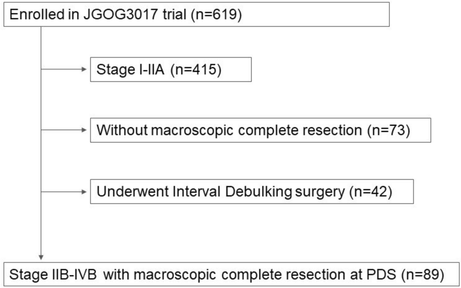
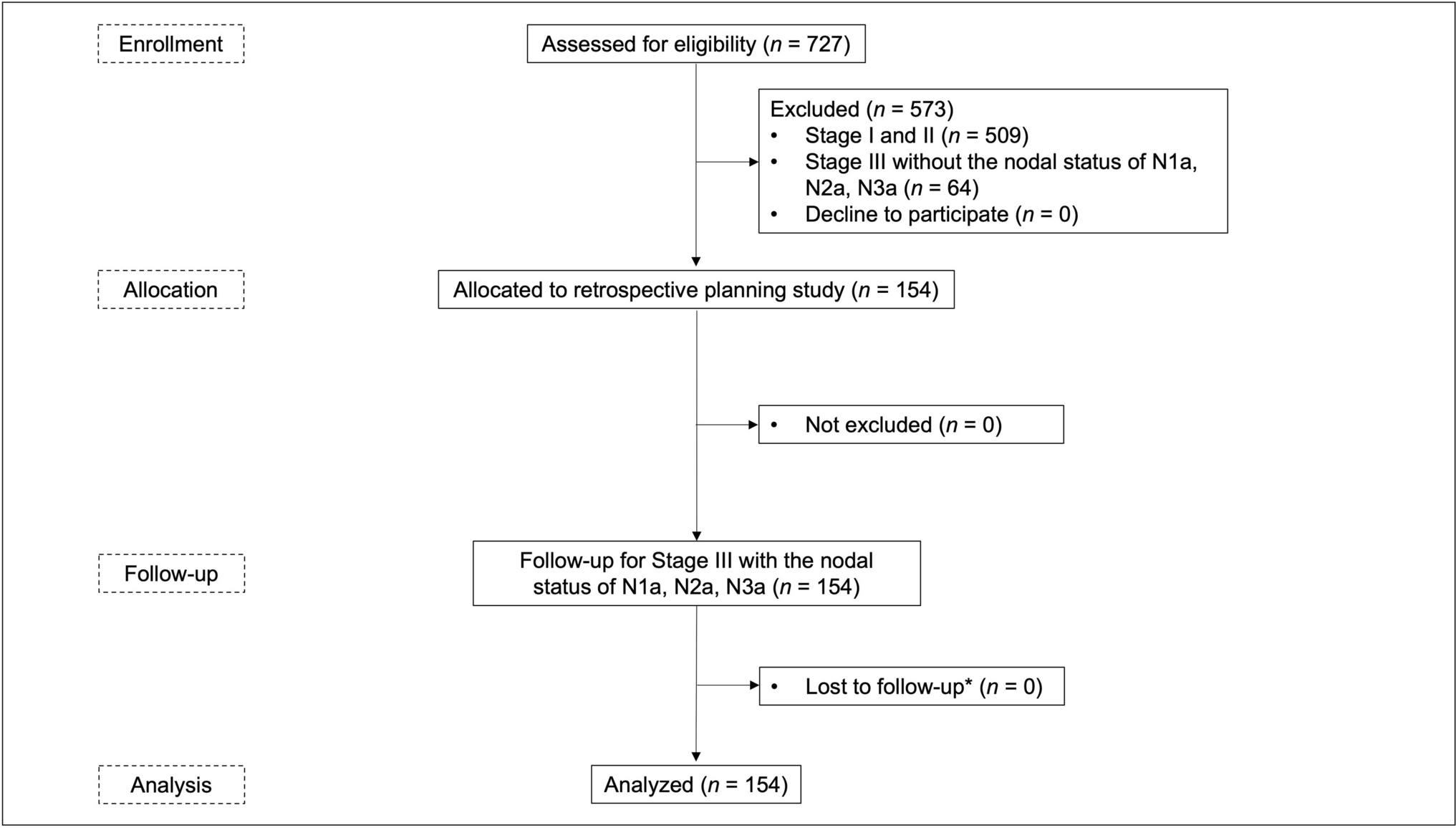
Importantly, our study focused on a subset of patients analogous to those included in the LION trial—those with FIGO stage IIB–IVB disease, no gross lymphadenopathy, and complete macroscopic resection. This was a deliberate design choice, as the applicability of the LION trial findings to OCCC remains uncertain given the limited representation of this histologic subtype. Therefore, our results should be interpreted in the context of this specific population, rather than generalized to all advanced OCCC patients. In addition, the limited sample size in this analysis may in part be attributed to the inherent clinical characteristics of OCCC. It is well established that OCCC is typically diagnosed at an early stage, and the proportion of patients with advanced-stage disease (FIGO stage III or higher) is relatively low, as previously reported [12]. This trend was also observed in the JGOG3017 phase III trial [11], despite its large-scale, multicenter design. Furthermore, when restricting the analysis to patients analogous to the LION trial cohort—those with advanced-stage disease, no gross lymphadenopathy, and complete macroscopic resection—the number of eligible patients became even more limited. Although our analysis used data from a randomized phase III trial, it should be emphasized that this ancillary study was retrospective in nature and not powered to detect small-to-moderate differences in survival outcomes. In particular, the number of patients in the non-lymphadenectomy group was only 12, which severely limits the statistical power and raises the possibility of a type II error. Therefore, the lack of statistically significant differences in survival should not be interpreted as definitive evidence of no benefit.
In conclusion, an ancillary analysis using data from JGOG3017 showed no statistically significant survival benefit from regional lymphadenectomy after complete resection in patients with advanced OCCC without clinically evident lymph node metastasis. However, the number of eligible cases was very small because advanced OCCC is rare. The limited cases in the non-dissection group and the retrospective design reduced statistical power, so the non-significant results may reflect a Type II error. These findings should not be interpreted as evidence against regional lymphadenectomy. Clarifying the role of lymphadenectomy in surgical management for advanced OCCC remains a critical research issue. This exploratory study represents a first step. The efficacy of node sampling for OCCC should be evaluated by a Japanese clinical trial group. Further retrospective data collection is ongoing, and a future prospective study in Japan is strongly encouraged.

Comments (0)